
Chronic Frontal sinusitis in detail for long question answer from standard ENT textbook in simplified manner
I'll read the medical library skill first, then search for comprehensive information on chronic frontal sinusitis.
Reading File
Now let me search the medical library for chronic frontal sinusitis content in parallel:
Finding Sources
Finding Sources
Excellent! Found the key sections. Now let me read all the relevant content in parallel - Scott-Brown's chronic frontal sinusitis section, Cummings indications/procedures sections, and frontal sinus surgery:
Reading File
Reading File
Reading File
Reading File
Reading File
Reading File
Reading File
Reading File
Reading File
Finding Sources
Reading File
Searching Images
frontal sinusitis CT scan endoscopy surgery
Reading File
Finding Sources
Reading File
Finding Sources
Reading File
Finding Sources
I now have comprehensive content from both Scott-Brown's and Cummings. Let me fetch a few key images from the textbook and compile the complete answer:
Loading Image
Loading Image
Loading Image
I now have comprehensive information from both Scott-Brown's and Cummings textbooks. Let me compose the complete, detailed answer.
Chronic Frontal Sinusitis - A Comprehensive Long-Answer Guide
Based on Scott-Brown's Otorhinolaryngology Head & Neck Surgery and Cummings Otolaryngology Head and Neck Surgery
1. INTRODUCTION AND DEFINITION
Chronic frontal sinusitis (CFS) is defined as persistent inflammation of the frontal sinus lasting more than 12 weeks. It usually arises as a component of chronic rhinosinusitis (CRS), which itself is described as multifactorial in aetiology and pathogenesis. CRS may be:
- CRSwNP - with nasal polyps
- CRSsNP - without nasal polyps
Both forms can obstruct the frontal sinus outflow tract and lead to chronic frontal sinusitis. Despite significant advances in medical and surgical therapy, chronic frontal sinusitis remains one of the most difficult diseases to manage in ENT practice.
2. APPLIED ANATOMY OF THE FRONTAL RECESS
The frontal recess is the key to understanding frontal sinus disease - it is a complex, narrow space and the problem most often lies here, not within the sinus itself.
Boundaries of the Frontal Recess:
| Wall | Structure |
|---|---|
| Anterior | Agger nasi cell / nasofrontal beak |
| Medial | Middle turbinate + lateral lamella |
| Lateral | Lamina papyracea / agger nasi air cells |
| Posterior | Ethmoid roof (anterior ethmoid artery runs across) |
Key Points:
- The agger nasi cell is the anteriormost ethmoid cell and largely determines the anteroposterior distance from skull base to frontal beak
- The superior attachment of the uncinate process determines frontal drainage direction
- The anterior ethmoid artery lies on a mesentery in some patients ("supraorbital ethmoid cell" variant) - a surgical hazard
- An intersinus septal cell may displace the frontal recess laterally
- Proper CT review (identifying agger nasi cell, lamina papyracea, lateral lamella height, anterior ethmoid artery) forms the surgical "roadmap"
3. AETIOLOGY AND PATHOGENESIS
The aetiology of CFS is multifactorial. The key factors include:
A. Anatomical Variations Obstructing the Frontal Recess
- Suprabulla cells, supraorbital ethmoid cells, frontal bulla cells, recessus terminalis
- Type 3 and 4 frontal ethmoidal cells are significantly associated with mucosal thickening
- Septal deviation, turbinate hypertrophy, middle turbinate pneumatization (concha bullosa)
- Agger nasi hyperplasia narrowing the frontal recess
However, the frequency of anatomical variations is no more prevalent in sinusitis patients than in controls - anatomy alone rarely causes CFS.
B. Mucosal and Inflammatory Factors
- Ongoing mucosal inflammation is considered the most important factor - more so than anatomy alone
- Mucosal oedema narrows the already-tight frontal recess
- Eosinophilic CRS (with polyps, asthma, aspirin intolerance - the "Samter's triad") carries a worse prognosis
- Biofilm formation by bacteria reduces antibiotic efficacy
C. Microbiology
The bacteriology of CRS differs importantly from acute rhinosinusitis:
- Higher prevalence of anaerobic organisms
- Common organisms: Streptococcus pneumoniae, Haemophilus influenzae, Moraxella catarrhalis
- Post-surgical cases: MRSA, Pseudomonas aeruginosa, Enterobacter species
D. Post-Surgical Factors
- Scarring and stenosis secondary to previous surgery
- Retained uncinate process or residual cells in frontal recess
- Lateralization of the middle turbinate (seen in 36-78% of revision cases)
- Osteoneogenesis - new bone formation secondary to mucosal stripping over bare bone, persistent infection, and surgical trauma
E. Systemic Factors
- Diabetes mellitus
- Immune deficiency (patients with 3+ acute episodes/year should have immunology checked)
- Aspirin-exacerbated respiratory disease (AERD)
- Allergy / allergic rhinitis
4. CLINICAL FEATURES
Symptoms
- Frontal headache / pressure - the cardinal symptom; felt over the forehead, often worse on bending forward
- Nasal obstruction / congestion
- Mucopurulent nasal discharge (anterior rhinorrhoea or post-nasal drip)
- Reduced or absent sense of smell (anosmia/hyposmia)
- Facial pain / pressure over the frontal region
Important caveat: "Sinus headaches" are frequently mislabelled - true frontal sinus headaches are less common than tension headaches presenting in the same location. A thorough history is essential to differentiate.
Signs
- Tenderness on palpation over the medial aspect of the supraorbital margin (floor of frontal sinus)
- Nasal endoscopy: mucopurulent discharge from the middle meatus, nasal polyps, mucosal oedema
- Pale/oedematous turbinates if allergic component
5. INVESTIGATIONS
A. Nasal Endoscopy
- Allows direct visualization of the middle meatus and frontal recess area
- Identifies polyps, mucosal disease, anatomical variants (e.g., concha bullosa)
- Guides microbiological sampling
B. CT Scan (Gold Standard Imaging)
- Coronal CT is the investigation of choice
- Shows mucosal thickening, opacification, osteomeatal complex obstruction, frontal recess anatomy
- Essential pre-operative "roadmap" - identify: agger nasi, uncinate process, lamina papyracea, lateral lamella height, anterior ethmoid artery position
- Evaluate for complications: bone erosion, orbital extension, intracranial involvement
C. Plain X-Ray (Occipitomental / Caldwell view)
- Now largely superseded by CT
- Still used as a template for the frontal sinus outline in osteoplastic flap surgery (occipitofrontal view at 100 cm distance is life-size)
D. MRI
- Superior for soft tissue characterisation
- Identifies mucocele (hyperintense T2 signal), differentiates from tumour, demonstrates intracranial/intraorbital extension
- Useful when malignancy or fungal disease suspected
E. Ultrasonography
- Simple, non-invasive bedside tool
- Can assess fluid within sinus but limited spatial resolution; rarely used now
F. Microbiological Cultures
- Endoscopically-guided swabs from middle meatus
- Guide targeted antibiotic therapy
6. MEDICAL MANAGEMENT
The mainstay of treatment for CFS is medical therapy, and the majority of patients respond to a combination of:
A. Antibiotics
- Empirical broad-spectrum antibiotics based on local resistance patterns
- Long-term macrolide therapy (erythromycin, clarithromycin) at low dose for 12 weeks to 6 months - works not just via antibacterial action but by downregulating mucosal inflammation. Studies show benefit in non-eosinophilic CRS subtypes particularly; less clear benefit in eosinophilic CRS.
- Macrolides also help disperse bacterial biofilms
B. Topical Intranasal Corticosteroids
- First-line anti-inflammatory treatment
- Reduce mucosal oedema and polyp size
- Used long-term
C. Systemic (Oral) Corticosteroids
- Short courses for acute exacerbations or significant polyposis
- Valuable "bridge" therapy pre-operatively
D. Saline Nasal Irrigations
- Isotonic and hypertonic saline nasal douches
- Mechanically clear secretions, improve mucociliary transport, reduce oedema
- Important adjunct at all stages
E. Treating Underlying Conditions
- Allergen avoidance / antihistamines for allergic rhinitis
- Aspirin desensitisation for AERD
- Optimizing immunity in immunodeficient patients
7. COMPLICATIONS
Table: Complications of Frontal Sinusitis
| Local (Frontal Sinus) | Orbital | Intracranial |
|---|---|---|
| Mucocele | Orbital cellulitis | Meningitis |
| Pyocele | Subperiosteal abscess | Epidural abscess |
| Osteomyelitis (Pott's Puffy Tumour) | Intra-orbital abscess | Subdural empyema |
| Intracerebral abscess | ||
| Superior sagittal sinus thrombosis | ||
| Cavernous sinus thrombosis |
Pott's Puffy Tumour
- Anterior spread of frontal sinusitis through the outer (anterior) table of the skull
- Causes a boggy subperiosteal abscess with osteomyelitis of the frontal bone
- A neurosurgical/ENT emergency
Mucocele / Pyocele
- Mucocele: epithelial-lined sac containing inspissated mucus, completely filling and expanding the sinus
- Caused by obstruction of the sinus ostium (inflammation, fibrosis, polyp, osteoma, previous surgery)
- Local bone resorption via prostaglandins, IL-1, TNF
- Can expand into orbit (proptosis/diplopia) or intracranially
- Treatment: endoscopic marsupialization (preferred) or open surgery
8. SURGICAL MANAGEMENT - INDICATIONS
Primary Indications for Surgery (before considering ESS)
- Fungal disease in the frontal sinus
- Barotrauma
- Mucocele or pyocele
- Osteoma causing obstruction with mucosal disease
- Disfigurement or exophthalmos
- Osteomyelitis, osteogenesis, tumour
Indication after Failed Medical Treatment
- Persistent symptoms despite maximal medical therapy (antibiotics + steroids + irrigation)
Key Principle (Modern Concept):
Treatment is directed towards the drainage pathway (frontal recess) rather than the sinus itself. Opacification of the frontal recess alone is not an indication for instrumentation - it often clears if the ostiomeatal complex is opened and disease treated medically.
Importantly: many patients appear to have had their disease initiated by unnecessary instrumentation of the frontal recess.
9. SURGICAL PROCEDURES - GRADUATED APPROACH
The best surgical approach to the frontal sinus is a graduated, stepwise one:
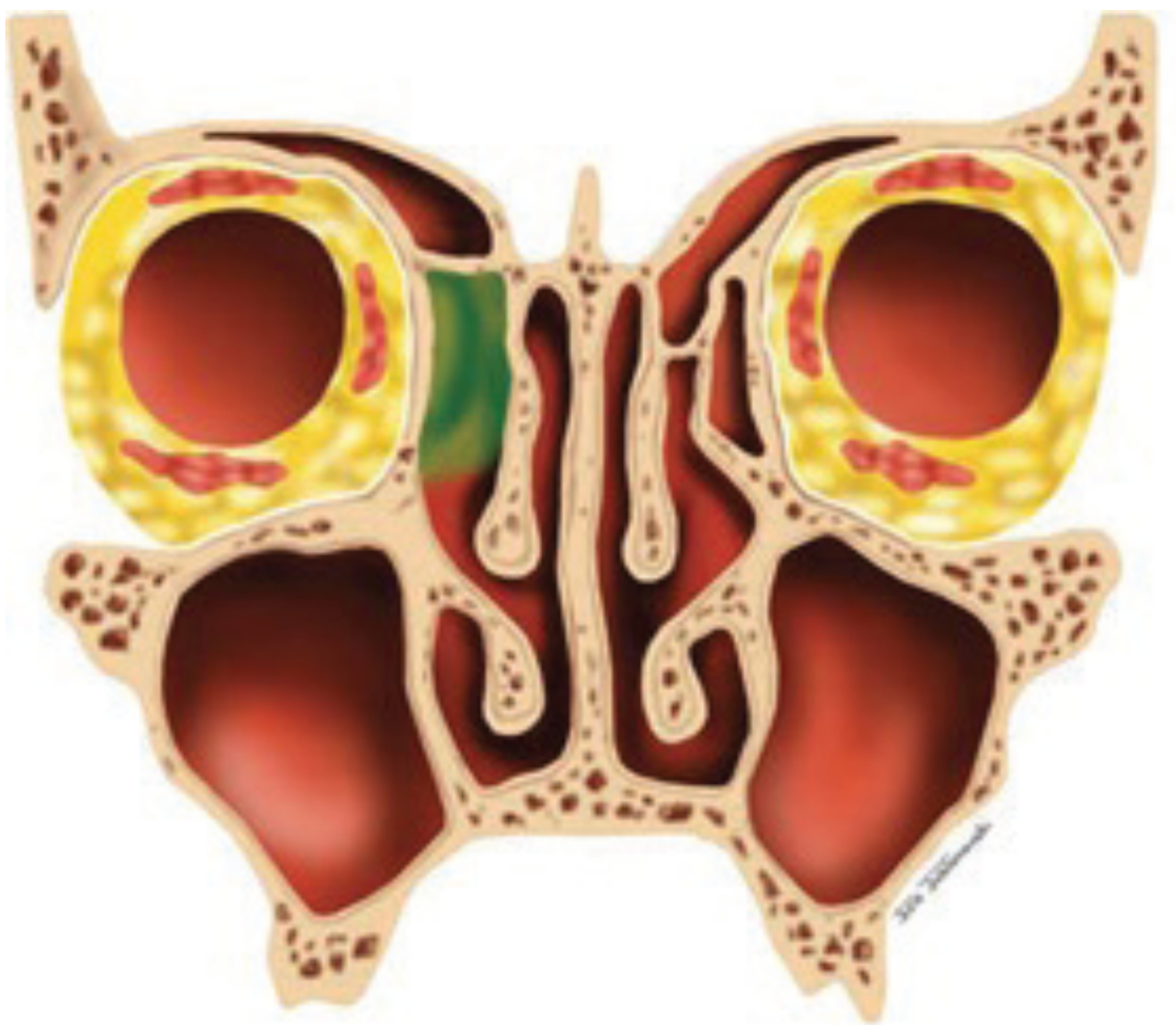
Draf type I frontal sinusotomy - removal of agger nasi and anterior ethmoid cells in the frontal recess (Scott-Brown's)
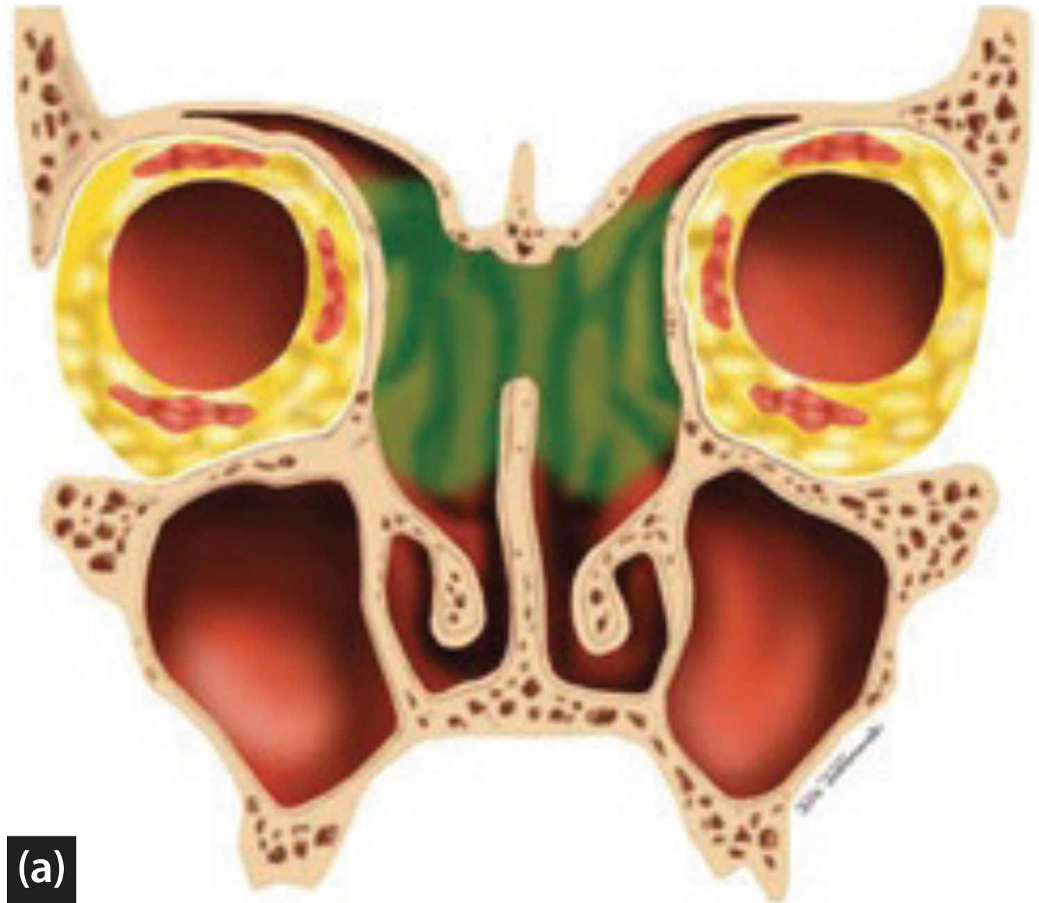
Draf type III (Modified Endoscopic Lothrop Procedure) - creates a single large common drainage pathway for both sinuses (Scott-Brown's)
Graduated Table (Scott-Brown's Table 99.3)
| Procedure | Indication / Description |
|---|---|
| No exploration | No disease present |
| Balloon sinuplasty | Limited disease; may have a role in certain situations |
| Draf Type I (Frontal sinusotomy + ethmoidectomy) | Remove cells in the frontal recess after FESS |
| Draf Type IIa | Widen frontal ostium from lamina papyracea to middle turbinate |
| Draf Type IIb | Extend opening to nasal septum; remove anterior middle turbinate, drill floor to midline |
| Draf Type III / MELP / Frontal Drillout | Resect floor of frontal sinus, superior nasal septum, interfrontal sinus septum |
| Osteoplastic flap + MELP | Above-and-below combined approach |
| Osteoplastic flap with obliteration | Remove all mucosa, obliterate with fat - gold standard for recalcitrant disease |
| Riedel's procedure | Removal of anterior table of frontal sinus |
| Cranialization | Remove sinus mucosa and posterior table |
A. Balloon Catheter Dilation (Balloon Sinuplasty)
- Concept: dilate the sinus ostium to restore aeration and mucociliary clearance, while preserving mucosa (reducing stenosis risk)
- May have a role in selected cases with limited disease
- Not appropriate for: nasal polyposis, osteoneogenesis, osteomyelitis, mucocele, or neoplasia
- Long-term data (>12 months) still limited; placebo effect not excluded
- Serious complications have been described
B. Draf Type I (Frontal Sinusotomy)
- Least invasive procedure
- Complete ethmoidectomy + removal of agger nasi, uncinate process, and anterosuperior ethmoid cells
- Clears obstruction inferior to the frontal ostium (within the frontal recess)
- Best when only minor frontal sinus pathology is present
- Insufficient for severe polyposis with asthma and aspirin intolerance
C. Draf Type II
- Type IIa: Widen the frontal ostium from lamina papyracea to the middle turbinate (using Kerrison or Hajek-Kofler punch)
- Type IIb: Extend to nasal septum; requires removing the anterior end of middle turbinate and drilling floor to midline
- Indications for IIb: unilateral frontal ostium osteoneogenesis, inverted papilloma, osteoma, medially placed mucocele
- Restenosis rate: IIb ~23% vs IIa ~3.6% (more drilling = more restenosis risk)
D. Draf Type III / Modified Endoscopic Lothrop Procedure (MELP)
- Also called: Frontal sinus drillout or Median drainage procedure
- Originally described by Lothrop in 1914 (external); Draf described the intranasal approach
- Creates a large common drainage pathway for both frontal sinuses by resecting:
- Superior nasal septum (septal window ≥2 x 2 cm)
- Frontal intersinus septum
- Floor of both frontal sinuses
Indications for MELP:
| Indication | Notes |
|---|---|
| Chronic frontal sinusitis refractory to previous ESS | Primary indication |
| CRS with NP + asthma + AERD | Controversial; may be primary procedure |
| Frontal mucocele | |
| Inverted papilloma | |
| Osteoneogenesis of frontal recess | |
| CSF leak repair | Sometimes needed for access |
| Failed previous frontal sinus surgery |
Surgical Steps (MELP):
- Complete sphenoethmoidectomy
- Clear frontal recess, identify frontal ostium (frontal mini-trephines may help)
- Create septal window (≥2 x 2 cm)
- Trim anterior end of middle turbinate to skull base
- Drill commences from one frontal recess, proceeds across midline to contralateral side
- Identify first olfactory neurons (posterior limit of dissection) - creates the "frontal T"
- Drill from contralateral side via septal window
- Reduce frontal intersinus septum and frontal beak to join both sides
- Preserve mucosa over posterior aspect of frontal ostium
- Lower bone over anterior skull base projection to primary olfactory neuron
The "Frontal T": The posterior margin of septectomy (vertical limb) and posterior margin of frontal sinus floor (horizontal limbs joining amputated middle turbinate ends) - critical safety landmark.
Complications of MELP: Injury to skin/nasal bones, orbital injury, skull base injury, CSF leak
Outcomes:
- Patency rate: ~95.9% (systematic review/meta-analysis)
- Failure rate: ~13.9% (80% of failures undergo revision MELP; remainder have obliteration)
E. Osteoplastic Flap Procedure (OPF)
Indication (Scott-Brown's Table 99.8):
- Wide exposure necessary, endoscopically inaccessible anatomy (large osteomas, lateral mucoceles, malignant tumours)
- Failed endoscopic procedures including MELPs
- Extensive frontal bone osteomyelitis
- Extensive neo-osteogenesis of frontal recess
- Diseased small underdeveloped sinuses (AP distance <8mm making MELP difficult)
- Frontal sinus fractures involving outflow tract
Procedure:
- Bicoronal (or brow / mid-forehead) incision
- Scalp flap reflected forward
- Template from plain X-ray (occipitofrontal view) used to outline the sinus
- Anterior table osteotomised and reflected inferiorly (hinge on periosteum)
- Full access to entire frontal sinus cavity
With obliteration (fat obliteration):
- All frontal sinus mucosa meticulously removed (residual mucosa leads to mucocele formation)
- Sinus obliterated with abdominal fat
- Gold standard for recalcitrant disease: success rates >90% at 8 years
- Long-term failure rate up to 25%
Complications of OPF with obliteration:
- CSF leak
- Persistent forehead numbness
- Severe headaches without evidence of recurrent disease
- Frontal bossing
- Supraorbital neuralgia
- Mucocele formation (from residual mucosa)
- Donor site complications (abdominal fat harvest)
- Difficulties with post-operative surveillance (CT follow-up needed)
In the modern endoscopic era, less than 2% of frontal sinus procedures in major centres are managed by OPF with obliteration. The ability to restore ventilation endoscopically from below while accessing from above via OPF has largely replaced pure obliteration.
10. REASONS FOR FAILURE OF FRONTAL SINUS SURGERY
Failure of frontal recess surgery may occur due to:
- Ongoing mucosal disease - persistent/recurrent nasal polyps, eosinophilic CRS, asthma
- Incomplete dissection - retained uncinate process and residual cells impeding drainage
- Lateralization of the middle turbinate - present in 36-78% of revision cases; the "frontal sinus rescue procedure" addresses this
- Scarring and synechia - common; not always symptomatic but can obstruct recess
- Osteoneogenesis - from mucosal stripping over bare bone; maintains inflammation → further stenosis
- Complex frontal recess anatomy - the most common reason for surgical failure overall
11. POST-OPERATIVE CARE
- Nasal saline irrigations from early post-operative period - important to clear debris
- Topical intranasal steroids - continued long-term
- Regular endoscopic debridement during early post-operative period
- Medical management of underlying CRS continues
- Long-term follow-up essential (frontal sinus patency assessed endoscopically)
12. OUTCOMES
| Procedure | Outcome Data |
|---|---|
| ESS (overall CRS) | Safe and effective when medical therapy fails |
| Frontal sinus surgery (endoscopic) | Level 3 evidence only; early patency ~90% at 12 months, falling to ~68% at 72 months |
| Non-eosinophilic CRS | 90% patency; eosinophilic CRS 85% (average follow-up 46 months) |
| MELP (systematic review) | 95.9% patency overall |
| OPF with obliteration | Success rate up to 93% at 8 years; long-term failure up to 25% |
An important limitation: some patients will continue to report symptoms despite objective evidence of a patent frontal recess - this mismatch between patient-reported outcomes and clinical/radiological findings is a major challenge in patient selection and outcome reporting.
SUMMARY: KEY POINTS FOR EXAM
- Chronic frontal sinusitis = >12 weeks persistent frontal sinus inflammation, usually part of CRS
- Frontal recess is the key - the problem is the drainage pathway, not usually the sinus itself
- Most important factor in aetiology: ongoing mucosal inflammation > anatomy alone
- Bacteriology: anaerobes more prevalent than in acute sinusitis; post-surgical adds MRSA/Pseudomonas
- Diagnosis: symptoms + endoscopy + coronal CT
- Treatment is stepwise: maximal medical therapy first, then graduated surgical escalation
- Medical therapy: antibiotics (macrolides for anti-inflammatory effect), intranasal steroids, saline irrigations
- Graduated surgical approach: Balloon → Draf I → Draf IIa → Draf IIb → MELP/Draf III → OPF ± obliteration
- MELP: patency ~96%, is minimally invasive alternative to obliteration - now preferred over OPF
- OPF with obliteration: gold standard for recalcitrant disease but falling out of favour in endoscopic era
- Mucosal preservation is paramount - bare bone → no cilia → osteoneogenesis → restenosis
- Complications: mucocele, pyocele, Pott's puffy tumour, meningitis, subdural empyema, cavernous sinus thrombosis
Sources: Scott-Brown's Otorhinolaryngology Head & Neck Surgery (Vol. 1, Chapter 99) and Cummings Otolaryngology Head and Neck Surgery (Chapter 46)
Csf rhinnorhea in similar manner
Finding Sources
Finding Sources
Excellent! Found comprehensive sections in both Scott-Brown's Vol 2 and Cummings. Reading them in parallel now:
Reading File
Reading File
The Scott-Brown's Vol 2 section is about post-surgical (acoustic neuroma) CSF rhinorrhoea - the main ENT topic on CSF rhinorrhoea is clearly in Cummings and Scott-Brown's Vol 1. Let me read the full Cummings section and also find the Scott-Brown's skull base chapter:
Reading File
Finding Sources
Reading File
Finding Sources
Reading File
Reading File
Reading File
Reading File
Reading File
Reading File
Reading File
Finding Sources
Reading File
Loading Image
I now have comprehensive, detailed content from Cummings Otolaryngology (Chapter 48) covering every aspect of CSF rhinorrhoea. Let me compile the complete long-answer response now.
CSF Rhinorrhoea - Comprehensive Long-Answer Guide
Based on Cummings Otolaryngology Head and Neck Surgery (Chapter 48) and Scott-Brown's Otorhinolaryngology Head & Neck Surgery
1. INTRODUCTION AND DEFINITION
CSF rhinorrhoea is defined as the leakage of cerebrospinal fluid (CSF) from the subarachnoid space into the sinonasal cavity, resulting in watery nasal discharge.
This occurs when there is a simultaneous defect in:
- The arachnoid and dura mater
- The skull base bone
- The paranasal sinus/nasal mucosa
...along with a pressure gradient sufficient to drive CSF flow.
CSF rhinorrhoea is clinically significant because it:
- May cause ascending meningitis (major cause of mortality/morbidity)
- May allow development of pneumocephalus (air entering through the defect → brain compression)
- May be the presenting sign of serious pathology (tumour, hydrocephalus)
Over the past three decades, management has evolved dramatically - endoscopic endonasal repair has largely supplanted transcranial open approaches.
2. CLASSIFICATION (Box 48.1 - Cummings)
CSF RHINORRHOEA
├── I. TRAUMATIC (>90%)
│ ├── A. Accidental
│ │ ├── 1. Immediate (within 48 hours of trauma)
│ │ └── 2. Delayed (days to months after trauma)
│ └── B. Surgical (Iatrogenic)
│ ├── 1. Complication of neurosurgical procedures
│ │ ├── Transsphenoidal hypophysectomy
│ │ ├── Frontal craniotomy
│ │ └── Other skull base procedures
│ └── 2. Complication of rhinologic procedures
│ ├── Sinus surgery (FESS)
│ ├── Septoplasty
│ └── Other combined skull base procedures
│
└── II. NONTRAUMATIC (<10%)
├── A. Elevated Intracranial Pressure
│ ├── 1. Intracranial neoplasm
│ ├── 2. Hydrocephalus (communicating/obstructive)
│ └── 3. Benign intracranial hypertension (BIH / pseudotumor cerebri)
└── B. Normal Intracranial Pressure
├── 1. Congenital anomaly
├── 2. Skull base neoplasm (NPC, sinonasal tumours)
├── 3. Skull base erosive process
│ ├── Sinus mucocele
│ ├── Osteomyelitis
│ └── Granulomatous diseases (e.g., GPA/Wegener's)
└── 4. Idiopathic (true "spontaneous")
Key Epidemiological Points:
-
90% of CSF rhinorrhoeas are traumatic
- ~80% of all traumatic leaks follow accidental closed head injury
- CSF rhinorrhoea is noted in only 2-3% of serious head trauma; but skull base fractures carry a CSF fistula in 12-30% of cases
-
50% of fistulas from accidental trauma are at the anterior cranial base (cribriform plate most common)
- Most traumatic leaks become evident within 2 days and nearly all within 3 months
- Iatrogenic CSF leaks are now more common than accidental trauma; 25% of ENT surgeons reported an intraoperative CSF leak in 5 years; rate during FESS = ~0.5%
3. ANATOMY OF THE SKULL BASE RELEVANT TO CSF RHINORRHOEA
The skull base barriers separating the subarachnoid space from the nose/sinuses include:
| Layer | Structure |
|---|---|
| Innermost | Arachnoid mater |
| Middle | Dura mater |
| Bony | Cribriform plate, ethmoid roof (fovea ethmoidalis), sphenoid planum, posterior wall frontal sinus, clivus |
| Outer | Sinus mucosa |
Common Sites of Skull Base Defects:
- Cribriform plate - most common; very thin (paper-thin laterally); passes olfactory nerve fibres
- Fovea ethmoidalis (ethmoid roof) - often asymmetric; injury risk during FESS
- Sphenoid sinus (planum sphenoidale, lateral recess, sella)
- Posterior wall of frontal sinus
- Temporal bone (tegmen tympani / mastoid) - presents as CSF otorrhoea → then rhinorrhoea via Eustachian tube
The lateral lamella of the cribriform plate is the thinnest bone in the skull base and is the most vulnerable area during endoscopic surgery.
4. PATHOPHYSIOLOGY
CSF Production and Dynamics:
- CSF is produced by the choroid plexus in the ventricles at 20 mL/hour in adults
- Circulates via foramina of Luschka and Magendie to subarachnoid spaces
- Total CSF volume = 140 mL (20 mL ventricles + 50 mL intracranial subarachnoid + 70 mL paraspinal)
- Normal CSF pressure: 4 cm H₂O (infants) to 14 cm H₂O (adults)
- Pressure maintained by balance between secretion (choroid plexus) and resorption (arachnoid villi)
- CSF secretion is constant → resorption rate determines pressure
For Active CSF Rhinorrhoea to occur, TWO conditions must coexist:
- Structural defect - disruption of arachnoid, dura, bone, and mucosa
- Pressure gradient - driving CSF outward (even low-pressure systems can leak if the defect is large)
Role of Elevated Intracranial Pressure (ICP) in "Idiopathic" CSF rhinorrhoea:
- A landmark finding: ALL patients who underwent lumbar puncture after successful endoscopic repair of nontraumatic CSF rhinorrhoea had elevated ICP (mean 26.5-32.5 cm H₂O)
- This implies occult ICP elevation is the driving force even in so-called "spontaneous" leaks
- The active CSF leak may actually be decompressing the raised ICP - so opening pressure at LP may be normal if actively leaking; it rises only after the leak is sealed
- Benign intracranial hypertension (BIH / idiopathic intracranial hypertension / pseudotumor cerebri):
- Increased ICP with NO intracranial mass/hydrocephalus/dural sinus thrombosis
- Features: headache, pulsatile tinnitus, papilloedema, visual disturbances, abducens palsy
- Demographics: typically obese, middle-aged women - same demographics as spontaneous CSF leak
- Associated with empty sella syndrome (pulsatile ICP transmitted to pituitary fossa → sella expansion)
- These three - nontraumatic CSF rhinorrhoea, BIH, and empty sella - may be manifestations of the same underlying pathophysiological process
5. CLINICAL FEATURES
Symptoms:
- Unilateral watery nasal discharge - the cardinal symptom
- Salty or metallic taste - characteristic (CSF dripping into posterior pharynx)
- Often positional / intermittent - worse on bending forward, straining, coughing
- Headache - may reflect elevated ICP or the original trauma
- History of relevant aetiology: trauma, surgery, symptoms of raised ICP (visual blurring, tinnitus)
Important Symptom Caveat:
- CSF rhinorrhoea is often intermittent - may be absent at time of examination
- Volume may be very small because CSF drains from a low-pressure system
- The diagnosis can therefore be elusive and easily missed
Signs:
1. Anterior rhinoscopy / Nasal Endoscopy:
- Glistening, moist nasal mucosa on the side of the leak
- A stream of clear fluid may be seen (active leak)
- Small meningocele may be visible at the junction of the nasal septum and cribriform plate
- A 3 mm telescope may be passed into the sphenoid os to look for clear fluid
2. The Lean Forward Test:
- Ask the patient to lean forward - CSF drips from the nostril (positive test)
- A negative test does NOT exclude CSF rhinorrhoea
3. The "Halo Sign" (Ring Test):
- Drop bloody nasal discharge onto filter paper/handkerchief/white tissue
- If CSF is present: clear ring (halo) surrounds the central bloody spot as CSF diffuses faster
- Limitation: False positive with tears or saliva (both also produce a halo)
- Not a reliable test; of historical interest
4. Signs of Elevated ICP (if spontaneous/nontraumatic):
- Papilloedema on fundoscopy
- Abducens (VI cranial nerve) palsy - false localizing sign of raised ICP
- Stigmata of maxillofacial trauma (traumatic causes)
6. DIAGNOSIS
Two-Step Diagnostic Process:
Step 1: Confirm the presence of CSF (biochemical markers) Step 2: Localise the skull base defect (imaging + intrathecal agents + endoscopy)
Step 1 - Biochemical Confirmation
| Test | Details | Comments |
|---|---|---|
| Glucose oxidase strips | Nasal secretion on strip; colour change = glucose present | High false-positive rate (reducing substances in tears and mucus react with strips). False-negative in bacterial meningitis (low CSF glucose). No longer recommended |
| β-2 Transferrin (Gold Standard) | Protein electrophoresis of nasal secretion | Found ONLY in CSF (not in serum, tears, nasal mucus, saliva). Very specific. A negative test is sufficient to avoid further invasive tests. False positive in: aqueous humour, chronic liver disease (alcohol). Limitation: needs adequate sample, central reference lab |
| β-Trace protein (βTP) | Prostaglandin D2 synthase; 2nd most common protein in CSF | 100% sensitivity and specificity for confirmed CSF rhinorrhoea. Rapid and cost-effective. NOT reliable in renal insufficiency (increases) or bacterial meningitis (decreases). Should be included in diagnostic battery |
| Transthyretin rapid chip immunosubtraction | Newer CSF marker | Results in 5-10 minutes vs 5-12 hours for β-2 transferrin/βTP |
| "Electronic nose" | Organic semiconductor volatile gas identification | Experimental; correctly identified CSF vs serum in 18/19 cases |
Step 2 - Localisation of the Defect
Imaging:
| Modality | Use |
|---|---|
| High-resolution CT (HRCT) - 1 mm coronal slices | Investigation of choice for bony detail. Identifies skull base bony dehiscences, fractures, pneumocephalus. Need high-quality reformatting algorithm. CT alone: a bony defect without positive β-2 transferrin does NOT confirm active leak |
| MRI (T2/FIESTA/CISS) | Superior soft tissue detail. Identifies meningoencephaloceles (T2 hyperintense), meningoceles, empty sella, intracranial masses. Does NOT require intrathecal injection (non-invasive cisternography) |
| CT Cisternography | Intrathecal injection of iodinated contrast via LP, then CT. High spatial resolution. Requires active/relatively large leak for reliable detection. Invasive |
| Radionuclide Cisternography | Intrathecal radionuclide tracer + pledgets in nose + nuclear scan. Poor sensitivity, poor spatial resolution. Largely superseded |
| MRI Cisternography (MR-C) | Non-invasive; uses specific MR protocols (FIESTA, CISS sequences). High resolution, no LP needed if flow is active |
| CT/MR Fusion | Combines excellent bony CT detail with MR soft tissue information in a single hybrid image. Excellent for complex cases |
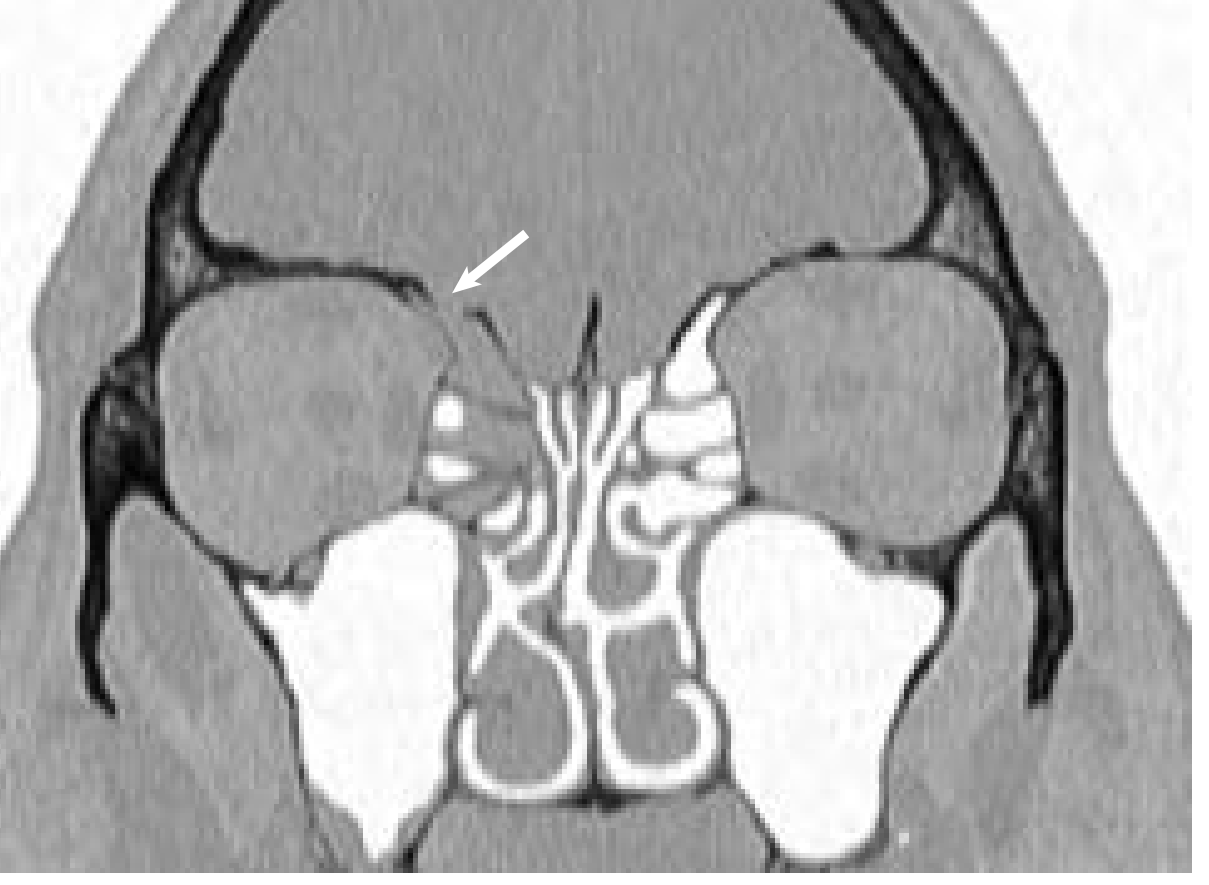
High-resolution coronal CT showing a bony skull base dehiscence (arrow) at the cribriform plate - likely site of an active CSF leak (Cummings)
Intrathecal Fluorescein:
- Dilute fluorescein injected intrathecally via lumbar puncture (0.1-0.5 mL of 10% solution diluted in 10 mL CSF → slow injection over 30 minutes)
- Active leak site glows bright green under blue-filtered endoscopy
- Allows precise localization and intraoperative confirmation
- Absence of fluorescein at end of repair confirms adequacy of closure
- CRITICAL WARNING: Serious neurological sequelae (seizures, paraplegia) have been reported with high doses - MUST use dilute solution only. Fluorescein is NOT FDA-approved for intrathecal use; used off-label.
7. DIFFERENTIAL DIAGNOSIS OF WATERY NASAL DISCHARGE
| Condition | Distinguishing Features |
|---|---|
| CSF rhinorrhoea | Unilateral, watery, salty taste, positional, positive β-2 transferrin |
| CSF otorrhoea presenting as rhinorrhoea | Skull base defect communicates with middle ear → CSF collects in middle ear → drains down Eustachian tube → unilateral rhinorrhoea; check middle ear for defect |
| Allergic rhinitis | Bilateral, seasonal, associated sneezing, itching, IgE-mediated |
| Vasomotor / perennial non-allergic rhinitis | Bilateral, watery; triggers (temperature, smells) |
| Retained saline irrigations | Post-FESS; resolves with stopping irrigations; does not persist |
| Ruptured sinus retention cyst | Yellow-tinged fluid (distinguishes from CSF); air-fluid level on imaging; resolves spontaneously |
| Lacrimal duct drainage | Associated with eye watering |
8. MANAGEMENT
A. Conservative (Non-Operative) Management
Indications: Most traumatic CSF leaks (particularly accidental trauma) - most resolve spontaneously
Measures (Box 48.2):
- Strict bed rest with head elevation for 1-2 weeks
- Lumbar catheter drainage (continuous, 10 mL/hour) OR serial spinal taps (intermittent LP)
- Goal: decompress ICP → reduces pressure driving the leak → allows healing at defect site
- Monitor: daily CSF cell count, protein, glucose, culture
- Complications of lumbar drain: meningitis risk, severe headache from low ICP, pneumocephalus (air drawn in through defect if ICP falls too low), catheter-related infection
- Avoid in markedly elevated ICP (herniation risk)
- Routine prophylactic antibiotics NOT warranted (ineffective and may induce resistance); skin flora coverage (1st-generation cephalosporin) for puncture site
- Avoid coughing, sneezing, nose blowing, and Valsalva manoeuvres
- Stool softeners (prevent straining/Valsalva)
Success: Most traumatic CSF leaks resolve within 7 days of conservative management
Indication for surgery: CSF leak persisting >7 days despite conservative management carries 8-10× increased risk of meningitis → surgical repair indicated
B. Surgical Management
Principle: Identify and seal the skull base defect with graft material
Three surgical routes:
1. Transcranial (Intracranial) Repair
- Frontal craniotomy for cribriform plate / anterior skull base
- Extended approaches for sphenoid sinus defects
- Grafts used: fascia lata, muscle plugs, pedicled galeal flaps, fibrin glue
- Limitations:
- Brain compression, hematoma, seizures, anosmia (olfactory nerve damage)
- Failure rates may exceed 25% despite direct access
- Now reserved for cases where endoscopic approach fails or anatomy precludes endonasal access
- Largely replaced by endoscopic approaches
2. External (Extracranial) Transcranial - Historical
- External incision + transsinus access (pre-endoscopic era)
- Now replaced by endoscopic approach
3. Endoscopic Endonasal Repair - Current Gold Standard
Introduced by Papay et al. (1989) and popularized in early 1990s by Kennedy, Mattox, Wigand, Stankewicz
Advantages:
- Excellent visualization (endoscope)
- Well tolerated by patient
- No external incision
- Post-repair monitoring via serial nasal endoscopy in office
- Outcomes excellent: 90% success rate (primary repair) and 97% success rate (secondary/revision repair) - large systematic review
Access:
- Pure endoscopic approach provides excellent access to:
- Ethmoid roof (fovea ethmoidalis)
- Cribriform plate (most common site)
- Most of the sphenoid sinus
- Lateral sphenoid leaks: may need extended approach through medial pterygomaxillary space
- Frontal sinus posterior table defects: pure endoscopic approach without compromising frontal sinus outflow tract is usually sufficient; a trephine or osteoplastic flap may be needed for far lateral or posterior table defects
Graft Materials (layered reconstruction):
| Layer | Material Options |
|---|---|
| Deepest (intradural/underlay) | Free fat graft, fascia lata (free), free bone graft |
| Middle | Acellular dermal allograft, xenogeneic collagen dural substitute |
| Surface | Free mucosal graft (most commonly from middle turbinate or nasal floor) |
| Sealant | Fibrin glue / tissue sealant |
| Packing | Resorbable (Nasopore/Gelfilm) + non-resorbable nasal packing |
Vascularized flaps: For high-flow leaks and/or large dural defects, vascularized mucosal flaps (e.g., Hadad-Bassagasteguy nasoseptal flap) are preferred over free grafts.
Underlay vs. Overlay Technique:
- Underlay: Graft is placed beneath the dura through the defect - best for larger defects, higher success rates
- Overlay: Graft placed on top of the bony defect edges - simpler, adequate for small defects
C. Postoperative Care
- Nasal packing removed several days after surgery
- Strict bed rest for several days post-op
- Antistaphylococcal antibiotics (prophylaxis against packing-related complications)
- Monitor for intracranial complications (haematoma) in ICU/stepdown for first 24 hours
- Patients instructed to avoid strenuous activity, sneezing, coughing for ~6 weeks
- Serial nasal endoscopy to monitor defect healing
Lumbar Drain Post-operatively:
- NOT routinely used (studies have NOT confirmed benefit in routine cases)
- Indicated selectively for:
- High-flow CSF leaks
- Suspected or confirmed elevated ICP
- Tenuous repair
- Continued for 1-5 days post-op if used
Long-term management of elevated ICP (spontaneous CSF rhinorrhoea):
- Acetazolamide (carbonic anhydrase inhibitor → reduces CSF production)
- Weight loss (in obese patients with BIH)
- CSF diversion procedures if above fail: ventriculoperitoneal shunt or lumboperitoneal drain
9. COMPLICATIONS OF CSF RHINORRHOEA
| Complication | Details |
|---|---|
| Bacterial Meningitis | Major cause of mortality and morbidity; most feared complication. Risk increases with duration of leak: >7 days leak = 8-10× increased meningitis risk. Reported incidence 2-50% |
| Pneumocephalus | Air enters through skull base defect (especially with low ICP from aggressive lumbar drainage); can cause brain compression |
| Recurrent meningitis | Repeated episodes may occur if defect not repaired (especially pneumococcal) |
| Intracranial abscess / Subdural empyema | Rare; ascending infection |
| Failure of repair | Requires revision (90% primary success → 97% revision success endoscopically) |
Prophylactic Vaccination:
For any patient with a history of CSF rhinorrhoea, consider immunization against:
- Streptococcus pneumoniae
- Haemophilus influenzae
- Meningococcus
These patients have a theoretically higher risk of acute bacterial meningitis (reconstructed barrier may be less robust). Low-risk intervention with potential high benefit.
Antibiotic Prophylaxis - Controversial:
- MacGee et al.: 14% meningitis WITH prophylaxis vs 5% WITHOUT (not statistically significant)
- Brodie meta-analysis (6 studies, 324 patients): 2.5% WITH prophylaxis vs 10% WITHOUT (P = 0.006)
- More recent large meta-analyses: no increased meningitis risk from skull base fractures with or without prophylaxis
- Current consensus: prophylactic antibiotics are NOT routinely recommended for post-traumatic CSF fistulae; may use 1st-generation cephalosporin for skin flora/wound coverage
10. SPECIAL SITUATIONS
Intraoperative CSF Leak During FESS:
- Recognized at the time: repair immediately using the above endoscopic techniques
- Small defect (<5mm): free mucosal graft + fibrin glue often sufficient
- Larger defect: formal repair with free graft layers + packing
Post-operative CSF Leak After FESS:
- May manage conservatively initially (bed rest, lumbar drainage)
- Most patients will require formal endoscopic repair
Nontraumatic / Idiopathic CSF Rhinorrhoea:
- Will NOT resolve spontaneously (unlike most traumatic leaks)
- Exclude underlying cause (brain tumour, hydrocephalus)
- Formal endoscopic repair warranted
- Elevated ICP must be addressed post-repair (acetazolamide, weight management, CSF diversion if needed)
- If not addressed → repair will fail due to ongoing high pressure
Frontal Sinus CSF Leaks:
- Most can be repaired endoscopically without compromising frontal sinus outflow tract
- Far lateral or posterior table defects: may need a trephine (small bur hole in anterior table) or osteoplastic flap for access
11. OUTCOMES
| Approach | Success Rate |
|---|---|
| Transcranial repair | ~75% (failure rates >25%) |
| Endoscopic repair (primary) | ~90% |
| Endoscopic repair (revision/secondary) | ~97% |
The excellent success rates and minimal morbidity of endoscopic repair have made it the preferred approach for the vast majority of CSF rhinorrhoea cases requiring surgery.
SUMMARY - KEY EXAM POINTS
- Definition: CSF leak from subarachnoid space to sinonasal cavity through simultaneous arachnoid/dural/bony/mucosal defect + pressure gradient
- Causes: Traumatic (>90%) - accidental > surgical; Nontraumatic (<10%) - BIH most important
- Most common site: Cribriform plate (anterior skull base)
- Key symptom: Unilateral watery nasal discharge with salty/metallic taste; positional/intermittent
- Halo sign: Clear ring around bloody spot on paper - unreliable (false positives with tears/saliva)
- Best biochemical marker: β-2 transferrin (specific, no false positives); β-trace protein (100% sensitivity and specificity)
- Glucose test strips: NOT reliable - high false positive rate; abandoned
- Imaging: HRCT (bony defect) + MRI (soft tissue, empty sella, meningoencephalocele)
- Intrathecal fluorescein: Precise localization intraoperatively; use DILUTE only - neurological complications with high doses
- Conservative treatment: Traumatic leaks - bed rest, head elevation, lumbar drainage, no straining
- Surgical repair: Endoscopic endonasal is gold standard - 90% primary success, 97% revision
- Graft: Free fat/fascia (underlay) + free mucosal graft (overlay) + fibrin glue + packing; vascularized flap for large/high-flow leaks
- Key risk: >7 days of leakage = 8-10× meningitis risk → operate if not resolved
- Prophylactic antibiotics: NOT routinely recommended for posttraumatic CSF fistulae
- Spontaneous CSF rhinorrhoea: Associated with BIH, empty sella, elevated ICP - will NOT resolve spontaneously; treat ICP post-repair with acetazolamide or shunt
- Vaccination: Pneumococcal, Hib, meningococcal - consider for all patients with CSF rhinorrhoea history
Sources: Cummings Otolaryngology Head and Neck Surgery, Chapter 48 (Cerebrospinal Fluid Rhinorrhea and Anterior Meningoencephalocele) and Scott-Brown's Otorhinolaryngology Head & Neck Surgery Vol 1 & 2
This is a shared conversation. Sign in to Orris to start your own chat.