
Diagram of forearm
Reading File
This is a visual/anatomy question. Let me search the medical library for forearm diagrams.
Finding Sources
Finding Sources
Great results. Let me read the Gray's Anatomy forearm section and the THIEME Atlas block to pull the diagrams.
Reading File
Reading File
Reading File
I found several excellent diagrams. Let me fetch the most relevant ones - the Gray's Anatomy forearm overview and cross-section, plus the THIEME Atlas muscle/bone diagrams.
Loading Image
Loading Image
Loading Image
Loading Image
Loading Image
Here are the forearm anatomy diagrams from the medical textbook library:
Forearm Anatomy
Fig. 1 - Overview & Cross-Section (Gray's Anatomy for Students)
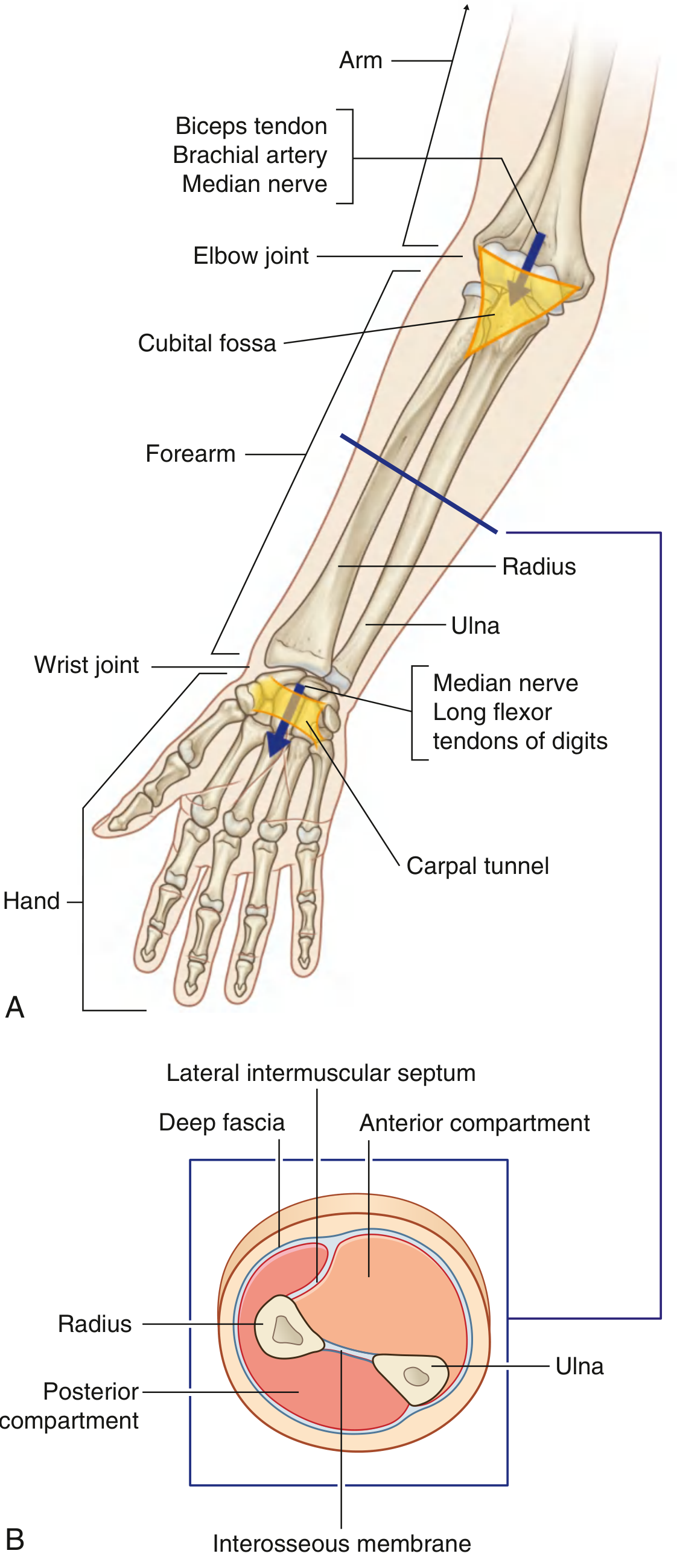
(A) Shows proximal and distal relationships: the biceps tendon, brachial artery, and median nerve enter via the cubital fossa at the elbow; distally, the median nerve and long flexor tendons of the digits pass through the carpal tunnel at the wrist. The radius (lateral) and ulna (medial) form the bony framework.
(B) Transverse cross-section through the mid-forearm showing the radius and ulna separated by the interosseous membrane, with the anterior (flexor) and posterior (extensor) compartments divided by the lateral intermuscular septum and deep fascia.
Fig. 2 - Right Radius (Gray's Anatomy for Students)
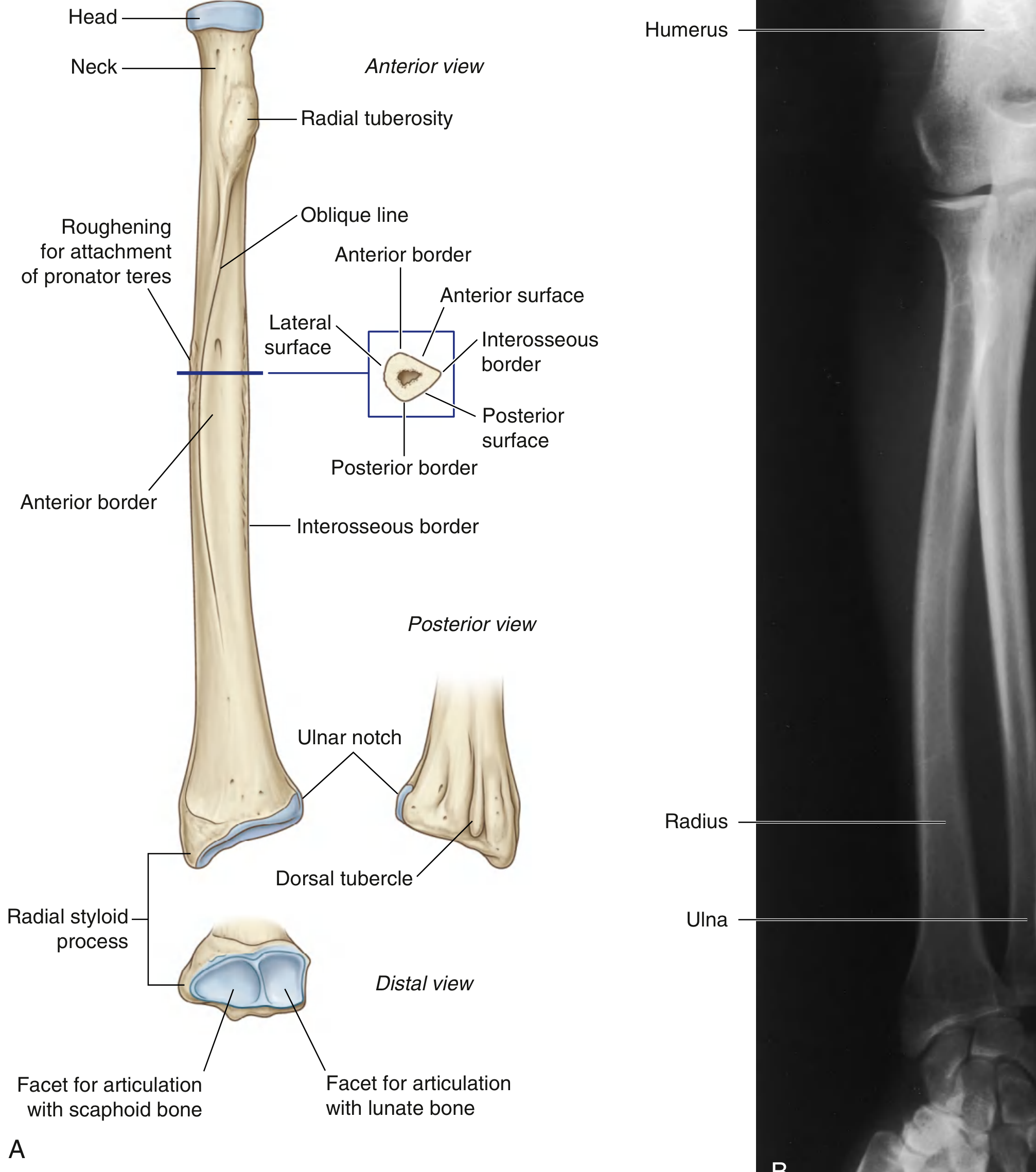
Key landmarks: radial tuberosity (proximal, for biceps insertion), oblique line, pronator teres attachment site on the shaft, radial styloid process and articular facets distally for scaphoid and lunate bones.
Fig. 3 - Right Ulna (Gray's Anatomy for Students)
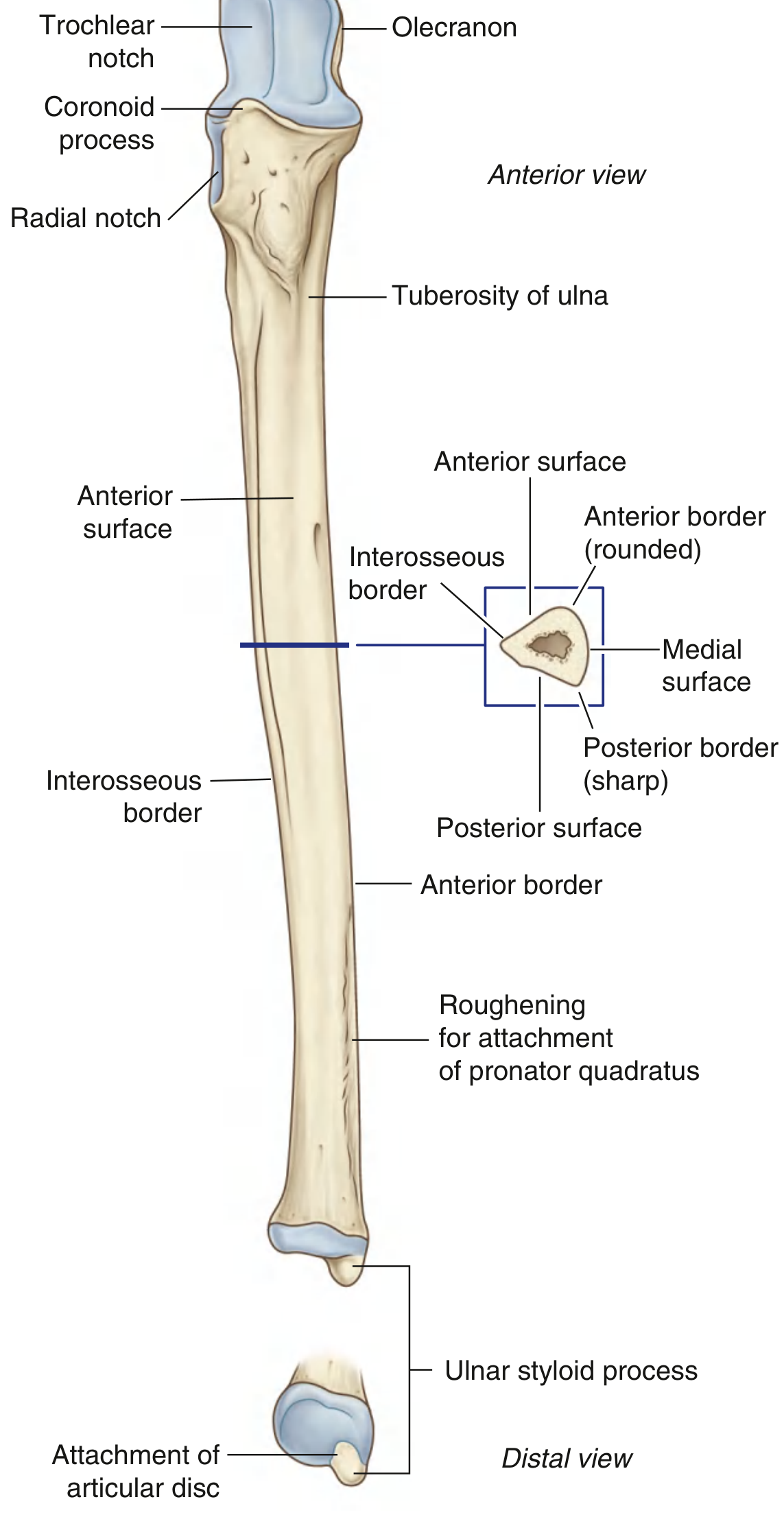
Key landmarks: olecranon and trochlear notch proximally (for elbow articulation), radial notch (proximal radioulnar joint), pronator quadratus attachment distally, ulnar styloid process.
Fig. 4 - Posterior (Extensor) Compartment Muscles (THIEME Atlas of Anatomy)
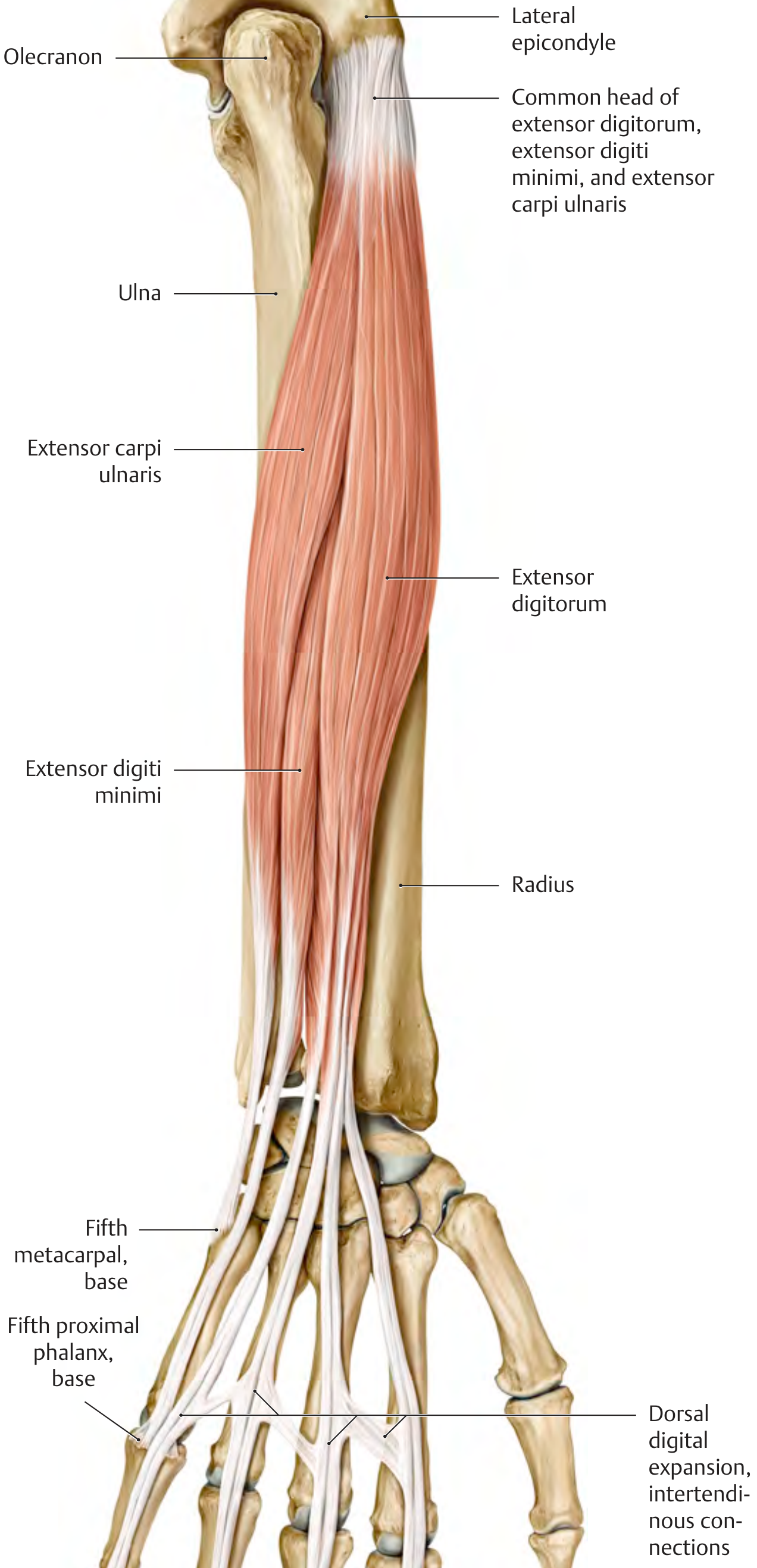
Shows the superficial extensor group arising from the common extensor origin at the lateral epicondyle, fanning out into tendons with dorsal digital expansions.
Fig. 5 - Posterior Forearm Arteries (THIEME Atlas of Anatomy)
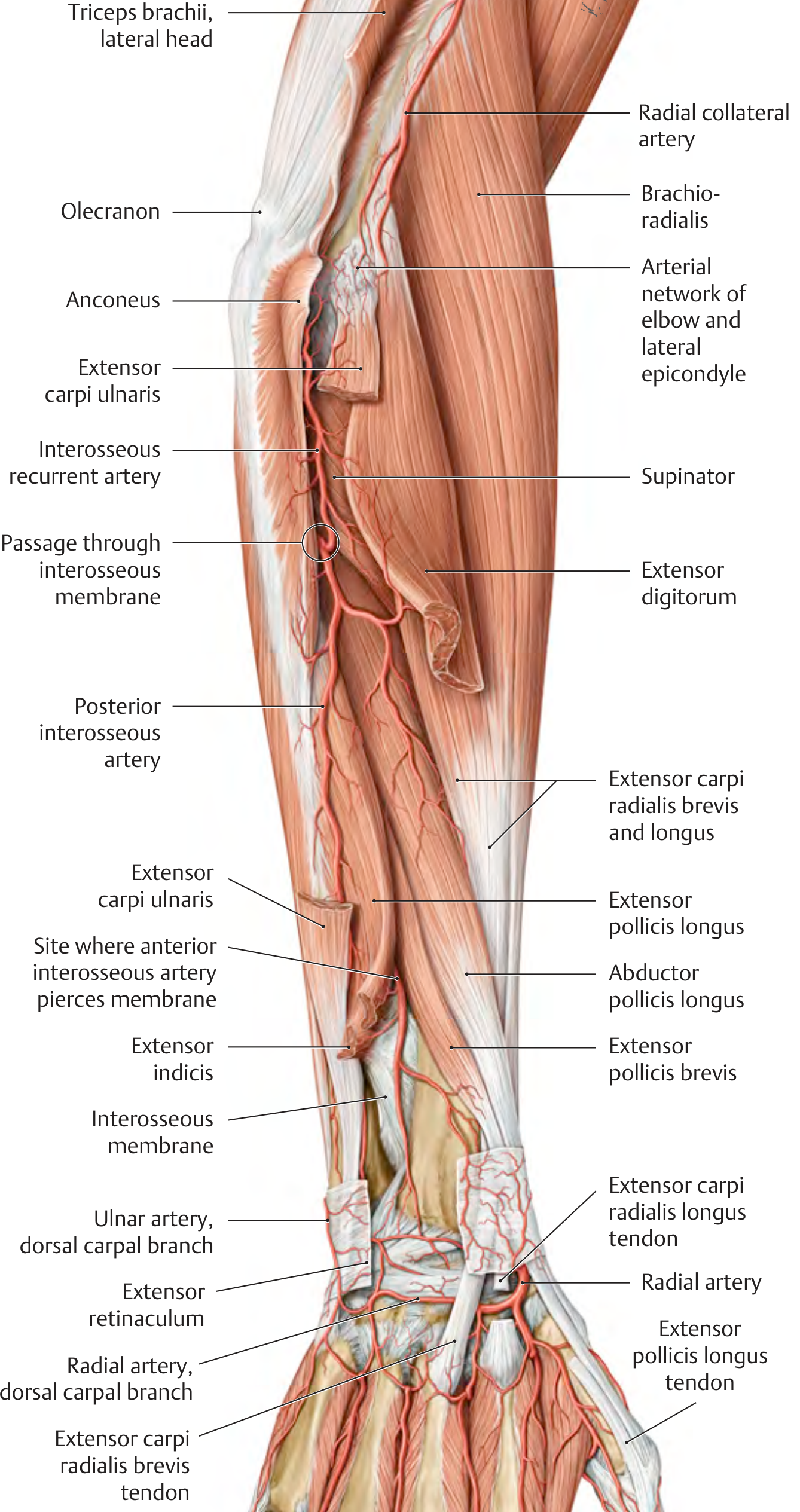
Shows the posterior interosseous artery entering the extensor compartment by piercing the interosseous membrane just below the supinator, and the anterior interosseous artery piercing distally to supply the dorsal wrist.
Key anatomical summary:
| Structure | Anterior (Flexor) Compartment | Posterior (Extensor) Compartment |
|---|---|---|
| Nerve supply | Median nerve (mainly), ulnar nerve | Radial nerve (posterior interosseous branch) |
| Function | Wrist/digit flexion, pronation | Wrist/digit extension, supination |
| Main artery | Anterior interosseous artery | Posterior interosseous artery |
| Bones | Radius (lateral), Ulna (medial), linked by interosseous membrane | Same |
Sources: Gray's Anatomy for Students, Fig. 7.79-7.81; THIEME Atlas of General Anatomy and Musculoskeletal System, pp. 339, 417
This is a shared conversation. Sign in to Orris to start your own chat.