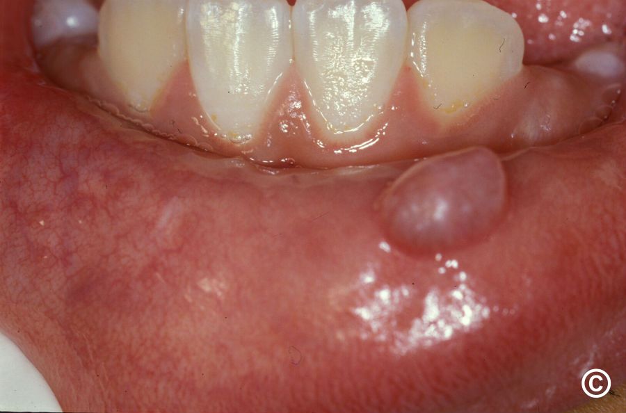
l l l Mucocoeles are nonpainful, potentially recurring swell ings. Some individuals report an increase in the size of their mucocoele during meals Extravasation-type mucocoeles are most often found on the lower lip and tongue, which are the most common places for injuries to the minor salivary glands Retention-type mucocoeles are uncommon and occur rarely on the lower lip. Instead, it manifests in the palate, cheek, gingiva and maxillary sinus Mucocoele typically appears as dome-shaped mucosal swellings ranging from one to several centimetres in size l l l l l They are most common in children and young adults The spilled mucin below the mucosal surface often im parts a bluish translucent hue to the swelling; however, deeper mucocoeles may be normal in colour The lesions are characteristically uctuant, but some long standing mucocoeles are rm on palpation (Fig. 31.10) Lower lip is the most common site for the mucocoeles, accounting for more than 60% of all the cases Mucocoeles are usually found lateral to the midline. Less common sites include the buccal mucosa, anterior, ven tral surface of tongue and the oor of the mouth, where it is known as ranula. simplify
- Painless, dome-shaped swellings of the mouth that can come back after treatment
- Range from 1 cm to several centimetres in size
- Most common in children and young adults
- Often bluish and semi-transparent (due to mucus near the surface)
- Deeper ones may look normal in colour
- Soft and fluid-filled to the touch; older ones may feel firm
- The lower lip is the most common site (over 60% of cases), usually off-centre
- Other sites: cheek, tongue (front/underside), and floor of the mouth
- When on the floor of the mouth, it is called a ranula
- Extravasation type (mucus leaks out due to injury) - found mainly on the lower lip and tongue
- Retention type (blockage of a gland duct) - rare on the lower lip; more common on the palate, cheek, gums, and maxillary sinus
- Some people notice the swelling gets bigger during meals (when saliva production increases)
Find clinical images of different mucocoeles
mucocoele clinical images oral lip ranula different types
1. Lower Lip Mucocoele (most common type)
2. Lower Lip Mucocoele - Clinical + Histology (Robbins Pathology)
3. Ranula - Floor of Mouth (Bailey & Love's Surgery)
- Top row: Characteristic bluish swelling on the floor of mouth resembling a frog's belly; surgical exposure; and intraoperative view
- Bottom row: Excised specimens showing the cystic sac and contained mucin
4. Ranula - MRI Imaging (Bailey & Love's Surgery)
Summary of types shown:
| Type | Site | Key Feature |
|---|---|---|
| Extravasation mucocoele | Lower lip | Bluish, soft, lateral to midline |
| Retention mucocoele | Palate, cheek, gingiva | Deeper, may be normal colour |
| Ranula | Floor of mouth | "Frog belly" appearance, can be large |
| Plunging ranula | Floor of mouth + neck | Dissects through mylohyoid, submental swelling |