
Lyons hypothesis 5 mark Short note
The Lyon's Hypothesis (also called X-inactivation or lyonization) states that in every somatic cell of a female mammal, one of the two X chromosomes is randomly and permanently inactivated early in embryonic development, forming a Barr body.
Searching sources...
Reading File
Finding Sources
Searching the Web
Lyon hypothesis X inactivation short note 5 marks genetics
Reading File
Reading File
Reading File
Loading Image
Here is a complete 5-mark short note on the Lyon Hypothesis, drawn from Robbins Pathology, Emery's Medical Genetics, and Basic Medical Biochemistry:
Lyon Hypothesis (X-Chromosome Inactivation / Lyonization)
Introduction
The Lyon Hypothesis was proposed by Dr. Mary Lyon in 1961 based on her observations of mosaic coat color patterns in mice heterozygous for X-linked genes. It describes the mechanism by which gene dosage equality between males (XY) and females (XX) is achieved through permanent inactivation of one X chromosome in each somatic cell of female mammals. This process is also called lyonization.
Postulates of the Lyon Hypothesis
As outlined in Robbins Pathology, the hypothesis states four key points:
- Only one X chromosome is genetically active in each somatic cell at any time.
- The other X chromosome - either of maternal or paternal origin - undergoes heteropyknosis (condensation) and is rendered genetically inactive.
- Inactivation is random: either the maternal (Xm) or the paternal (Xp) X can be inactivated in any given cell. This occurs in cells of the blastocyst around day 5.5 of embryonic life (approximately the 16-cell stage).
- Inactivation is permanent and clonal: once an X is inactivated in a cell, all daughter cells derived from that cell carry the same inactive X.
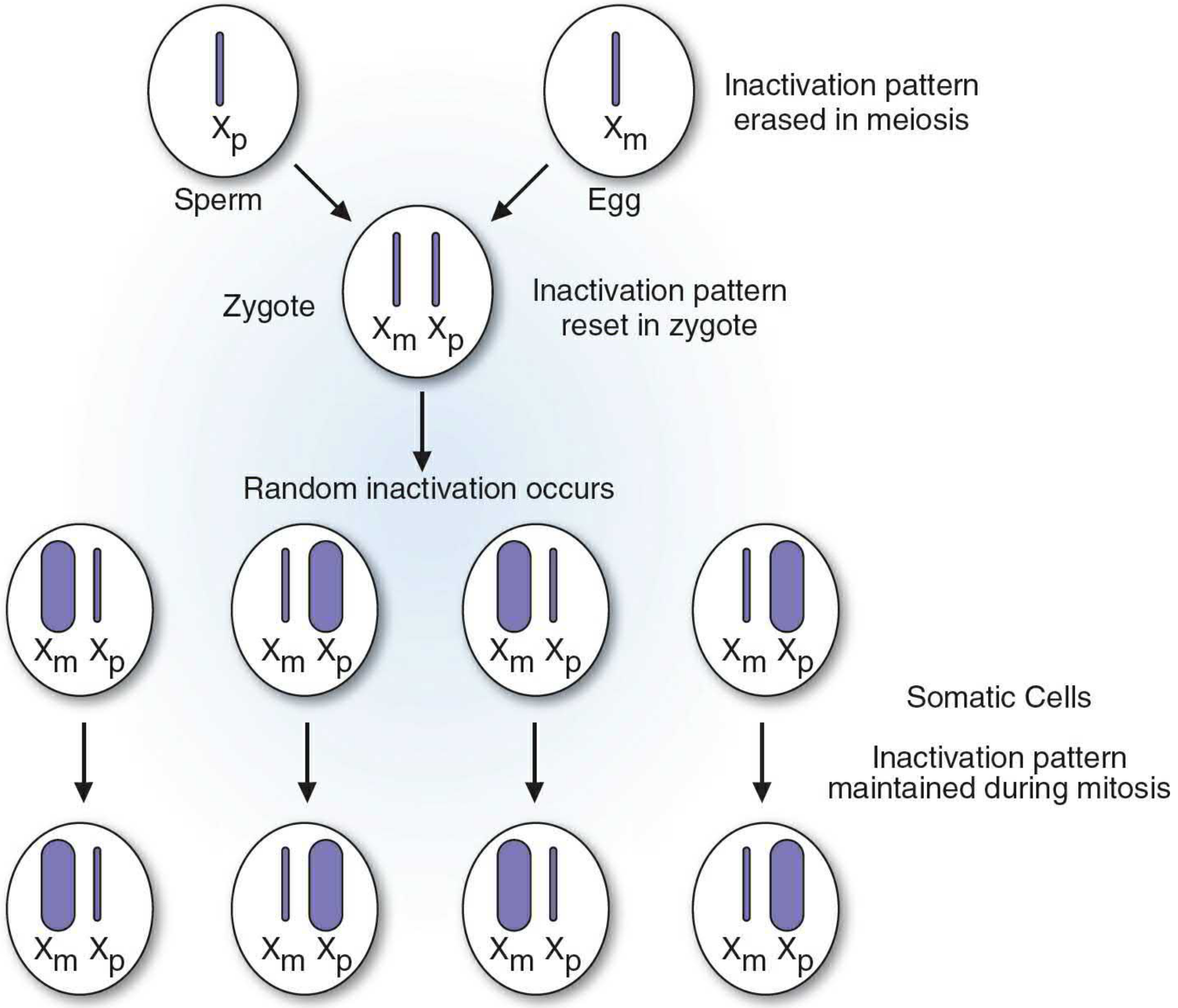
The Barr Body
The inactive X chromosome exists in a condensed form during interphase as a darkly staining mass called the Barr body (sex chromatin), visible at the inner nuclear membrane. The number of Barr bodies = (number of X chromosomes - 1).
| Karyotype | Barr Bodies |
|---|---|
| 46, XX (normal female) | 1 |
| 46, XY (normal male) | 0 |
| 47, XXY (Klinefelter) | 1 |
| 47, XXX (Triple X) | 2 |
| 45, X (Turner) | 0 |
Molecular Mechanism
The molecular basis of X-inactivation involves a long non-coding RNA (lncRNA) called XIST (X-inactive specific transcript), encoded on the X chromosome. XIST RNA is transcribed from and "coats" the X chromosome to be inactivated. It initiates gene silencing through chromatin modification and DNA methylation. XIST expression is switched off on the active X chromosome. - Robbins Pathologic Basis of Disease
Female as a Mosaic
Because inactivation is random, normal females are mosaics - they possess two populations of cells: one with an active maternal X and the other with an active paternal X. This explains why carrier females of X-linked recessive disorders (e.g., hemophilia A, Duchenne muscular dystrophy) are generally unaffected but may occasionally show mild features depending on which X is inactivated in a greater proportion of cells.
Modifications to the Original Hypothesis
The hypothesis has been subsequently refined:
- Initially it was assumed that all genes on the inactive X are switched off. This is incorrect.
- Approximately 30% of genes on Xp and ~3% on Xq escape X-inactivation and remain expressed from both X chromosomes.
- The inactive X is selectively reactivated in oogonia before the first meiotic division - both X chromosomes are needed for normal oogenesis. This explains the gonadal abnormalities in Turner syndrome (45,X), where a single X dose is insufficient.
- The pseudoautosomal regions at the tips of X and Y also escape inactivation.
Significance
- Explains dosage compensation between XX females and XY males.
- Explains the Barr body as a morphological correlate of the inactive X.
- Explains variable expressivity in female carriers of X-linked disorders.
- Underlies disorders like Turner syndrome and Klinefelter syndrome.
Sources: Robbins & Cotran Pathologic Basis of Disease, 10e | Robbins & Kumar Basic Pathology | Emery's Elements of Medical Genetics and Genomics | Basic Medical Biochemistry: A Clinical Approach, 6e
This is a shared conversation. Sign in to Orris to start your own chat.