
Clubbing pathophysiology
digital clubbing fingers nail pathology
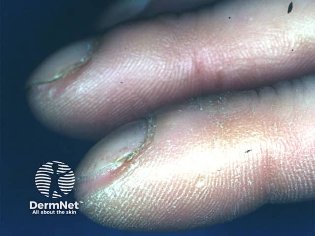
Clinical photograph of the distal digits demonstrating digital clubbing. Modality: Clinical photography; technique: macro close‑up digital image with bright‑field illumination and high resolution to emphasize nail bed morphology. Anatomical location: fingers (distal phalanges) with visible nail plates, nail beds, and periungual tissue; dorsal view highlighting the nail–bed interface. Visual features: bulbous, expanded distal digits; nail plates show increased curvature with convex nail beds; reduced angle between the nail plate and the adjacent skin; glossy, smooth periungual skin; absence of cutaneous ulcers in view; subtle subungual fullness may be present. Pathologic/clinical findings: clubbing is a clinical sign characterized by hyperplastic nail bed change; not a histologic lesion, but correlates with chronic hypoxia and systemic disease. Diagnostic significance: digital clubbing signals potential underlying cardiopulmonary pathology or hepatobiliary disease; commonly associated conditions include interstitial lung disease (idiopathic pulmonary fibrosis, bronchiectasis), chronic obstructive pulmonary disease, cyanotic heart disease, liver cirrhosis, and inflammatory bowel disease; warrants systemic workup. Differential considerations: idiopathic clubbing; secondary clubbing from pulmonary/vascular disease or congenital heart disease; alternatively pseudoclubbing from nail dystrophy. Clinical correlation: correlate with oxygen saturation, chest imaging, echocardiography, hepatic panel; multidisciplinary assessment recommended. Documented progression can guide treatment decisions and patient counseling in dermatology, pulmonology, and internal medicine.
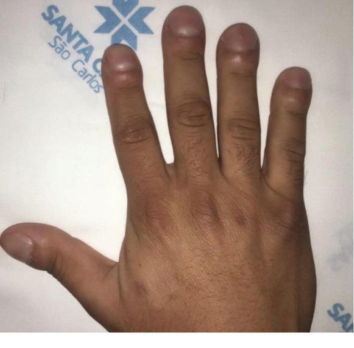
A clinical photograph of a human hand demonstrating classic digital clubbing (Hippocratic fingers). The image shows the dorsal view of the fingers, where there is significant bulbous enlargement and rounding of the distal phalanges. Key visual features include an increased longitudinal and transverse curvature of the nails, resulting in a 'watch-glass' appearance. The Lovibond angle (the angle between the nail plate and the proximal nail fold) is visibly obliterated and exceeds 180 degrees, a hallmark sign of the condition. The soft tissue at the nail base appears thickened and spongy. This finding is clinically significant as an indicator of chronic hypoxemia often associated with primary ciliary dyskinesia (PCD), bronchiectasis, or cyanotic cardiovascular diseases. The photograph is an educational example used in internal medicine and pulmonology to demonstrate physical examination signs of systemic or pulmonary pathology.
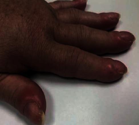
This clinical photograph shows a close-up view of a human hand demonstrating classic digital clubbing (Hippocratic fingers). The distal phalanges of the thumb and fingers are visibly bulbous and enlarged, losing their normal distal tapering. Key morphological features include a distinct convexity of the fingernails and an increased Lovibond angle (the angle between the nail plate and the proximal nail fold). The surrounding soft tissue appears edematous and erythematous, particularly around the nail beds, which exhibit a characteristic shiny, thickened skin texture. This finding is a critical clinical sign often associated with chronic hypoxemia, underlying pulmonary or cardiovascular disease, or as a component of hypertrophic osteoarthropathy in the context of malignancy. The image serves as a high-fidelity visual reference for medical students and clinicians for identifying systemic manifestations of internal pathology during a physical examination.
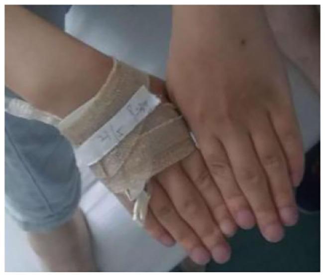
This clinical photograph displays the hands of a patient exhibiting clear signs of chronic hypoxemia and inflammatory changes. The primary visible pathology includes marked digital clubbing, characterized by the bulbous enlargement of the distal phalanges and a loss of the normal angle between the nail plate and the proximal nail fold. The fingernails and distal fingertips demonstrate a bluish-purplish discoloration consistent with peripheral cyanosis. Additionally, there is noticeable fusiform swelling of the proximal interphalangeal (PIP) joints, particularly prominent on the middle fingers, suggesting an associated inflammatory or rheumatic process. One hand is partially obscured by a tan elastic bandage and adhesive tape around the wrist and dorsum, with a visible intravenous (IV) catheter line, indicating acute medical management. The combination of digital clubbing and cyanosis is clinically significant for underlying chronic respiratory or cardiovascular disease, such as the pulmonary interstitial fibrosis mentioned in the associated clinical context.
Pathophysiology of Digital Clubbing
Definition and Morphology
- Obliteration of the Lovibond angle - normally ~160-165°; with clubbing it becomes 180° or greater
- Sponginess at the nail base - the nail "floats" on a softened, vascular nailbed
- Increased nail curvature in both longitudinal and coronal planes
- Loss of the Schamroth sign - no diamond-shaped window when dorsal surfaces of opposing fingers are pressed together
- Increased capillary density in the stroma of the distal digit
- Fishman's Pulmonary Diseases and Disorders, p. 447; Murray & Nadel's Textbook of Respiratory Medicine
Pathogenesis - Current Hypotheses
1. Vasodilation and Interstitial Edema (Central Mechanism)
- Fishman's, p. 447
2. The "Humoral Bypass" Hypothesis (Most Widely Accepted)
| Mechanism | Examples |
|---|---|
| Right-to-left cardiac shunt | Cyanotic congenital heart disease, patent ductus arteriosus with reversed shunt |
| Bronchial circulation proliferation | Suppurative lung disease, bronchiectasis |
| Hepatic arteriovenous shunts | Liver cirrhosis, hepatopulmonary syndrome |
- Fishman's, p. 448; Murray & Nadel's
3. Platelet Aggregates and Megakaryocyte Fragments
- Normally, megakaryocytes shed platelets in the lung capillaries; large platelet/megakaryocyte particles are fragmented during passage through the pulmonary microcirculation
- In patients with right-to-left shunts (cyanotic heart disease, intrapulmonary shunts), these large platelet particles bypass the lung and reach distal extremities intact
- There, they interact with vascular endothelium and release growth factors that drive connective tissue proliferation
- Harrison's Principles of Internal Medicine 22E, p. 3013; Firestein & Kelley's Textbook of Rheumatology
4. Growth Factor Cascade - VEGF and PDGF
- VEGF (vascular endothelial growth factor) - the primary driver
- PDGF (platelet-derived growth factor)
- Hepatocyte growth factor (HGF)
- Murray & Nadel's; Firestein & Kelley's
5. Prostaglandin E2 (PGE2) Pathway - Genetic Insight
- Mutations in HPGD (encoding 15-hydroxyprostaglandin dehydrogenase) - the enzyme that degrades PGE2
- Mutations in SLCO2A1 (encoding the prostaglandin transporter)
- Andrews' Diseases of the Skin; Firestein & Kelley's
Integrated Mechanism
Underlying disease (lung cancer, CHD, cirrhosis, etc.)
         ↓
Shunting / bypass of pulmonary capillary bed
         ↓
Intact platelet aggregates + VEGF/PDGF/HGF reach digit vasculature
         ↓
Platelet-endothelial interaction → growth factor release
         ↓
VEGF → vasodilation + new capillary formation + AV anastomoses
PDGF/HGF → connective tissue proliferation
         ↓
↑ Hydrostatic pressure → interstitial fluid accumulation
         ↓
Bulbous soft tissue swelling, nail bed thickening → CLUBBING
Why COPD Does NOT Cause Clubbing
- Fishman's, p. 448
Clinical Correlates
-
Clubbing can develop in ~2 weeks (e.g., new empyema) and reverse in ~2 weeks (e.g., after corrective cardiac surgery) - consistent with a reversible vascular/humoral mechanism
-
Unilateral clubbing: localized vascular abnormality - ipsilateral pulmonary sulcus tumor invading the brachial plexus, Takayasu arteritis, sarcoidosis
-
Differential clubbing (e.g., toes only in patent ductus arteriosus with reversed shunt): reflects differential blood flow patterns
-
Found in 1% of internal medicine admissions; associated with serious disease in 40% of those patients
-
Murray & Nadel's; Fishman's