
Anatomy of lungs
Reading File
Now let me search the medical library for lung anatomy content in parallel.
Finding Sources
Finding Sources
Finding Sources
Reading File
Good. Now let me get the main structural anatomy sections - the gross anatomy and the Robbins pathology lung section:
Finding Sources
Reading File
Reading File
Excellent - Murray & Nadel's block 1 has the full anatomy chapter. Let me read more of it and also get Gray's Anatomy content on bronchopulmonary segments:
Reading File
Reading File
Let me now get the Color Atlas content on bronchopulmonary segments and fetch key images:
Reading File
Loading Image
Loading Image
Excellent images obtained. Now let me get the key sections on airways, alveoli, and blood supply from Murray & Nadel:
Reading File
I now have rich content from Murray & Nadel's Respiratory Medicine and Gray's Anatomy for Students to compile a thorough answer. Let me also quickly fetch the left lung image and the bronchial tree diagram:
Loading Image
I now have comprehensive content and excellent diagrams to produce a detailed, well-illustrated answer on lung anatomy.
Anatomy of the Lungs
Sources: Murray & Nadel's Textbook of Respiratory Medicine | Gray's Anatomy for Students | Color Atlas of Human Anatomy, Vol. 2
Overview and Gross Anatomy
The lungs are paired, spongy, cone-shaped organs occupying most of the thoracic cavity. In life they weigh approximately 900-1000 g combined (nearly 40-50% of which is blood). At end-expiration (FRC) the gas volume is about 2.5 L, rising to approximately 6 L at total lung capacity. Lung density is not uniform - it is highest (~1 g/mL) near the hilum and lowest (~0.1 g/mL) in the periphery, and increases from apex to base due to gravitational effects on blood distribution.
Each lung has:
- An apex projecting above the clavicle into the root of the neck
- A base resting on the diaphragm
- Three surfaces: costal, mediastinal, and diaphragmatic
- A hilum on the mediastinal surface where the bronchus, pulmonary artery, and pulmonary veins enter/exit
Lobes and Fissures
| Feature | Right Lung | Left Lung |
|---|---|---|
| Lobes | 3 (superior, middle, inferior) | 2 (superior, inferior) |
| Fissures | Oblique + horizontal | Oblique only |
| Special feature | - | Lingula (tongue-like projection of superior lobe, homolog of middle lobe) |
| Bronchopulmonary segments | 10 | 9 (apical and posterior fuse into apicoposterior) |
Bronchial Tree
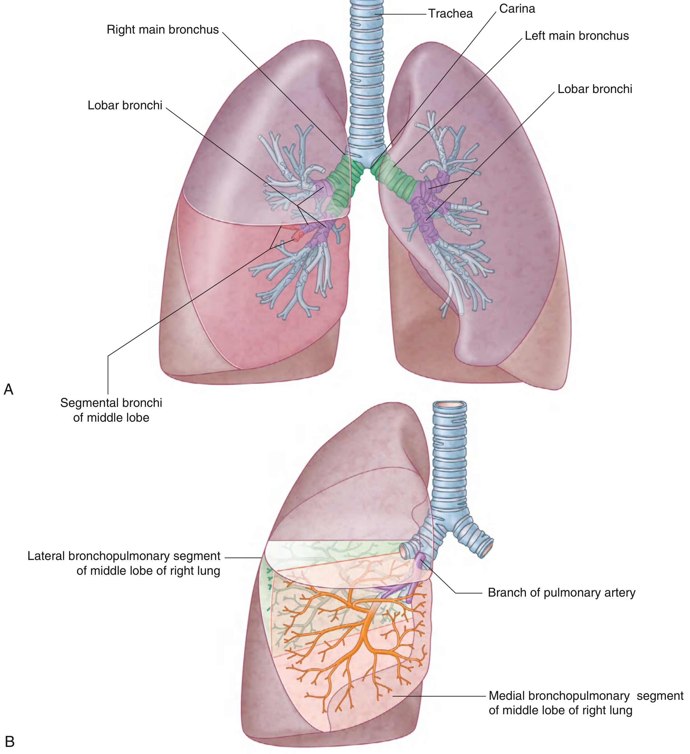
The airway generations from trachea to alveolus:
- Trachea - bifurcates at the carina at vertebral level T4/T5
- Main bronchi - right and left; the right is shorter, wider, and more vertical (clinically important: aspirated objects lodge more easily in the right)
- Lobar bronchi - right: 3 lobar bronchi; left: 2 lobar bronchi (8-12 mm diameter). The right superior lobar bronchus branches ~1-2.5 cm from the carina; middle and inferior branch ~5 cm from it
- Segmental bronchi - right: 10; left: 9 (from Color Atlas of Human Anatomy, Vol. 2, p. 214)
- Subsegmental bronchi - medium and small bronchi, then bronchioles
- Terminal bronchioles - last purely conducting generation; no alveoli in walls
- Respiratory bronchioles - begin gas exchange; have alveoli in walls
- Alveolar ducts - all walls consist of alveoli
- Alveolar sacs and alveoli - terminal units of gas exchange
Bronchopulmonary Segments
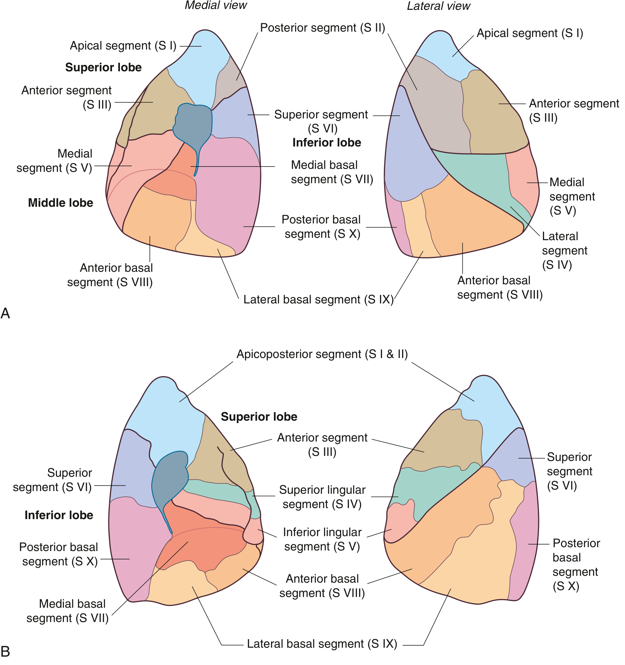
A bronchopulmonary segment is the smallest functionally independent region of the lung - the smallest area that can be surgically isolated and removed without affecting adjacent regions. Each segment is:
- Supplied by one segmental bronchus and an accompanying pulmonary artery branch (bronchoarterial unit)
- Drained by pulmonary veins that travel intersegmentally along segment boundaries
- Shaped like an irregular cone (apex toward hilum, base at lung surface)
Right lung - 10 segments:
- Superior lobe: Apical (S1), Posterior (S2), Anterior (S3)
- Middle lobe: Lateral (S4), Medial (S5)
- Inferior lobe: Superior (S6), Medial basal (S7), Anterior basal (S8), Lateral basal (S9), Posterior basal (S10)
Left lung - 9 segments:
- Superior lobe: Apicoposterior (S1+2), Anterior (S3), Superior lingular (S4), Inferior lingular (S5)
- Inferior lobe: Superior (S6), Medial basal (S7), Anterior basal (S8), Lateral basal (S9), Posterior basal (S10)
Left Lung and Mediastinal Relations
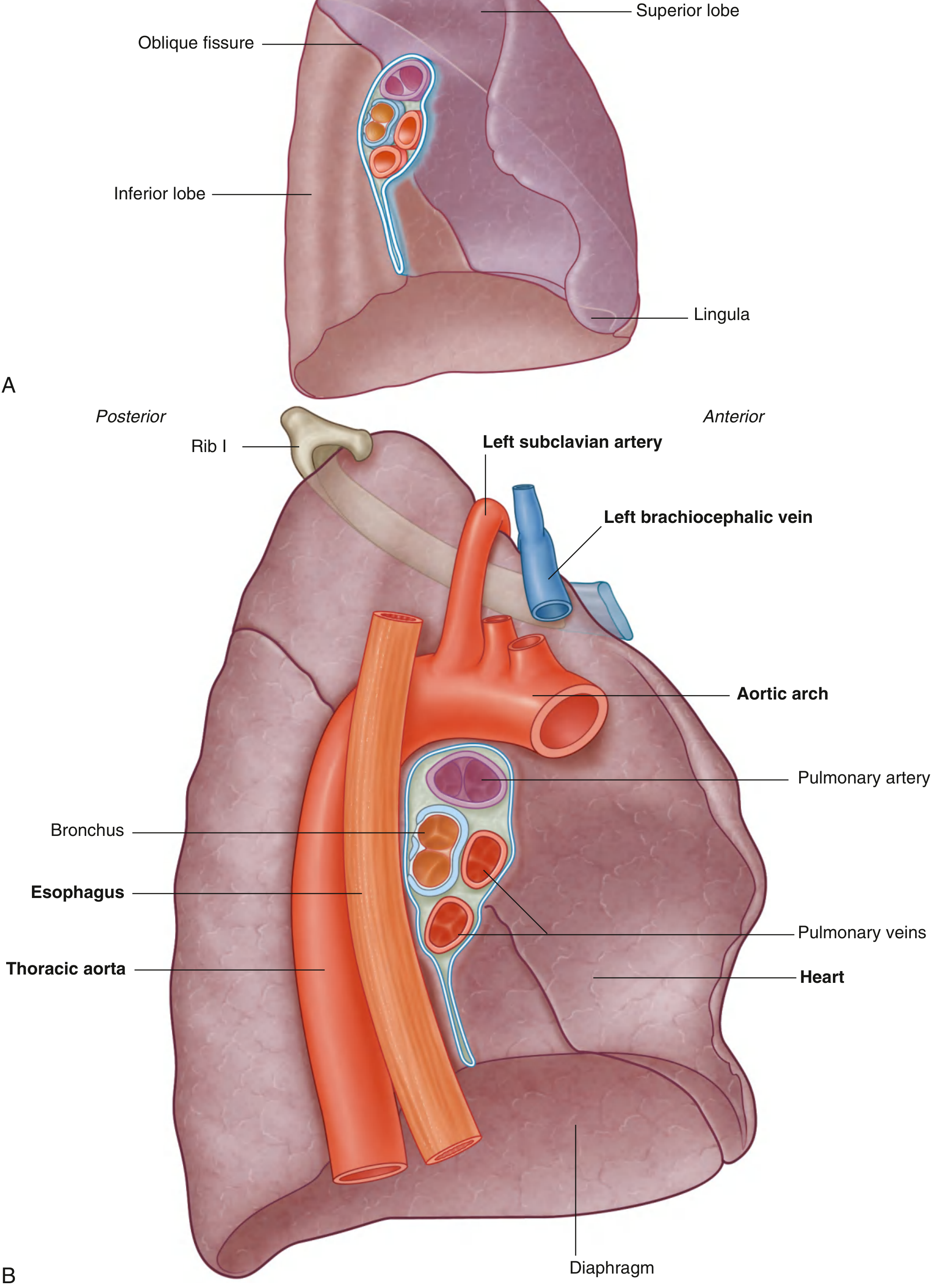
Key structures related to the lung hilum and mediastinal surface include the aortic arch, esophagus, thoracic aorta, heart, pulmonary artery, pulmonary veins, and the phrenic nerve (runs anterior to hilum on both sides).
Pulmonary Circulation
- Pulmonary trunk bifurcates at vertebral level T4/T5, slightly left of midline
- Right pulmonary artery - longer, passes horizontally across the mediastinum, anterior to the right main bronchus
- Left pulmonary artery - shorter, arches over the left main bronchus
- Pulmonary arteries carry deoxygenated blood to the lungs; pulmonary veins carry oxygenated blood back to the left atrium
- Vascular distending pressures increase at the rate of 1 cm H₂O per cm height downward in the lung, which is why vessels appear more distended in the lung bases on chest X-ray
Bronchial Circulation
The bronchial arteries (branches of the thoracic aorta) supply oxygenated blood to the airway wall, connective tissue, and visceral pleura. This is a separate, smaller circuit from the pulmonary circulation.
Alveoli and Gas Exchange
The lung contains approximately 300 million alveoli, providing a total gas exchange surface area of ~70 m². Key points:
- More than 50% of all alveoli are in the outer 30% of the lung radius (hilum to chest wall)
- Alveolar walls are extremely thin to minimize the diffusion distance for O₂
- Two alveolar lining cell types (from Murray & Nadel, p. 44+):
- Type I pneumocytes - large, flat cells covering ~95% of the alveolar surface; primary gas-exchange cells
- Type II pneumocytes - cuboidal; produce surfactant; serve as progenitor cells for repair after injury
- The blood-air barrier consists of: alveolar epithelium + basement membrane + capillary endothelium (average thickness ~0.5 μm)
Airway Lining (Mucosal Architecture)
The conducting airway epithelium is pseudostratified columnar and contains:
- Basal cells - progenitor cells; renew epithelium after injury
- Ciliated cells - ~half of all epithelial cells; each cell has ~200 cilia beating at 8-15 Hz to drive mucociliary clearance centrally
- Goblet cells - secrete mucus
- Club (Clara) cells - increase distally; secretory and detoxification functions
- Submucosal glands - serous and goblet secretory cells; decrease toward periphery
Lymphatics
The lung has a rich lymphatic network that drains toward the hilum. Two main compartments:
- Deep (peribronchial) lymphatics - follow airways and vessels inward
- Superficial (subpleural) lymphatics - under the visceral pleura
Lymph drains to hilar nodes, then to mediastinal nodes, and ultimately into the thoracic duct or right lymphatic duct.
Pleura
- Visceral pleura - covers the lung surface directly; innervated by autonomic fibers (not pain-sensitive)
- Parietal pleura - lines the thoracic wall, diaphragm, and mediastinum; innervated by somatic fibers (pain-sensitive)
- Pleural space - a potential space containing only a few microliters of fluid in life; the apparent separation seen in cadaver sections is a fixation artifact
Connective Tissue Framework
The lung has two interstitial connective tissue compartments:
- Parenchymal (alveolar wall) interstitium - collagen, elastin, and reticulin fibrils arranged in a basket-like structure around alveoli, allowing expansion in all directions without excessive recoil
- Loose-binding (extra-alveolar) connective tissue - peribronchovascular sheaths, interlobular septa, and visceral pleura
The interlobular septa form visible boundaries between secondary lobules (0.5-3 cm polygonal units), most prominent on the lung surface; they often contain accumulated carbon pigment, giving them a dark outline.
Innervation
The lungs receive both sympathetic and parasympathetic innervation via the pulmonary plexus at the hilum:
- Parasympathetic (vagus nerve, CN X) - bronchoconstriction, increased mucus secretion
- Sympathetic (T2-T5) - bronchodilation, vasoconstriction
- Murray & Nadel's Textbook of Respiratory Medicine, pp. 32-134 | Gray's Anatomy for Students, pp. 208-211 | Color Atlas of Human Anatomy Vol. 2, pp. 213-215
This is a shared conversation. Sign in to Orris to start your own chat.