
TA rupture management for TA Repair or EHL transfer approaches classifications from campbell and hoppenfield
Reading File
Now let me search the medical library for TA (Tibialis Anterior) rupture management, TA repair, EHL transfer, and relevant classifications from Campbell's and Hoppenfield.
Finding Sources
Finding Sources
Finding Sources
Found key blocks. Let me read the Campbell's sections on TA rupture management and classification in parallel.
Reading File
Reading File
Finding Sources
Good - I have the EHL transfer technique. Now let me get the classification section and the full TA rupture management content from Campbell's, and also search Hoppenfield.
Reading File
Finding Sources
Finding Sources
Excellent. Let me now read the complete rupture repair technique section, Miller's TA section (likely contains classification), and the TA rupture management section together.
Reading File
Reading File
Reading File
Now let me get the remaining part of the complete repair technique and search for Ouzounian/Myerson classification of TA rupture which Campbell's and Hoppenfield typically reference.
Reading File
Finding Sources
Finding Sources
Now let me get the postoperative care section for the complete repair (Technique 88.20) and check Hoppenfield's book for TA rupture.
Reading File
Finding Sources
Finding Sources
Searching the Web
tibialis anterior tendon rupture classification Ouzounian Myerson type acute chronic management EHL transfer
I now have comprehensive information from Campbell's Operative Orthopaedics 15th Ed and supplementary sources. Hoppenfield is not in this library. Let me compile the full answer.
Tibialis Anterior (TA) Tendon Rupture: Management, Classification, TA Repair & EHL Transfer
Sources: Campbell's Operative Orthopaedics 15th Ed 2026; Miller's Review of Orthopaedics 9th Ed; Ouzounian & Anderson 1995; Vosoughi et al. 2019
Epidemiology & Anatomy
TA rupture is the third most common tendon rupture in the lower extremity (after Achilles and patellar tendons). It is still rare - less than 200 cases were reported in Western literature up to the mid-2000s.
The tendon typically ruptures at one of two locations:
- At its insertion into the medial cuneiform (Fig. 88.59 in Campbell's)
- Beneath the superomedial limb of the inferior extensor retinaculum - this zone is relatively avascular and is associated with attritional ruptures
Two mechanisms:
- Attritional/spontaneous - in patients >45 years with predisposing conditions (inflammatory arthritis, steroid injections, diabetes)
- Acute traumatic - forceful dorsiflexion of an acutely plantarflexed foot against resistance (soccer, American football)
- Campbell's Operative Orthopaedics 15th Ed, p. 4884
Classification of TA Rupture
There is no single universally accepted eponymous classification from Campbell's or Hoppenfield for TA rupture. The working classification used in clinical practice (referenced by Ouzounian & Anderson 1995, Markarian et al. 1998, and adopted in treatment algorithms) is based on:
A. Chronicity (Ouzounian/Clinical Classification)
| Category | Timeframe | Characteristics |
|---|---|---|
| Acute | < 4 weeks | Minimal retraction, good tissue quality |
| Subacute | 4-12 weeks | Moderate retraction, may still allow direct repair |
| Chronic | > 12 weeks | Significant retraction, muscle atrophy, fatty infiltration, degenerated stumps |
B. Gap Size After Debridement (Vosoughi et al. 2019 - Treatment Algorithm)
| Gap | Acute Traumatic | Chronic |
|---|---|---|
| ≤ 2.5 cm | Direct repair | Direct repair or reconstruction |
| 2.5 - 5 cm | Free sliding lengthening / turn-down flap | Tendon reconstruction |
| > 5 cm | Tendon reconstruction | Tendon reconstruction (EHL transfer if fatty infiltration) |
C. Aetiology
- Traumatic (laceration, acute overload)
- Atraumatic/Spontaneous (attritional, degenerative - more common)
Diagnosis (Campbell's)
- Presenting complaint: weakness of dorsiflexion and (to lesser degree) anterior ankle pain
- Physical exam can be misleading: extrinsic toe extensors compensate for dorsiflexion, masking the degree of deficit
- Palpable tendon defect may be absent in chronic cases (scar healing in continuity with lengthening)
- Chronic ruptures: contracture of long toe extensors causing toe clawing
- Beischer et al. test for distal tendinosis: ankle plantarflexed + hindfoot everted + midfoot abducted/pronated to passively stretch TA - sensitivity 90%, specificity 95%
- Campbell's, p. 4884-4885
Treatment
Non-operative
- Reserved for low-demand patients or those with surgical contraindications
- Short leg brace with 90-degree downstop for 3-6 months
- NSAIDs + immobilization for tenosynovitis (removable walking cast, 6 weeks total)
- Note: in more active patients, surgical management of both acute and chronic ruptures showed better outcomes than non-operative management
- Campbell's, p. 4887; Miller's Review, p. 591
Surgical Techniques (Campbell's)
Technique 88.18: Synovectomy of the Anterior Tibial Tendon
- Incision begins just distal to the ankle joint, courses medially and posteriorly to the anterior tibial tendon (does not cross the ankle joint)
- Open sheath, excise synovium; one or both limbs of the inferior extensor retinaculum can be preserved
- Postoperative: cast 4-6 weeks; compressive ankle corset for additional 4-6 weeks
Technique 88.19: Debridement and Repair of Distal Anterior Tibial Tendon (Grundy et al.)
For insertional tendinosis with >50% involvement where non-operative treatment has failed
- Supine positioning, thigh tourniquet; curvilinear incision over TA tendon
- Open tendon sheath, examine for tendinosis and split tears
- Excise degenerated tendon, exostosis of medial cuneiform, and midfoot osteophytes
- If >50% of normal tendon remains: repair longitudinal split with No. 0 Ethibond; Bio-Corkscrew FT suture anchor with No. 2 FiberWire at medial cuneiform insertion
- If <50% of normal tendon remains: augment with EHL transfer (see EHL Transfer steps below)
- Campbell's, p. 4885-4886
EHL Transfer Technique (Augmentation - within Technique 88.19)
This is indicated when <50% of normal tendon remains after debridement (insertional tendinosis) or when the primary tendon cannot be primarily repaired:
- Make a second longitudinal incision over the dorsolateral aspect of the 1st MTP joint
- Identify extensor hallucis brevis (EHB) and extensor hallucis longus (EHL); scrape their surfaces with No. 15 blade to facilitate adhesion
- Suture EHL to EHB with No. 0 PDS and No. 0 Vicryl, with hallux IP joint held in 20 degrees of dorsiflexion
- Identify EHL tendon proximally and transect it immediately proximal to the EHB tenodesis site
- Pull EHL tendon into proximal wound; tag cut end with Krackow suture
- Close the distal incision
- Drill a 4.5-mm hole in the medial cuneiform from dorsal to plantar
- Pass EHL tendon through the split in the remaining anterior tibial tendon, then through the drill hole from plantar to dorsal
- Secure EHL tendon with a 5.5-mm Bio-Interference screw under firm (not maximal) tension, ankle held in plantigrade position
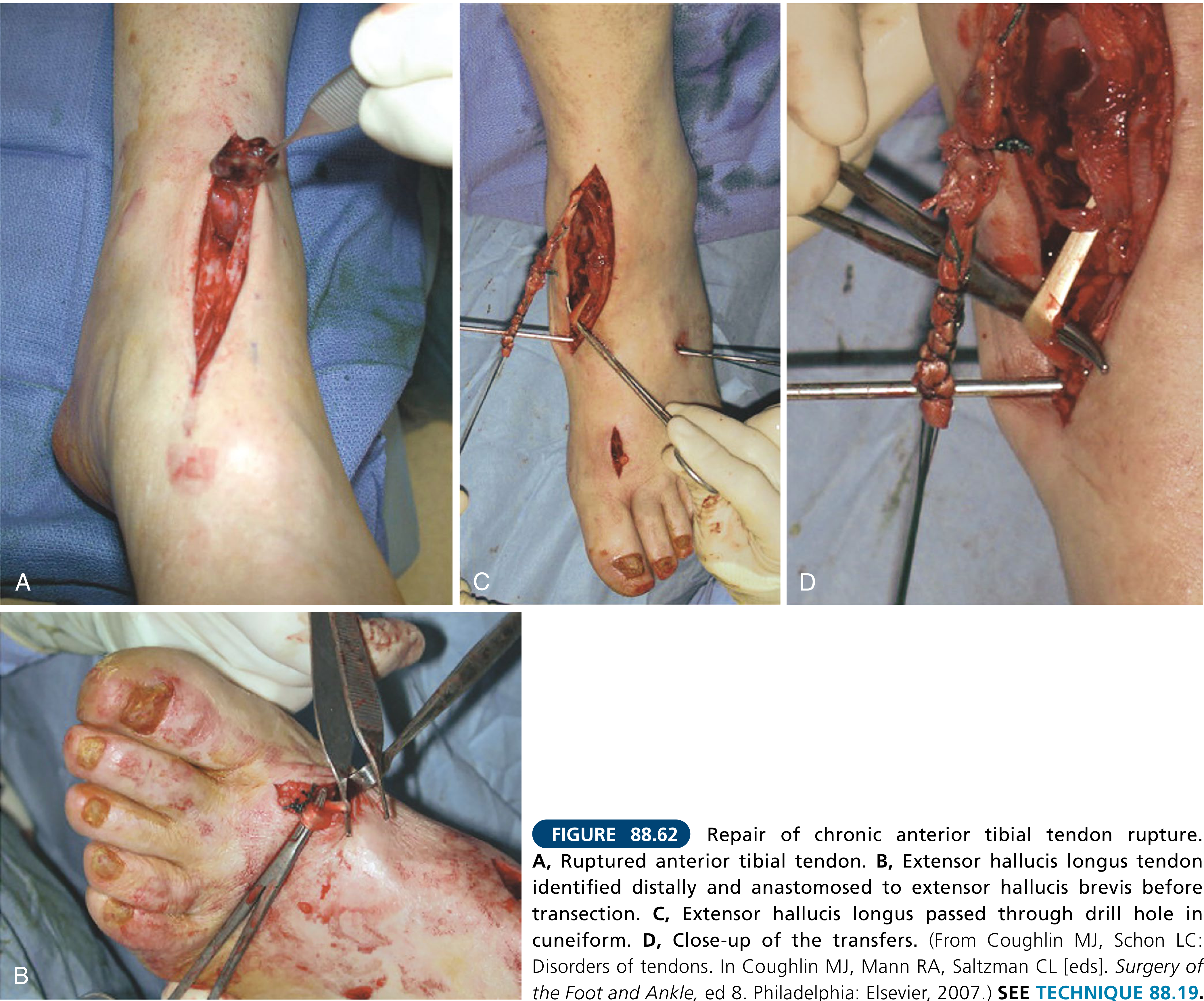
- Campbell's, p. 4886
Technique 88.20: Repair of Complete Rupture of the Anterior Tibial Tendon
(Uses same incision as Technique 88.18 - medial to anterior tibial tendon, does not cross ankle joint)
For rupture beneath the INFEROMEDIAL limb of inferior extensor retinaculum:
- Incise the inferomedial band of inferior extensor retinaculum
- Advance the tendon as far distally as possible
- Drill a hole in the medial cuneiform from dorsal to plantar (enlarge incrementally to 0.635-2.5 cm)
- Pass tendon through the hole with a Bunnell weave of No. 0 nonabsorbable suture on free straight needles; pull tendon into hole while dorsiflexing ankle 20-30 degrees; suture tendon edge to adjacent periosteum and deep fascia
For rupture beneath the SUPEROMEDIAL limb:
- Incise the superomedial limb of inferior extensor retinaculum
- If tendon cannot be advanced: make a second small incision (2-3 cm) anteromedially above the ankle to retrieve the proximal stump
- Pass a long curved clamp along the tendon proximal-to-distal; use a rubber urinary catheter sutured to tendon to pull it distally if needed
- If tendon cannot reach medial cuneiform: use navicular as the bony insertion instead
If gap cannot be bridged by primary repair (Sammarco et al. - Tendon Reconstruction):
- Interpositional graft sources (preferred order): plantaris, extensor digitorum, peroneus tertius, Achilles; semitendinosus allograft
- Fold grafts 2-3 times to match TA tendon diameter
- Repair extensor retinaculum to prevent bowstringing
- Campbell's, p. 4887-4888
Technique 88.21: Minimally Invasive Tendon Reconstruction with Semitendinosus Autograft (Michels et al.)
- If 5 degrees of dorsiflexion cannot be achieved preoperatively: perform gastrocnemius recession first
- Harvest semitendinosus with a tendon stripper (as in ACL reconstruction)
- Small longitudinal incision above superior extensor retinaculum, leaving extensor retinaculum intact (minimizes adhesions, decreases recovery time)
- Evidence shows this results in minimal disturbance of the extensor retinaculum, decreasing recovery time, adhesions, and wound problems
- Campbell's, p. 4888
Postoperative Care Summary
| Procedure | Cast | Weight Bearing | Boot / Return |
|---|---|---|---|
| Synovectomy (88.18) | Short leg, 4-6 weeks | Immediate (to tolerance, crutches day 1-7) | Compressive ankle corset for additional 4-6 weeks |
| Debridement ± EHL transfer (88.19) | Boot with heel wedges | Gradual | Shoe wear ~8 weeks; full activities 3-4 months |
| Complete repair (88.20) | Short leg cast, ankle at 0°, 4-6 weeks | Allowed after 3 weeks | Hinged boot (full DF, no PF) until 10-12 weeks; autograft cases wear cast 6 weeks |
Evidence Summary (Campbell's Systematic Review)
In a systematic review cited in Campbell's 15th Ed, treatment outcomes ranked as follows (best to less optimal):
- Direct repair, turn-down tibialis anterior flap, semitendinosus autograft, EDL autograft, plantaris autograft - all showed better outcomes than EHL autograft reconstruction techniques
- Allograft vs. autograft reconstruction: both achieved good patient-reported outcomes and both graft types remained intact in comparison studies
- Key principle: when TA muscle shows fatty infiltration on MRI, free tendon graft is not useful - EHL transfer is preferred in this scenario
Miller's Review Key Points (Orthopaedic Board Summary)
- Tenosynovitis: uncommon; seen in inflammatory arthritis; NSAIDs + walking cast/boot; corticosteroid injections may provide relief but increase risk of tendon rupture
- Complete ruptures: rare, mainly older patients; commonly missed diagnosis (presents as painless anterior ankle mass; foot drop subtle due to toe extensor recruitment)
- Primary repair generally improves functional results regardless of age
- Tendon grafting augmentation if adequate excursion and healthy muscle
- Interpositional graft required in delayed cases
- Consider adding gastrocnemius recession/Achilles lengthening to take tension off the TA reconstruction
- Miller's Review of Orthopaedics 9th Ed, p. 591
Note on Hoppenfield
Hoppenfield is not available in this textbook library. The classification and management principles attributed to "Hoppenfield" in the context of TA rupture most commonly refer to the anatomical/surgical approach descriptions in Surgical Exposures in Orthopaedics (Hoppenfield & deBoer), which describes the anterior approach to the ankle and dorsum of the foot - the same approach used for TA repair. The incision is made medial to the tibialis anterior tendon, staying lateral to the great saphenous vein and medial to the superficial peroneal nerve branches, consistent with what Campbell's describes in Technique 88.18-88.20.
This is a shared conversation. Sign in to Orris to start your own chat.