

1000307531.jpg
Explain this condition
analyse_media
Reading File
Now let me search the medical library for relevant content on the key finding - poor inspiratory effort / suboptimal chest X-ray, bilateral bronchovascular markings, and potential underlying pathology in a 63-year-old male.
Finding Sources
Finding Sources
Finding Sources
Finding Sources
Reading File
Finding Sources
I now have all the information I need to give a comprehensive explanation. Let me also fetch the reference CXR image from the textbook.
Loading Image
Reading File
I now have comprehensive information to provide a thorough explanation. Here is a complete breakdown:
Chest X-Ray Analysis - Mr. Sehatlal Nagesh, 63 yrs Male
Patient details on the film: Mr. Sehatlal Nagesh, 63-year-old male, chest X-ray dated 27.07.2026, Ayushman Hospital, Balaghat. Reporting physician: Dr. B.M. Sharnagat.
Key Radiological Findings
1. Suboptimal Inspiratory Effort (Most Prominent Finding)
The most striking technical issue on this film is poor inspiratory effort. Only about 5-6 anterior rib interspaces are visible above the diaphragm (a normal full-inspiration PA chest X-ray should show at least 10 posterior ribs or 6 anterior ribs). Both hemidiaphragms are elevated. This single factor is responsible for several apparent abnormalities:
- It makes the lung tissue look crowded and denser than it really is
- It causes the heart to appear wider and more horizontal ("pseudo-cardiomegaly")
- It obscures the costophrenic angles at the lung bases
- It makes bronchovascular markings appear more prominent
2. Bilateral Prominent Bronchovascular Markings
There are diffuse, increased bronchovascular markings visible bilaterally, especially in the perihilar and mid-to-lower zones. This is likely at least partly artifactual from the poor inspiration (tissue crowding). However, when seen in a 63-year-old male, genuine increased markings can be a sign of:
- Chronic Bronchitis / COPD - The "blue bloater" pattern of COPD (centrilobular emphysema) classically shows increased lung markings rather than arterial deficiency. As noted in Fishman's Pulmonary Diseases and Disorders, "patients who have the pattern of increased lung markings often have centrilobular emphysema and manifest the 'blue bloater' syndrome."
- Cardiac failure - Pulmonary venous congestion from left heart failure increases perihilar and basal vascular markings
- Chronic infections - Recurrent pneumonia, bronchiectasis
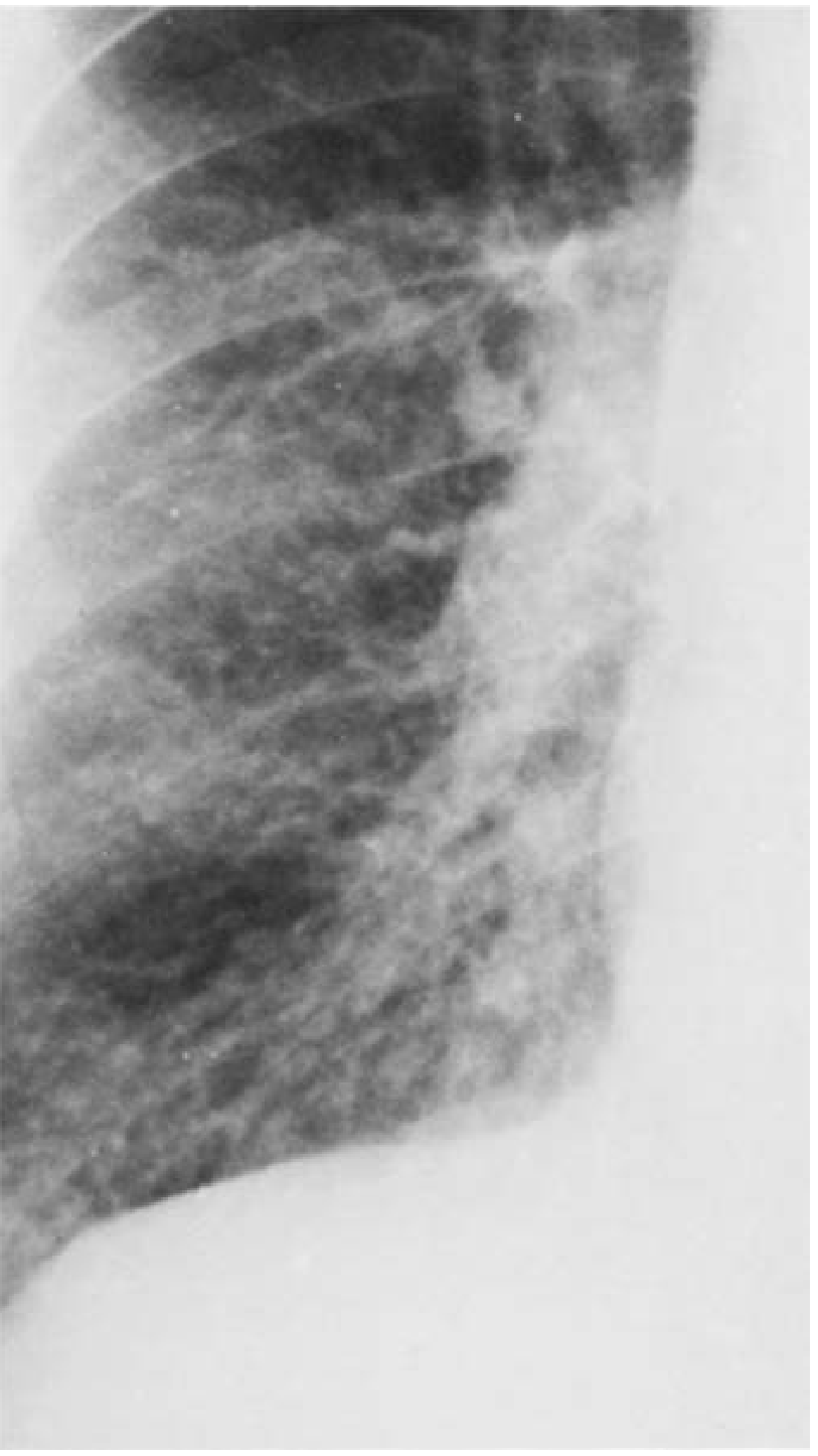
3. Cardiac Silhouette
The heart size cannot be accurately assessed on this film because of the poor inspiration. A falsely wide cardiac shadow is a well-known artifact of expiratory-phase or inadequate inspiration films. Formal assessment of cardiomegaly requires the cardiothoracic ratio (CTR) on a good inspiratory PA film.
4. Costophrenic Angles
Both costophrenic angles are partly obscured by elevated diaphragms. No large free-flowing pleural effusion is clearly seen, though a small/subpulmonic effusion cannot be excluded.
5. Mediastinum and Trachea
The trachea is midline. No mediastinal shift or widening is noted.
6. Bones and Soft Tissues
No rib fractures, lytic lesions, or bony pathology detected.
Summary / Radiological Impression
| Finding | Interpretation |
|---|---|
| Elevated diaphragms, only 5-6 anterior ribs | Poor inspiratory effort - limits full evaluation |
| Bilateral increased bronchovascular markings | Could be artifact (poor inspiration) or real pathology |
| Heart silhouette | Cannot assess accurately - film is suboptimal |
| Costophrenic angles | Partially obscured; no obvious large effusion |
| No focal consolidation | Pneumonia less likely; no mass seen |
Clinical Correlation Needed
Given the patient's age (63 years), a repeat PA chest X-ray with full inspiration is strongly recommended before drawing firm conclusions. The treating physician should correlate with:
- Smoking history (key risk factor for COPD)
- Symptoms: chronic cough, sputum production, exertional breathlessness (pointing to COPD/chronic bronchitis)
- Symptoms: orthopnoea, ankle swelling, paroxysmal nocturnal dyspnoea (pointing to cardiac failure)
- SpO2, spirometry (FEV1/FVC ratio), and ECG would be the next appropriate investigations
As stated in Fishman's Pulmonary Diseases and Disorders: "The radiograph rarely provides substantive help [in chronic bronchitis]. Vascular markings throughout the lung fields are sometimes prominent, but this finding is nonspecific."
Disclaimer: This is an educational interpretation for learning purposes. A formal radiological report by a qualified radiologist, correlated with the patient's full clinical history, is required for any clinical decision-making.
This is a shared conversation. Sign in to Orris to start your own chat.