
17 yr old female with pou since 1 month her CBC,lft,esr,crp, procalcitonin,ure,urine tb pcr,lepto ,dengue , chikengunya,Widal weil felix test, malaria , ana, ra factor , aso , chest xray and usg abdomen with pelvis all are normal , blood culture normal no bacterimia, tsh normal what is the next step
"fever of unknown origin" adolescent diagnosis workup
"fever of unknown origin" diagnosis next steps CT PET
PUO pyrexia unknown origin adolescent next investigation CT scan PET lymphoma autoimmune 2024
https://www.pediatriconcall.com/pediatric-journal/view/fullt…
PUO in a 17-Year-Old Female - Next Steps After Negative Tier-1 & Tier-2 Workup
Immediate Gaps to Fill First
| Missing Investigation | Why It Matters |
|---|---|
| EBV serology (monospot/EBV VCA IgM/IgG) | Infectious mononucleosis is the most common missed cause in adolescents |
| CMV serology / CMV PCR | Second most common missed viral cause in this age group |
| HIV ELISA | Mandatory in any PUO - must not be missed |
| Parvovirus B19 IgM | Can cause prolonged fever in adolescents |
| Serum ferritin | If markedly elevated (>10,000 ng/mL), strongly suggests systemic JIA (Still's disease) or macrophage activation syndrome (MAS) |
| Peripheral blood smear | Atypical lymphocytes (EBV/CMV), blasts (leukemia), or parasites |
| Urine routine/microscopy repeat | Renal TB, glomerulonephritis |
| LDH + uric acid | If elevated, raises concern for occult lymphoma |
| Serum ACE level | Sarcoidosis |
| IGRA (Quantiferon Gold) or Mantoux | TB even with normal CXR and negative urine TB PCR |
Tier-3: Next Investigations (Now Indicated)
1. CECT Chest, Abdomen and Pelvis (HIGH PRIORITY)
- USG can miss deep lymphadenopathy, mediastinal nodes, early solid tumors, and retroperitoneal masses
- Rules out occult lymphoma (Hodgkin's is classic in adolescent females), solid tumors, occult abscess, hepatosplenic TB, and mesenteric adenitis
- Must be done before bone marrow or biopsy
2. Echocardiography (HIGH PRIORITY)
- Blood cultures were negative, but culture-negative endocarditis (Bartonella, Coxiella/Q-fever, HACEK organisms, fungi) is a real entity
- Also screens for atrial myxoma - a rare but important cause of PUO with normal inflammatory markers
- Also request Coxiella burnetti (Q-fever) serology and Bartonella serology if echo shows any vegetation
3. 18F-FDG PET/CT (TIER-3 GOLD STANDARD if CT is inconclusive)
- Has the highest diagnostic yield (50-60% positive findings) in classic PUO after negative initial workup
- Identifies occult infections, deep lymph node disease, granulomatous inflammation, and malignancy simultaneously
- The EANM 2024 consensus guidelines recommend FDG-PET/CT early in the PUO workup to guide targeted biopsy
- Recent Frontiers in Medicine 2024 cohort (n=284) confirmed strong diagnostic value particularly for infection/inflammation and lymphoma
4. Bone Marrow Aspirate + Biopsy (if CT/PET non-revealing)
- Detects: leukemia, lymphoma with marrow infiltration, hemophagocytic lymphohistiocytosis (HLH), disseminated TB, leishmaniasis, histoplasmosis
- Send for culture (AFB + fungal), histology, and flow cytometry
5. Repeat Detailed Physical Examination
- Check specifically for: evanescent salmon-colored rash (Still's disease), lymph nodes (especially epitrochlear, posterior cervical), splenomegaly, subtle joint swelling, oral ulcers (Behcet's), rashes of SLE/vasculitis, fundoscopy for uveitis
Differential Diagnoses to Actively Rule Out in a 17-Year-Old Female
| Category | Specific Diagnosis | Key Test |
|---|---|---|
| Infection | EBV, CMV mononucleosis | EBV/CMV serology |
| Infection | Culture-negative endocarditis | Echo + Bartonella/Coxiella serology |
| Infection | Disseminated TB (normal CXR possible) | IGRA, CECT, bone marrow culture |
| Infection | Visceral leishmaniasis (kala-azar) | rK39 rapid test, bone marrow |
| Malignancy | Hodgkin's lymphoma | CECT / PET-CT, node biopsy |
| Malignancy | Acute leukemia | Blood smear + bone marrow |
| Autoimmune | Systemic JIA / Adult-onset Still's disease | Ferritin >10,000, rash, arthritis |
| Autoimmune | SLE (incomplete/early form) | ANA was done - but add anti-dsDNA, C3/C4 |
| Autoimmune | Vasculitis (Takayasu's in this age) | CECT angio, ESR |
| Autoinflammatory | Familial Mediterranean fever | MEFV gene mutation testing |
| Hematologic | HLH | Ferritin, triglycerides, fibrinogen, NK cell activity |
| Drug fever | Any concurrent medication | Stop all non-essential drugs |
| Factitious fever | Psychological cause | Observe temperature under supervision |
Practical Step-by-Step Priority Order
- Send immediately: EBV/CMV serology, HIV, serum ferritin, LDH, peripheral smear, IGRA, Bartonella/Coxiella serology, anti-dsDNA + C3/C4, rK39
- Imaging: CECT chest/abdomen/pelvis + Echocardiography
- If CT inconclusive: FDG-PET/CT
- If PET/CT inconclusive or lymph node identified: Lymph node biopsy (do not empirically treat)
- If all above negative: Bone marrow aspirate + biopsy
- Do NOT start empirical steroids or antibiotics unless diagnosis is confirmed or there is high clinical suspicion with imminent deterioration - this can mask lymphoma and infectious causes
17 yr old female with pou since 1 month her CBC,lft,esr,crp, procalcitonin,ure,urine tb pcr,lepto ,dengue , chikengunya,Widal weil felix test, malaria , ana, ra factor , aso , chest xray and usg abdomen with pelvis all are normal , LDH normal, blood culture normal no bacterimia, peripheral smear shows iron deficiency anemia , serum ferritin is normal (on lower side) tsh normal what is the next step
PUO iron deficiency anemia adolescent female IBD Crohn celiac GI cause workup 2024
PUO + Iron Deficiency Anemia (Normal-Low Ferritin) in a 17-Year-Old Female - Next Steps
What the New Finding Tells You
- Normal ferritin in IDA = ferritin was not elevated by acute-phase response, suggesting the anemia is from true iron depletion, NOT anemia of chronic disease
- In a 17-year-old female, IDA has three main causes: menstrual blood loss, GI blood loss/malabsorption, or dietary deficiency
- The fever + IDA combination strongly points to a GI source - particularly inflammatory bowel disease (Crohn's), celiac disease, or intestinal tuberculosis / parasitic infection
Step 1: Critical Questions to Ask (History Gaps)
| Question | Why It Matters |
|---|---|
| Menstrual history - heavy periods, cycle duration, clot passage? | If menorrhagia is present, this could explain IDA without needing further GI workup first |
| Any abdominal pain, diarrhea, blood/mucus in stool? | Even intermittent or mild - Crohn's can be subtle |
| Weight loss, anorexia? | IBD, intestinal TB, celiac |
| Mouth ulcers, perianal disease, skin rashes? | Crohn's extraintestinal features |
| Diet history - vegetarian/vegan, poor intake? | Dietary IDA |
| Any medications including NSAIDs? | NSAID-induced GI erosions causing occult blood loss + drug fever |
Step 2: Immediate Blood Tests to Add
| Test | Rationale |
|---|---|
| Anti-tissue transglutaminase IgA (TTG-IgA) + total serum IgA | Screen for celiac disease - can present as isolated IDA without diarrhea (Harrison's, p. 2588) |
| Anti-deamidated gliadin peptide IgG (DGP-IgG) | If IgA-deficient (check total IgA first) |
| Serum iron + TIBC + transferrin saturation | Confirm true IDA vs. mixed picture |
| EBV IgM/IgG + CMV serology | Still not sent - most commonly missed viral cause in this age |
| HIV ELISA | Mandatory in any PUO |
| Stool for occult blood (FOBT x 3 samples) | Screen for GI blood loss |
| Stool for ova, cysts, parasites + concentration technique | Hookworm (Ancylostoma), Trichuris, Strongyloides - all cause IDA + fever |
| Stool for Helicobacter pylori antigen | H. pylori causes IDA via chronic gastric blood loss and impairs iron absorption |
| IGRA (Quantiferon Gold) | Intestinal TB can have normal CXR and normal urine TB PCR |
| Serum calprotectin (or fecal calprotectin) | If elevated, strongly suggests IBD |
Step 3: Imaging - CECT Abdomen and Pelvis (NOW INDICATED)
- USG abdomen was normal but cannot reliably detect bowel wall thickening, mesenteric fat stranding, skip lesions of Crohn's, or ileocecal TB
- CECT specifically evaluates: terminal ileum, ileocecal junction, mesenteric lymph nodes, bowel wall enhancement
- Request CT enterography (CTE) if bowel disease is suspected - this is more sensitive for small bowel Crohn's than standard CECT
Step 4: Endoscopy (HIGH PRIORITY - TIER 3)
A. Upper GI Endoscopy (OGD) with duodenal biopsy
- Confirms celiac disease (Marsh classification on biopsy - villus blunting, crypt hyperplasia, intraepithelial lymphocytes)
- Detects H. pylori gastritis, erosive esophagitis, peptic ulcer disease causing occult blood loss
- Per Harrison's: "Diagnosis [of celiac] in adults with positive antibody levels is confirmed by endoscopy with small-intestinal biopsy"
B. Ileocolonoscopy with terminal ileum intubation + biopsies
- Detects Crohn's disease (cobblestoning, skip lesions, aphthous ulcers, ileal involvement)
- Detects ulcerative colitis, intestinal TB (ileocecal area), CMV colitis
- Intestinal TB can look exactly like Crohn's on colonoscopy - send biopsies for AFB culture, PCR, and histology (caseating vs. non-caseating granulomas)
Step 5: Gynecological Evaluation (Parallel Track)
- Refer to gynecology for assessment of menstrual blood loss as a contributing cause of IDA
- Pelvic USG was normal (rules out structural causes like fibroids, ovarian cysts)
- Assess for anovulatory cycles - common in adolescent females - which can cause heavy dysfunctional uterine bleeding without structural abnormality
- If menorrhagia confirmed: assess for von Willebrand disease (coagulation screen, VWF antigen + activity) - an important and commonly missed cause of heavy periods + IDA in teenage girls
Revised Differential Diagnosis with IDA as a Clue
| Diagnosis | Probability | Key Features Supporting It |
|---|---|---|
| Crohn's disease | HIGH | Fever + IDA + normal CRP (early/mild Crohn's can have normal CRP) + adolescent female |
| Celiac disease | HIGH | IDA as sole presentation (no diarrhea in 30-40% of celiac cases); per Harrison's: "isolated iron-deficiency anemia due to duodenal disease" is a recognized presentation |
| Intestinal TB | HIGH (in Indian context) | Fever + IDA + normal CXR (extrapulmonary TB); ileocecal region most common |
| Hookworm / intestinal parasites | MODERATE | Chronic GI blood loss + malabsorption + fever |
| H. pylori gastritis | MODERATE | Common, causes iron malabsorption + occult GI blood loss + fever |
| Menorrhagia + coincidental viral fever | MODERATE | Must be excluded gynecologically |
| IBD-associated systemic JIA/Still's | LOW (ferritin would be high) | Ferritin low-normal makes this less likely |
| Lymphoma | LOW (LDH normal) | Normal LDH + normal USG makes this less likely for now |
Practical Priority Order
TODAY:
1. Detailed menstrual history + gynecology referral
2. Send: TTG-IgA + total serum IgA, EBV/CMV serology, HIV, IGRA,
Stool FOBT x3, stool OCP, stool H. pylori antigen,
serum iron + TIBC + transferrin saturation, fecal calprotectin
NEXT 48-72 HOURS:
3. CECT abdomen/pelvis (or CT enterography if bowel disease suspected)
4. Echocardiography (still needed - culture-negative endocarditis)
AFTER IMAGING RESULTS:
5. OGD with duodenal biopsy (celiac) + H. pylori CLO test
6. Ileocolonoscopy with terminal ileum intubation + biopsies
(Crohn's vs. intestinal TB - send AFB culture, PCR, histology)
IF ALL ABOVE NEGATIVE:
7. Bone marrow aspirate + biopsy
8. FDG-PET/CT
How to read igra test
how to interpret IGRA QuantiFERON TB Gold Plus result positive negative indeterminate cutoff 2024
How to Read an IGRA Test
What IGRA Measures
- QuantiFERON-TB Gold Plus (QFT-Plus) - ELISA-based, measures IFN-γ in IU/mL
- T-SPOT.TB - ELISPOT-based, counts spot-forming cells (SFCs)
Platform 1: QuantiFERON-TB Gold Plus (QFT-Plus)
The Four Tubes
| Tube | Contains | Purpose |
|---|---|---|
| Nil | Nothing | Baseline background IFN-γ (internal negative control) |
| TB1 | ESAT-6 + CFP-10 peptides | CD4+ T-cell response |
| TB2 | ESAT-6 + CFP-10 + additional CD8 peptides | CD4+ + CD8+ T-cell response |
| Mitogen | Phytohaemagglutinin (PHA) | Positive control - confirms cells are alive and responsive |
How to Calculate Values
- TB1 - Nil = TB1 response
- TB2 - Nil = TB2 response
- Mitogen - Nil = Mitogen response
Official QFT-Plus Interpretation Table
| Nil (IU/mL) | TB1 - Nil (IU/mL) | TB2 - Nil (IU/mL) | Mitogen - Nil (IU/mL) | Result | Meaning |
|---|---|---|---|---|---|
| ≤8.0 | ≥0.35 AND ≥25% of Nil | Any | Any | POSITIVE | M. tuberculosis infection likely |
| ≤8.0 | Any | ≥0.35 AND ≥25% of Nil | Any | POSITIVE | M. tuberculosis infection likely |
| ≤8.0 | <0.35 OR <25% of Nil | <0.35 OR <25% of Nil | ≥0.50 | NEGATIVE | M. tuberculosis infection NOT likely |
| ≤8.0 | <0.35 OR <25% of Nil | <0.35 OR <25% of Nil | <0.50 | INDETERMINATE | Cannot be determined |
| >8.0 | Any | Any | Any | INDETERMINATE | Cannot be determined |
The "25% of Nil" Rule - Why It Exists
- Nil = 4.0 IU/mL (already high baseline)
- TB1 - Nil = 0.40 IU/mL (meets ≥0.35 cutoff)
- But 0.40 is only 10% of 4.0 → does NOT meet the 25% rule → NOT positive
Platform 2: T-SPOT.TB (ELISPOT)
Four Wells
| Well | Contents |
|---|---|
| Nil control | No antigen (background) |
| Panel A | ESAT-6 peptides |
| Panel B | CFP-10 peptides |
| Mitogen control | Positive control |
T-SPOT Interpretation
| Adjusted Spot Count | Result |
|---|---|
| ≥8 SFCs in Panel A OR Panel B | POSITIVE |
| 5-7 SFCs | BORDERLINE (repeat or use clinical judgment) |
| ≤4 SFCs | NEGATIVE |
| Nil >10 SFCs OR Mitogen <20 SFCs | INDETERMINATE |
What Each Result Means Clinically
✅ POSITIVE
- TB infection (latent or active) is likely
- Does NOT differentiate between latent TB infection (LTBI) and active TB disease
- Does NOT tell you when infection occurred (recent vs. old)
- Magnitude of the value (e.g., 5.0 vs. 0.5 IU/mL) does NOT correlate with disease stage, activity, or risk of progression
- False positives with: M. kansasii, M. marinum, M. szulgai, M. gordonae (NOT with BCG vaccination)
❌ NEGATIVE
- TB infection is not likely
- But a negative result does NOT exclude active TB, especially in:
- Immunocompromised patients (HIV, on steroids, biologics)
- Very early infection (window period: 8-12 weeks after exposure)
- Miliary TB or severely immunosuppressed (reduced sensitivity)
- Goldman-Cecil states: "The sensitivity of these tests is reduced in immunocompromised persons, persons with sarcoidosis, after live virus vaccination, and in persons with active TB"
⚠️ INDETERMINATE
- Nil >8.0 IU/mL - High background IFN-γ (patient has active inflammatory/autoimmune process, or non-specific stimulation of cells). Blood may have been incubated too long.
- Mitogen - Nil <0.5 IU/mL - Cells failed to respond to the positive control = immunosuppression, poor sample handling, improper tube filling, delay in transport
Key Advantages Over TST (Mantoux)
| Feature | IGRA | TST/Mantoux |
|---|---|---|
| BCG cross-reactivity | No - preferred in BCG-vaccinated patients | Yes - false positive common |
| Requires return visit | No (single blood draw) | Yes (48-72 hr reading) |
| Reader subjectivity | None | High |
| NTM cross-reactivity | Only 4 specific NTMs | Many NTMs |
| Best use | BCG-vaccinated, serial testing, adults | Children <5 years, limited lab access |
Critical Points to Remember
- IGRA cannot distinguish LTBI from active TB - a positive result needs clinical context, CXR, and symptoms evaluation
- Do not repeat IGRA after treatment - it usually stays positive even after successful treatment for LTBI; this is expected and not a sign of failure
- Low-positive values (0.35-1.0 IU/mL) are in a "gray zone" - serial testing in this range can fluctuate between positive and negative; always interpret with clinical risk
- In high-risk patients (HIV, household contacts), if IGRA is negative but clinical suspicion is high, treat for LTBI anyway - both TST and IGRA can be false negative
- Dual testing (IGRA + TST together) is recommended only in high-risk + high-progression-risk patients (e.g., household contact + HIV); if either is positive, treat
Interpret this report
python3 -c " import pdfplumber with pdfplumber.open('/tmp/workspace/attachments/e5d8a0c2-ed11-4de1-85b0-b56cb46901bf/IGRA.pdf') as pdf: for i, page in enumerate(pdf.pages): print(f'=== PAGE {i+1} ===') text = page.extract_text() print(text) print() tables = page.extract_tables() for j, table in enumerate(tables): print(f'--- TABLE {j+1} ---') for row in table: print(row) print() "
IGRA Report Interpretation - Ms. Jumana, 15F
Raw Values from the Report
| Parameter | Value (IU/mL) |
|---|---|
| Gamma Interferon, Antigen tube | 0.95 |
| Gamma Interferon, Nil tube (background) | 0.69 |
| TB Ag minus Nil | 0.25 |
| Final Result | NEGATIVE |
Step-by-Step Interpretation
Step 1: Is the Nil valid?
- Nil = 0.69 IU/mL → ≤8.0 IU/mL ✅
- The background is acceptable. The test is valid.
Step 2: Apply the two-part positivity criteria to TB Ag - Nil (0.25):
- 0.25 < 0.35 → ❌ FAILS
- 25% of 0.69 = 0.17 IU/mL
- 0.25 > 0.17 → ✅ Would pass this part...
- But since Criterion A already fails (<0.35), this is moot.
✅ RESULT: NEGATIVE
M. tuberculosis infection is NOT likely
Important Clinical Caveats for This Patient
1. This is a "Gray Zone Negative"
- TB Ag - Nil = 0.25 is close to the 0.35 cutoff but below it
- The Nil (background) is relatively high at 0.69 (normal is usually <0.2-0.3)
- A high Nil can represent non-specific immune activation from an active inflammatory or infectious process - which is consistent with her ongoing fever
2. IGRA Does NOT Rule Out Active TB or Intestinal TB
- IGRA is designed to detect latent TB infection (LTBI)
- In active TB (especially extrapulmonary/intestinal TB), IGRA sensitivity drops to 60-80%
- The test cannot exclude intestinal TB, which remains in her differential
- Per Goldman-Cecil: "The sensitivity of these tests is reduced in... persons with active TB"
3. No Mitogen Control Reported
- This lab did not include a mitogen tube (positive control for immune function)
- Without the mitogen result, we cannot confirm that her immune cells were functioning properly
- The lab note acknowledges this: "Where a poor response due to an immunosuppressive condition is suspected, a Mitogen control may be used"
- This is a significant limitation of this particular report
4. Window Period
- If she was exposed to TB within the last 8-12 weeks, IGRA can be falsely negative (pre-seroconversion window period)
What to Do Clinically
| Action | Rationale |
|---|---|
| Do NOT use this result alone to exclude TB | Intestinal TB remains a possibility with PUO + IDA + high Nil background |
| Proceed with CECT abdomen + ileocolonoscopy | The definitive investigation for intestinal TB |
| If colonoscopy shows ileocecal lesion | Send biopsy for AFB culture, TB PCR, and histology (caseating granulomas) |
| Consider repeat IGRA with mitogen tube | If immune suppression is suspected, mitogen control is needed to validate the negative result |
17 yr old female with pou since 1 month her CBC,lft,esr,crp, procalcitonin,ure,urine tb pcr,lepto ,dengue , chikengunya,Widal, weil felix test, malaria , ana, ra factor , aso , chest xray and usg abdomen with pelvis all are normal , blood culture normal no bacterimia, tsh normal igra negative , stool calprotectin negative what is the next step
PUO negative calprotectin negative IGRA next step CT echocardiography EBV CMV Still disease approach 2024
PUO - 17F - Updated Workup After Negative IGRA + Negative Stool Calprotectin
What the New Negatives Tell You
| Result | What It Effectively Rules Out |
|---|---|
| Stool calprotectin negative | Active intestinal inflammation - IBD (Crohn's, UC) is now very unlikely (NPV >93%). Per Frameworks for Internal Medicine: FUO from IBD is uncommon and requires mucosal inflammation to cause calprotectin elevation |
| IGRA negative | Latent TB (LTBI) unlikely - but does NOT exclude active/extrapulmonary TB in a symptomatic patient |
| Negative ANA, RA factor, ASO | Common autoimmune causes (SLE, RA, rheumatic fever) screened but note: ANA alone has a high false-positive rate in FUO; early SLE can have negative ANA |
What Is Still NOT Ruled Out
🔴 Critical Missing Tests (Should Have Been Done Already)
| Test | Why Still Urgently Needed |
|---|---|
| EBV VCA IgM + IgG, EBNA IgG | Most common missed cause in 15-25 year age group; heterophile antibody (monospot) can be negative in early infection |
| CMV IgM + IgG / CMV PCR | Second most common missed viral cause in this age; can cause 4-6 week febrile illness |
| HIV ELISA + p24 antigen | Mandatory in any PUO; primary HIV infection causes febrile illness lasting weeks |
| Serum ferritin | Single most important test now: if markedly elevated (>2000 ng/mL), this is virtually diagnostic of Adult-onset Still's disease (AOSD) - peaks at ages 15-25 years |
| Anti-dsDNA + C3 + C4 | ANA was negative but early SLE can be ANA-negative; anti-dsDNA is more specific; low C3/C4 with leucopenia suggests SLE |
| Serum ACE level | Sarcoidosis - presents with bilateral hilar lymphadenopathy (can be missed on plain CXR) |
| Bartonella henselae serology | Cat scratch disease - causes FUO with or without visible lymphadenopathy; transmitted by cat scratch/bite |
| Coxiella burnetii (Q fever) serology | Blood culture-negative fever; occupational/animal exposure |
| Brucella serology | Important in India; can cause subacute fever for weeks; Widal was done but not Brucella agglutination |
Tier-3 Investigations Now Indicated
1. CECT Chest + Abdomen + Pelvis (TOP PRIORITY)
- Detects mediastinal and hilar lymphadenopathy (missed on plain CXR) - Hodgkin's lymphoma, sarcoidosis
- Detects retroperitoneal adenopathy - lymphoma
- Identifies occult solid tumors, splenic lesions, hepatic lesions
- Diagnoses intestinal TB (ileocecal thickening, mesenteric nodes) even with negative IGRA and normal CXR
- Per Frameworks for Internal Medicine: "The posterior cervical, epitrochlear, supraclavicular, hilar, mediastinal, and retroperitoneal lymph nodes are most likely to provide a diagnosis in patients with FUO"
2. Echocardiography
- Rules out culture-negative endocarditis (Bartonella, Coxiella, HACEK organisms, fungi)
- Rules out atrial myxoma - causes constitutional symptoms, fever, emboli - often missed as it can be normal on USG
- Blood cultures were negative but this does not exclude culture-negative endocarditis
3. Detailed Repeat Clinical Examination (Every Day)
- Salmon-pink evanescent rash appearing during fever - pathognomonic of AOSD (Systemic JIA)
- Per Frameworks: "AOSD is associated with bimodal age distribution with peaks between ages 15-25 years... The rash often appears when the patient is febrile and disappears during afebrile periods... Markedly elevated serum ferritin levels often >2000 ng/mL are characteristic"
- Lymphadenopathy (epitrochlear nodes especially - cat scratch disease, lymphoma)
- Pharyngitis (AOSD, EBV)
- Joint swelling/tenderness (AOSD)
- Any skin lesion, oral ulcer, perianal lesion
Revised Priority Differential Diagnosis
| Rank | Diagnosis | Key Supporting Feature | Confirmatory Test Needed |
|---|---|---|---|
| 1 | EBV / CMV infection | Most common cause in 15-25 age group; can last 4-6 weeks | EBV/CMV serology (not yet done) |
| 2 | Adult-onset Still's Disease (AOSD) | Peak age 15-25; quotidian fever; evanescent rash; pharyngitis | Serum ferritin >2000, Yamaguchi criteria |
| 3 | Hodgkin's Lymphoma | Classic "B symptoms" fever in adolescent; LDH was normal but can be normal early | CECT chest (mediastinal nodes), biopsy |
| 4 | Sarcoidosis | Hilar adenopathy on CXR/CT; ACE level elevated | CT chest, serum ACE, tissue biopsy |
| 5 | Intestinal TB | Normal IGRA does NOT exclude it; normal CXR is compatible | CECT abdomen, colonoscopy + biopsy |
| 6 | Culture-negative endocarditis | Bartonella, Q-fever, HACEK - all blood culture negative | Echo + specific serology |
| 7 | Cat scratch disease | Bartonella henselae - FUO without obvious lymphadenopathy possible | Bartonella serology |
| 8 | Early/incomplete SLE | ANA can be negative early; add anti-dsDNA + C3/C4 | Anti-dsDNA, complement levels |
| 9 | Familial Mediterranean fever (FMF) | Recurrent episodes (check pattern) + serositis | MEFV gene mutation testing |
| 10 | Drug fever | Any concurrent medications? | Stop all non-essential drugs and observe |
Practical Step-by-Step Plan RIGHT NOW
SEND TODAY (Blood Tests):
✅ EBV VCA IgM + IgG + EBNA IgG
✅ CMV IgM + IgG (or CMV PCR)
✅ HIV ELISA + p24 antigen
✅ SERUM FERRITIN (critical - if >2000 = AOSD)
✅ Anti-dsDNA + C3 + C4
✅ Serum ACE
✅ Bartonella henselae IgM + IgG
✅ Brucella agglutination test (Rose Bengal + Standard Agglutination Test)
✅ Serum LDH + uric acid (if not done)
IMAGING (Within 48 Hours):
✅ CECT chest + abdomen + pelvis (with IV contrast)
✅ Echocardiography (transthoracic)
CLINICAL:
✅ Observe patient during fever spikes for evanescent rash
✅ Document fever pattern - quotidian (once daily) or double-quotidian (twice daily) = AOSD
✅ Ask about cat contact (Bartonella), animal contact (Brucella, Q-fever)
✅ Thorough drug history - rule out drug fever
IF CT SHOWS LYMPHADENOPATHY:
✅ Lymph node excision biopsy
(posterior cervical > epitrochlear > supraclavicular preferred over axillary/inguinal)
IF FERRITIN >2000 + QUOTIDIAN FEVER + RASH + PHARYNGITIS:
✅ Apply Yamaguchi criteria for AOSD diagnosis
✅ Refer rheumatology
IF ALL ABOVE NEGATIVE:
✅ 18F-FDG PET/CT (highest yield in unexplained PUO after 2nd tier workup)
✅ Bone marrow aspirate + biopsy
17 yr old female with pou since 1 month her CBC,lft,esr,crp, procalcitonin,ure,urine tb pcr,lepto ,dengue , chikengunya,Widal, weil felix test, malaria , ana, ra factor , aso , chest xray and usg abdomen with pelvis all are normal , blood culture normal no bacterimia, tsh normal igra negative , stool calprotectin negative , serum ferritin normal (on lower side) , peripheral smear shows iron deficiency anemia what is the next step
PUO iron deficiency anemia negative calprotectin negative IGRA 17 year old female next investigation EBV CMV celiac endoscopy
PUO + IDA (Low-Normal Ferritin) + Negative Calprotectin + Negative IGRA - What Now?
Synthesizing the Picture
| Finding | Interpretation |
|---|---|
| IDA on smear + low-normal ferritin | True iron depletion (NOT anemia of chronic disease - which would show high ferritin) |
| Negative stool calprotectin | Active mucosal intestinal inflammation (IBD) is unlikely |
| Negative IGRA | LTBI unlikely - but does NOT exclude intestinal/active TB |
| Normal ESR, CRP, procalcitonin | Low-grade or smoldering process; does NOT exclude early Crohn's, celiac, or malignancy |
| Normal USG abdomen + normal CXR | Deep structures not yet assessed |
| Normal LFT, CBC (apart from IDA) | Liver, bone marrow not grossly infiltrated |
| Normal ferritin (low-normal) | AOSD (Still's disease) effectively excluded - would expect ferritin >2000 ng/mL |
- GI malabsorption (celiac disease - most likely)
- Occult GI blood loss (H. pylori gastritis, peptic ulcer, small bowel lesion)
- Menstrual blood loss (heavy periods as IDA source, with a separate/coincidental fever cause)
- Occult malignancy (lymphoma - even with normal LDH, early Hodgkin's possible)
NEXT STEP 1 (URGENT - Send Today): Blood + Stool Tests
For the IDA:
| Test | Rationale |
|---|---|
| Anti-TTG IgA + total serum IgA | Single most important test now. Celiac disease classically presents as isolated IDA with NO diarrhea in up to 30-40% of cases; per Harrison's: "isolated iron-deficiency anemia due to duodenal disease" is a recognized presentation. Screening test of choice is TTG-IgA |
| Serum iron + TIBC + transferrin saturation | Confirm and quantify true IDA vs. mixed picture |
| Stool H. pylori antigen | H. pylori causes IDA via chronic gastritis + iron malabsorption; very prevalent in India |
| Stool for occult blood (FOBT x3) | Screen for GI blood loss - note: negative calprotectin does not exclude FOBT-positive blood loss |
| Stool for ova, cysts, parasites | Hookworm (Ancylostoma duodenale) - causes IDA + can cause fever; stool examination is essential |
For the PUO (still missing):
| Test | Rationale |
|---|---|
| EBV VCA IgM + IgG + EBNA | Most common missed cause in this age group; can cause 4-6 week PUO |
| CMV IgM + IgG | Second most common viral cause |
| HIV ELISA | Mandatory |
| Brucella agglutination (SAT + Rose Bengal) | Widal done but Brucella not mentioned - subacute febrile illness |
| Anti-dsDNA + C3 + C4 | ANA done but anti-dsDNA is more specific for SLE |
| Serum ACE | Sarcoidosis |
| LDH + uric acid | Lymphoma screen (even if previously "normal" - trend matters) |
NEXT STEP 2: Imaging - CECT Abdomen + Pelvis + Chest
- Deep retroperitoneal/mesenteric lymph nodes
- Mediastinal/hilar lymph nodes (Hodgkin's lymphoma, sarcoidosis)
- Bowel wall thickening (intestinal TB, early Crohn's - even with negative calprotectin)
- Hepatic/splenic micro-lesions
NEXT STEP 3: Echocardiography
NEXT STEP 4: Endoscopy (After Serology Results)
If TTG-IgA positive OR high clinical suspicion for celiac:
- OGD (Upper GI endoscopy) with 4-6 duodenal biopsies - Marsh classification (villus blunting, crypt hyperplasia, increased intraepithelial lymphocytes = Celiac)
- Also do CLO test for H. pylori during same scope
Even if TTG-IgA negative:
- OGD is still indicated in this case - a young female with PUO + IDA + fever where the GI source is not identified
- Ileocolonoscopy to assess terminal ileum (intestinal TB, Crohn's, even with negative calprotectin - early/patchy disease can give false-negative calprotectin)
Menstrual history first:
- Before ordering endoscopy, clarify menstrual blood loss - if heavy periods are the IDA source, the GI workup priority changes (though the fever still needs explaining)
- Ask specifically: clot passage, pad/tampon count per day, duration of heavy flow
- If menorrhagia confirmed: screen for von Willebrand disease (coagulation screen, VWF antigen + activity) - commonly missed in teenage girls with heavy periods
Revised Differential (After All Negatives So Far)
| Rank | Diagnosis | Why Still in Differential | Key Test to Confirm/Exclude |
|---|---|---|---|
| 1 | Celiac disease | IDA as sole presentation (no diarrhea needed); fever from nutritional/immune activation | TTG-IgA + duodenal biopsy |
| 2 | EBV/CMV infection | Not yet tested; most common cause in this age group | EBV/CMV serology |
| 3 | H. pylori gastritis | IDA + chronic gastric inflammation → fever; very common in India | Stool H. pylori antigen + OGD |
| 4 | Hookworm / intestinal parasites | IDA + GI blood loss + possible fever; common in India | Stool OCP (concentration method) |
| 5 | Hodgkin's lymphoma | Classic PUO in adolescent; normal LDH possible early; needs CT | CECT chest (mediastinal nodes), biopsy |
| 6 | Intestinal TB | IGRA negative but does NOT exclude active/intestinal TB; normal CXR compatible | CECT + colonoscopy + biopsy (AFB culture) |
| 7 | Sarcoidosis | Hilar adenopathy on CT; systemic granulomatous disease | CT chest + serum ACE + biopsy |
| 8 | Menorrhagia + coincidental viral fever | 17-year-old female with IDA - heavy periods very common cause | Gynecology referral + menstrual history |
| 9 | Culture-negative endocarditis | Bartonella, Q-fever | Echo + specific serology |
| 10 | SLE (early/incomplete) | ANA negative but anti-dsDNA may be positive | Anti-dsDNA + C3/C4 |
Summary Action Plan
IMMEDIATE (TODAY):
1. TTG-IgA + total serum IgA ← most important single test
2. Serum iron + TIBC + transferrin saturation
3. EBV VCA IgM/IgG + CMV IgM/IgG
4. HIV ELISA
5. Stool H. pylori antigen + stool OCP (ova, cysts, parasites)
6. Stool FOBT x3
7. Anti-dsDNA + C3 + C4
8. Serum ACE + LDH + uric acid
9. Brucella agglutination
WITHIN 48-72 HOURS:
10. CECT chest + abdomen + pelvis (with IV contrast)
11. Echocardiography
12. Gynecology referral - menstrual history + VWF screen if menorrhagia
AFTER RESULTS:
- If TTG-IgA positive → OGD with duodenal biopsy
- If CT shows lymphadenopathy → lymph node biopsy
(prefer posterior cervical / supraclavicular / epitrochlear)
- If CT normal + all serology negative → Ileocolonoscopy + OGD
- If all above negative → FDG-PET/CT + bone marrow biopsy
17 yr old female with pou since 1 month her CBC,lft,esr,crp, rft , procalcitonin,ure,urine tb pcr,lepto ,dengue , chikengunya,Widal, weil felix test, malaria , ana, ra factor , aso , chest xray and usg abdomen with pelvis all are normal , blood culture normal no bacterimia, tsh normal igra negative , stool calprotectin negative , LDH normal what is the next step
fever unknown origin extensive negative workup adolescent next step CECT PET echocardiography EBV CMV 2024 guidelines
https://www.merckmanuals.com/professional/infectious-disease…
PUO - 17F - Tier-1 + Tier-2 Fully Negative - Structured Next Steps
Where We Stand: Mapping the Completed Workup
✅ Done and Negative (What Has Been Ruled Out)
| Category | Tests Done | What Is Excluded |
|---|---|---|
| Common infections | Dengue, Chikungunya, Malaria, Leptospirosis, Widal (Typhoid), Weil-Felix (Rickettsiae) | All major tropical fevers |
| TB | IGRA negative, Urine TB PCR negative | Latent TB unlikely; active/extrapulmonary TB NOT fully excluded |
| Bacteremia | Blood cultures x3 negative | Septicemia, standard SBE |
| Autoimmune | ANA, RA factor, ASO all negative | SLE (screen), RA, rheumatic fever |
| Organ function | CBC, LFT, RFT, TSH all normal | Organ failure, thyroid disease |
| Inflammation markers | ESR, CRP, Procalcitonin normal | Major bacterial/inflammatory process suppressed/absent |
| Malignancy screen | LDH normal, USG abdomen/pelvis normal | Lymphoma less likely (normal LDH); no abdominal mass |
| Bowel inflammation | Stool calprotectin negative | Active IBD (Crohn's/UC) very unlikely |
| Systemic | TSH normal | Thyroid cause excluded |
🔴 NOT Yet Done - Critical Gaps
THE NEXT STEPS - Organized by Priority
PRIORITY 1: Blood Tests to Send TODAY
Group A - Viral Serology (Most Likely Diagnosis in This Age Group)
| Test | Why Critical |
|---|---|
| EBV VCA IgM + IgG + EBNA IgG | EBV (infectious mononucleosis) is the single most commonly missed diagnosis in 15-25 year olds with PUO; can cause 4-6 weeks of fever; monospot/heterophile can be false negative early - need specific EBV serology |
| CMV IgM + IgG (or CMV PCR) | CMV mononucleosis-like syndrome causes prolonged fever 2-6 weeks; second most common missed viral cause in this age |
| HIV ELISA + p24 antigen | Mandatory in every PUO regardless of perceived risk; primary HIV infection (acute retroviral syndrome) causes 3-6 week febrile illness; completely treatable if caught |
| Parvovirus B19 IgM | Causes prolonged fever + arthralgia in adolescents; often missed |
Group B - Autoimmune (Incomplete Screen So Far)
| Test | Why Critical |
|---|---|
| Serum ferritin | Single most important rheumatologic test now. If >2000 ng/mL: Adult-onset Still's disease (AOSD) - peaks exactly at ages 15-25 years. Per Frameworks for Internal Medicine: "AOSD has bimodal age distribution with peaks between ages 15-25 years... markedly elevated serum ferritin levels often >2000 ng/mL are characteristic" |
| Anti-dsDNA antibody | ANA was negative but anti-dsDNA is more specific for SLE; early SLE can be ANA-negative |
| Serum C3 + C4 complement | Low C3/C4 + leukopenia + fever = SLE until proven otherwise |
| Anti-cyclic citrullinated peptide (anti-CCP) | More specific than RA factor for seronegative RA presenting as FUO |
Group C - Infection (Missed/Uncommon)
| Test | Why Critical |
|---|---|
| Brucella agglutination test (SAT + Rose Bengal) | Widal was done (typhoid) but NOT Brucella; subacute brucellosis causes 3-8 week PUO in India; animal/dairy contact history |
| Bartonella henselae IgM + IgG | Cat scratch disease causes FUO with OR without visible lymphadenopathy; history of cat contact? |
| Coxiella burnetii (Q-fever) serology Phase I + II IgG | Zoonotic - can cause chronic culture-negative PUO; very important given blood cultures are negative |
| Serum ACE (Angiotensin Converting Enzyme) | Elevated in sarcoidosis - causes PUO + bilateral hilar lymphadenopathy often missed on plain CXR |
| Toxoplasma IgM + IgG | Acquired toxoplasmosis - lymphadenopathy + prolonged fever in young adults |
PRIORITY 2: Imaging - WITHIN 48 HOURS
CECT Chest + Abdomen + Pelvis (with IV contrast) - NON-NEGOTIABLE
- Plain CXR misses: mediastinal adenopathy (Hodgkin's lymphoma), hilar adenopathy (sarcoidosis), subtle pulmonary nodules (miliary TB, histoplasmosis)
- USG misses: retroperitoneal nodes, deep mesenteric adenopathy, bowel wall thickening (intestinal TB even with negative IGRA), splenic micro-lesions, hepatic focal lesions
| Finding | Diagnosis |
|---|---|
| Mediastinal/hilar lymphadenopathy | Hodgkin's lymphoma, sarcoidosis, TB |
| Retroperitoneal lymphadenopathy | Lymphoma, TB, Castleman's disease |
| Ileocecal thickening + mesenteric nodes | Intestinal TB (note: IGRA negative does NOT exclude it) |
| Splenic lesion/splenomegaly | Lymphoma, EBV, visceral leishmaniasis |
| Liver lesion/hepatomegaly | Lymphoma, abscess, leishmaniasis |
| Pericardial/pleural effusion | SLE, TB, Still's disease |
Echocardiography (Transthoracic)
- Rules out culture-negative endocarditis - Bartonella, Coxiella/Q-fever, HACEK organisms, fungi - ALL blood culture negative
- Rules out atrial myxoma - a rare but classic cause of PUO that mimics infection and connective tissue disease; USG abdomen will NOT detect this
- Per Merck Manual FUO guidelines: echocardiography is a standard Tier-3 investigation in all unexplained PUO
PRIORITY 3: Clinical Re-examination (Every Encounter)
| Finding | Diagnosis |
|---|---|
| Salmon-pink evanescent macular rash (appears with fever, fades when afebrile) | AOSD (systemic JIA) - PATHOGNOMONIC |
| Quotidian fever pattern (one or two spikes per day at predictable times) | AOSD |
| Nonsuppurative pharyngitis | AOSD, EBV |
| Joint swelling/tenderness - especially wrists, MCPJs, PIPs | AOSD, reactive arthritis |
| Lymphadenopathy - especially posterior cervical, epitrochlear, supraclavicular | EBV, CMV, lymphoma, cat scratch disease, toxoplasmosis |
| Splenomegaly (may have developed since last exam) | EBV, CMV, lymphoma, visceral leishmaniasis |
| Oral ulcers | SLE, Behcet's disease |
| Relative bradycardia with high fever | Typhoid (Widal can be false negative early), Brucella, Q-fever |
PRIORITY 4: If CT Shows Lymphadenopathy - Lymph Node Biopsy
- Prefer excisional biopsy over needle aspiration (higher diagnostic yield)
- Send for: Histology (Reed-Sternberg cells for Hodgkin's), Flow cytometry (lymphoma panel), AFB culture + TB PCR (TB), Fungal culture (histoplasmosis, cryptococcus)
PRIORITY 5: If ALL of the Above Is Negative
| Investigation | When to Use |
|---|---|
| 18F-FDG PET/CT | After CT + echocardiography negative; endorsed by EANM 2024 consensus and SNMMI/IDSA 2024 AUC guidelines for FUO; diagnostic yield 60-80%; identifies occult infection, malignancy, vasculitis simultaneously |
| Bone marrow aspirate + trephine biopsy | If PET shows marrow uptake or if all above negative; send for histology, AFB + fungal culture, flow cytometry, leishmaniasis smear |
| Exploratory laparoscopy / CT-guided biopsy | If CT identifies a lesion but non-invasive diagnosis not possible |
Complete Diagnostic Checklist - Prioritized
TODAY - BLOOD:
□ EBV VCA IgM + IgG + EBNA IgG
□ CMV IgM + IgG
□ HIV ELISA + p24 antigen
□ Parvovirus B19 IgM
□ SERUM FERRITIN (most important single rheumatology test)
□ Anti-dsDNA + C3 + C4
□ Anti-CCP antibody
□ Brucella agglutination (SAT + Rose Bengal)
□ Bartonella henselae IgM + IgG
□ Q-fever (Coxiella) Phase I + II IgG/IgM
□ Serum ACE
□ Toxoplasma IgM + IgG
□ Peripheral blood smear (atypical lymphocytes, blasts)
WITHIN 48 HOURS - IMAGING:
□ CECT chest + abdomen + pelvis (IV contrast)
□ Echocardiography (transthoracic)
AFTER CT RESULTS:
□ Lymph node excisional biopsy (if adenopathy found)
□ Upper GI endoscopy + ileocolonoscopy (if bowel lesion found)
IF ALL NEGATIVE:
□ 18F-FDG PET/CT
□ Bone marrow aspirate + trephine biopsy
DO NOT:
✗ Start empirical steroids before excluding lymphoma
✗ Start empirical anti-TB before tissue diagnosis
✗ Start empirical antibiotics without a microbiological target
17 yr old female with pou since 1 month her CBC,lft,esr,crp, rft, procalcitonin,ure,urine tb pcr,lepto ,dengue , chikengunya,Widal, weil felix test, malaria , ana, ra factor , aso , chest xray and usg abdomen with pelvis all are normal , blood culture normal no bacterimia, tsh normal, igra negative , stool calprotectin negative ,Brucella test negative , serum ferritin normal , peripheral smear shows iron deficiency anemia what is the next step
iron deficiency anemia fever adolescent female celiac disease EBV CMV next investigation TTG IgA endoscopy 2024
PUO + IDA + Normal Ferritin + Brucella Negative - What Now?
Synthesizing What the Combination Tells You
| Finding | Clinical Interpretation |
|---|---|
| IDA on smear + normal ferritin | True iron store depletion, NOT anemia of chronic disease. Body is genuinely iron-depleted. |
| Normal ESR + CRP + procalcitonin | No major acute/chronic systemic inflammatory response - argues against active bacterial infection and classic autoimmune flares |
| Normal LFT, RFT, CBC | Bone marrow, liver, kidneys not grossly affected |
| Negative calprotectin | No active mucosal intestinal inflammation - classic IBD (Crohn's/UC) effectively excluded |
| Negative IGRA | Latent TB unlikely - active/extrapulmonary TB still NOT excluded |
| Normal ferritin | Still's disease (AOSD) effectively excluded - would require ferritin >2000 ng/mL |
| Negative Brucella | Brucellosis excluded |
| Normal LDH + normal USG | Lymphoma/malignancy less likely (not excluded) |
| All standard tropical fevers negative | Dengue, malaria, leptospirosis, typhoid, rickettsia excluded |
- Malabsorption (celiac disease - iron absorbed in the duodenum/proximal jejunum)
- Chronic GI blood loss (H. pylori gastritis, intestinal parasites, occult bleeding)
- Menstrual blood loss (heavy dysfunctional uterine bleeding in adolescence)
THE NEXT STEPS - In Order of Priority
🔴 STEP 1: These Blood Tests Must Be Sent TODAY
A. For the IDA (GI/Malabsorption Workup)
| Test | Rationale | Expected Finding |
|---|---|---|
| Anti-TTG IgA + total serum IgA | #1 priority test. Celiac disease is the most underdiagnosed cause of isolated IDA in young females with NO diarrhea. Per Harrison's (22nd ed): "patients may be identified after presenting with osteoporosis, iron-deficiency anemia, or detection of abnormal liver enzymes" and "isolated iron-deficiency anemia due to duodenal disease" is a recognized presentation. TTG-IgA sensitivity is 93%, specificity 96%. Always measure total IgA simultaneously to detect IgA deficiency (which gives false-negative TTG-IgA) | Positive = proceed to OGD + duodenal biopsy |
| Stool H. pylori antigen | H. pylori causes iron malabsorption via atrophic gastritis + competes for luminal iron; very prevalent in India; can cause low-grade systemic immune activation = fever | Positive = eradication therapy + repeat iron studies |
| Stool for ova, cysts, parasites (OCP) - concentration method | Hookworm (Ancylostoma duodenale) is a leading cause of IDA + occult GI blood loss in India; also consider Trichuris trichiura, Strongyloides | Eggs/larvae = anthelminthic treatment |
| Stool for occult blood (FOBT x3 samples) | Quantifies ongoing GI blood loss even in absence of overt bleeding | Positive = endoscopy mandatory |
| Serum iron + TIBC + transferrin saturation | Quantifies severity and confirms IDA (transferrin sat <16%, TIBC elevated) | Confirms IDA pattern |
B. For the PUO (Still-Untested Causes)
| Test | Rationale |
|---|---|
| EBV VCA IgM + IgG + EBNA IgG | Most critical missing test for the fever. EBV mononucleosis is the #1 missed cause in ages 15-25. Can cause 4-8 weeks of fever. NOT yet tested in this workup. |
| CMV IgM + IgG | Second most common viral cause of prolonged fever in this age group |
| HIV ELISA + p24 antigen | Mandatory in every PUO. Primary HIV infection causes 3-6 week febrile illness. |
| Toxoplasma IgM + IgG | Acquired toxoplasmosis - lymphadenopathy + prolonged fever in young adults, often missed |
| Anti-dsDNA + C3 + C4 | ANA was negative but anti-dsDNA is more specific; early SLE can have normal ANA; low complement with fever = SLE |
| Serum ACE | Sarcoidosis - hilar adenopathy missed on plain CXR; granulomatous disease with fever |
| Bartonella henselae IgM + IgG | Cat scratch disease causes FUO with or without lymphadenopathy; ask about cat contact |
| rK39 rapid test (Leishmania) | Visceral leishmaniasis (kala-azar) - endemic in Bihar/UP India; causes prolonged fever + splenomegaly; USG can be subtle early |
🔴 STEP 2: Gynecology Referral + Menstrual Assessment
- Heavy periods with clot passage, pad/tampon count >5/day, duration >7 days?
- If menorrhagia confirmed: this explains the IDA, but the fever still needs a separate explanation
- Screen for von Willebrand disease (coagulation screen + VWF antigen + VWF activity) - the most common inherited bleeding disorder in teenage girls; frequently undiagnosed
- If menstrual loss is the IDA cause AND fever remains unexplained → continue PUO workup separately
🟡 STEP 3: Imaging - Within 48-72 Hours
CECT Chest + Abdomen + Pelvis (with IV contrast)
- USG cannot detect mediastinal/hilar adenopathy (Hodgkin's lymphoma, sarcoidosis)
- USG cannot detect bowel wall thickening reliably (intestinal TB despite negative IGRA, early Crohn's despite negative calprotectin)
- USG cannot detect retroperitoneal adenopathy, mesenteric nodes, or early splenic infiltration
- Request CT enterography (CTE) specifically if small bowel disease suspected
Echocardiography
- Culture-negative endocarditis (Bartonella, Q-fever, HACEK) - all blood culture negative
- Atrial myxoma - classic cause of PUO mimicking infection
🟡 STEP 4: Endoscopy (After Serology Results)
OGD (Upper GI Endoscopy) + Duodenal Biopsy
- TTG-IgA positive → confirm celiac (Marsh classification on biopsy: villus blunting + crypt hyperplasia + increased intraepithelial lymphocytes)
- OR clinical suspicion remains high despite negative serology (IgA-deficient patients can have false-negative TTG-IgA → check total IgA first)
- Also perform CLO test / rapid urease test for H. pylori during same scope
Ileocolonoscopy with Terminal Ileum Intubation
- CT shows ileocecal abnormality
- FOBT positive
- Biopsy: send for histology + AFB culture + TB PCR (intestinal TB vs Crohn's)
🟢 STEP 5: If All Above Negative
| Investigation | Trigger |
|---|---|
| 18F-FDG PET/CT | After CT + echo negative; 2024 EANM/SNMMI guidelines endorse early use in PUO; diagnostic yield 60-80% |
| Bone marrow aspirate + trephine biopsy | If PET shows marrow uptake OR all investigations unrevealing; rule out leukemia, lymphoma marrow involvement, HLH, leishmaniasis |
Concise Priority Action List
TODAY:
1. Anti-TTG IgA + total serum IgA ← single most impactful test
2. EBV VCA IgM/IgG + EBNA IgG ← most common missed fever cause
3. CMV IgM/IgG + HIV ELISA
4. Stool H. pylori antigen + stool OCP + FOBT x3
5. Serum iron + TIBC + transferrin saturation
6. Anti-dsDNA + C3/C4 + serum ACE
7. Toxoplasma IgM/IgG + Bartonella IgM/IgG
8. rK39 for visceral leishmaniasis (if Bihar/UP origin)
WITHIN 48 HOURS:
9. Gynecology referral - menstrual history + VWF screen if menorrhagia
10. CECT chest + abdomen + pelvis
11. Echocardiography
AFTER RESULTS:
12. OGD + duodenal biopsy if TTG-IgA positive or IgA-deficient
13. Ileocolonoscopy if CT/FOBT positive
14. Lymph node biopsy if CT shows adenopathy
IF STILL NEGATIVE:
15. FDG-PET/CT
16. Bone marrow aspirate + biopsy
- Anti-TTG IgA - for the IDA (celiac disease presenting as isolated IDA without diarrhea)
- EBV serology - for the fever (most common missed cause in a 17-year-old female, not yet tested)
Interpret this result
python3 -c " import pdfplumber with pdfplumber.open('/tmp/workspace/attachments/e5d8a0c2-ed11-4de1-85b0-b56cb46901bf/NEDA.pdf') as pdf: for i, page in enumerate(pdf.pages): print(f'=== PAGE {i+1} ===') text = page.extract_text() print(text) print() tables = page.extract_tables() for j, table in enumerate(tables): print(f'--- TABLE {j+1} ---') for row in table: print(row) print() "
Aldosterone / Renin Ratio - Fathima Neda, 12F
Raw Values
| Parameter | Result | Units | Reference Range (Upright) |
|---|---|---|---|
| Aldosterone | 8.23 | ng/dL | 2.21 - 35.30 |
| Renin, Direct | 46.08 | mIU/L | 4.40 - 46.10 |
| Aldosterone-Renin Ratio (ARR) | 1.786 | ng/mIU | <20.60 = Normal |
Result: ✅ NORMAL
ARR = 1.786 (Reference: <20.6)
| Component | Value | Interpretation |
|---|---|---|
| Aldosterone (8.23 ng/dL) | Within normal range (2.21-35.30) | Not elevated |
| Renin, Direct (46.08 mIU/L) | At the upper limit of normal (4.40-46.10) | Not suppressed |
| ARR (1.786) | Well below the cutoff of 20.6 | Normal ratio |
What This Means Clinically
Primary Aldosteronism (PA) is EXCLUDED
- High aldosterone (autonomous overproduction from adrenal adenoma or bilateral hyperplasia)
- Low/suppressed renin (because the adrenal is producing aldosterone independently, bypassing the RAAS feedback)
- High ARR (>20.6, and particularly >45 = markedly elevated)
- Aldosterone is normal
- Renin is normal (not suppressed)
- ARR is very low at 1.786 - far below the cutoff
- This pattern rules out primary aldosteronism with confidence
Important Pre-Analytical Considerations
| Condition Required | Clinical Significance |
|---|---|
| Patient ambulatory/upright for ≥2 hours before draw | Upright posture stimulates renin and aldosterone; if drawn supine, renin may be falsely low → falsely elevated ARR. Reference range used (upright) assumes this was met. |
| No spironolactone or eplerenone for 4-6 weeks | These drugs raise renin → lower ARR → false negative |
| No other antihypertensives for 2 weeks | Beta-blockers suppress renin (false positive ARR); ACE inhibitors/ARBs raise renin (false negative ARR) |
| Hypokalemia corrected | Hypokalemia suppresses aldosterone → false negative ARR |
| Not on sodium-restricted diet | Low sodium raises renin → lower ARR → false negative |
Clinical Context - Why Was This Test Ordered?
| Cause | Status Based on ARR |
|---|---|
| Primary aldosteronism (Conn's syndrome, bilateral adrenal hyperplasia) | ✅ Excluded by this result |
| Renovascular hypertension (renal artery stenosis) | Renin would be HIGH → ARR low → this result is actually consistent; needs renal Doppler |
| Renal parenchymal disease | Normal LFT/RFT reportedly normal |
| Pheochromocytoma | Requires separate workup (24hr urine catecholamines/metanephrines) |
| Cushing's syndrome | Separate workup (24hr urinary free cortisol, midnight salivary cortisol) |
| Coarctation of aorta | Clinical exam + echo |
Next Steps Based on This Result
- Renal Doppler ultrasound - renovascular hypertension (renal artery stenosis) is the most common cause of secondary HTN in children; renin is normal-high (46.08 at upper limit), which is compatible
- 24-hour urine catecholamines + metanephrines - rule out pheochromocytoma
- 24-hour urinary free cortisol + ACTH - rule out Cushing's syndrome
- Renal function + urinalysis + urine protein:creatinine ratio - renal parenchymal disease (glomerulonephritis, reflux nephropathy)
- Echocardiography - rule out coarctation of aorta, assess for end-organ damage (LVH)
- Thyroid function (TSH) - hypothyroidism causes diastolic HTN
This result in relation with low cortisol and low acth indicates
python3 -c " import pdfplumber with pdfplumber.open('/tmp/workspace/attachments/e5d8a0c2-ed11-4de1-85b0-b56cb46901bf/NEDA (1).pdf') as pdf: for i, page in enumerate(pdf.pages): print(f'=== PAGE {i+1} ===') text = page.extract_text() print(text) print() "
Combined Interpretation: Low Cortisol + Low ACTH + Normal ARR (Low-Normal Aldosterone + Normal Renin) in a 12-Year-Old Female
The Hormone Pattern at a Glance
| Hormone | Status | Value (this report) |
|---|---|---|
| Cortisol | LOW | (per your description) |
| ACTH | LOW | (per your description) |
| Aldosterone | Normal (8.23 ng/dL) | Within range |
| Renin | Normal-high (46.08 mIU/L) | At upper limit of normal |
| ARR | Normal (1.786) | Well below 20.6 cutoff |
Diagnosis: SECONDARY ADRENAL INSUFFICIENCY
The Logic - Step by Step
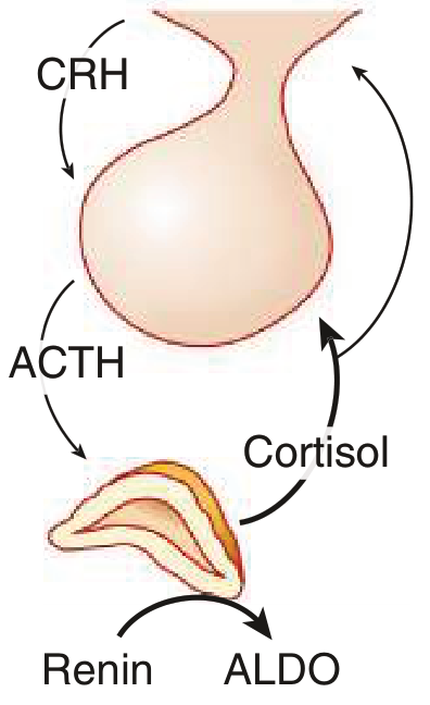
- Hypothalamus releases CRH → stimulates pituitary to release ACTH → ACTH stimulates adrenal cortex to produce cortisol
- If cortisol is low AND ACTH is also low, the pituitary is failing to drive the adrenal → this is secondary (pituitary) or tertiary (hypothalamic) insufficiency
- If this were primary adrenal insufficiency (Addison's disease), cortisol would be low BUT ACTH would be HIGH (pituitary screaming at a failing adrenal gland)
"Unlike primary adrenal insufficiency, the adrenal gland is not destroyed in secondary adrenal insufficiency. Since aldosterone secretion is primarily under control of the renin-angiotensin system, it remains unaffected."
- In primary adrenal insufficiency (Addison's): The entire adrenal cortex is destroyed → BOTH cortisol AND aldosterone are low → renin is HIGH (compensating for low aldosterone)
- In secondary adrenal insufficiency: Only the ACTH-dependent zona fasciculata (cortisol) is affected → aldosterone (zona glomerulosa, RAAS-dependent) is PRESERVED → aldosterone is normal, renin is normal
Comparison Table: Primary vs Secondary Adrenal Insufficiency
| Feature | Primary (Addison's) | Secondary (this patient) |
|---|---|---|
| Cortisol | LOW | LOW |
| ACTH | HIGH (compensatory) | LOW (the problem) |
| Aldosterone | LOW (adrenal destroyed) | NORMAL ✓ |
| Renin | HIGH (low aldosterone → RAS activated) | Normal ✓ |
| ARR | Low or indeterminate | Normal ✓ |
| Hyperpigmentation | YES (high ACTH stimulates melanocytes) | NO |
| Hyperkalemia | YES (no aldosterone) | NO |
| Hyponatremia | YES (severe, salt-wasting) | Mild (SIADH-type from ADH) |
| Volume depletion/shock | More severe | Less severe |
| Site of problem | Adrenal gland | Pituitary / Hypothalamus |
In a 12-Year-Old Girl - What Causes Secondary Adrenal Insufficiency?
Most Important Causes in a Child:
| Cause | Key Features | Investigation |
|---|---|---|
| Craniopharyngioma | Most common pituitary tumor in children; often presents with growth failure, headache, visual field defects, diabetes insipidus + multiple pituitary hormone deficiencies | MRI pituitary with gadolinium - URGENT |
| Pituitary adenoma | Less common in children but possible; may cause mass effect | MRI pituitary |
| Exogenous steroid use (iatrogenic - most common overall) | Long-term oral/inhaled/topical steroids suppress HPA axis; up to 50% of patients on prednisone >5mg/day for >3 months develop adrenal suppression | Detailed drug/medication history |
| Autoimmune hypophysitis | Lymphocytic infiltration of pituitary; may have other autoimmune conditions | MRI (enlarged pituitary), anti-pituitary antibodies |
| Traumatic brain injury | Head trauma history | History + MRI |
| Langerhans cell histiocytosis | Can infiltrate pituitary stalk; also causes diabetes insipidus | MRI, skeletal survey |
| Sarcoidosis | Granulomatous infiltration of pituitary stalk | MRI, serum ACE |
| Empty sella syndrome | Incidental finding; partial pituitary insufficiency | MRI |
| Septo-optic dysplasia | Congenital; visual problems + pituitary deficiency | MRI, visual fields |
Clinical Features to Look for in This Child
| Feature | Hormone Deficient |
|---|---|
| Short stature / growth failure | GH deficiency |
| Delayed puberty | LH/FSH deficiency |
| Polyuria + polydipsia | ADH deficiency (diabetes insipidus) |
| Hypothyroidism symptoms | TSH deficiency |
| Headaches, vomiting, visual changes | Mass lesion (craniopharyngioma) |
| Bitemporal hemianopia | Optic chiasm compression |
| No hyperpigmentation | Confirms secondary (not primary) adrenal insufficiency |
| No severe hyperkalemia | Confirms aldosterone intact = secondary |
What to Do Next - Urgent Investigations
URGENT:
1. MRI pituitary gland with gadolinium contrast
(rule out craniopharyngioma, pituitary tumor, hypophysitis, LCH)
2. Full pituitary hormone panel:
- GH + IGF-1 (growth hormone deficiency)
- LH + FSH (gonadotropin deficiency)
- TSH + Free T4 (central hypothyroidism)
- Prolactin (elevated in stalk compression, low in hypopituitarism)
- ADH/urine osmolality (diabetes insipidus)
- Morning testosterone/estradiol (sex hormone axis)
3. Synacthen (ACTH stimulation) test:
- Give 250 mcg cosyntropin IV
- Measure cortisol at 0, 30, 60 minutes
- Cortisol <18 mcg/dL (immunoassay) or <14 mcg/dL (mass spectrometry)
at 60 min confirms adrenal insufficiency
- In secondary AI, adrenal glands may still respond acutely to exogenous
ACTH (as they are not destroyed), so a low-dose (1 mcg) test is preferred
4. Visual field testing (perimetry)
- Bitemporal hemianopia = optic chiasm compression by craniopharyngioma
5. Ophthalmology referral
- Papilloedema (raised ICP), optic atrophy
Immediate Management
- Hydrocortisone (glucocorticoid) replacement - weight-based dosing (~8-12 mg/m²/day)
- No mineralocorticoid (fludrocortisone) needed - aldosterone is normal (this is the key clinical difference from primary AI)
- Stress dosing education (3x normal dose during illness/surgery/"sick day rules")
- Medical alert bracelet
- Endocrinology referral urgently
Summary
MRI pituitary with gadolinium meaning
MRI Pituitary with Gadolinium - What It Means
What Is an MRI?
What Is Gadolinium?
"Gadolinium remains excluded from the brain by the blood-brain barrier, except in areas where this barrier breaks down, such as inflammation or tumor. On T1 images, gadolinium-enhanced structures appear white."
- Healthy brain tissue has an intact blood-brain barrier → gadolinium cannot enter → stays dark
- Tumors, inflammation, abnormal blood vessels → barrier is disrupted → gadolinium leaks in → appears bright white on T1 images
Why Is the Pituitary Special?
| Structure | Enhancement Pattern |
|---|---|
| Normal pituitary tissue | Enhances brightly and uniformly |
| Pituitary adenoma (tumor) | Enhances later than normal gland → appears as a dark spot within bright gland on early images |
| Craniopharyngioma | Often has a cystic + solid + calcified component; cyst wall enhances, solid part enhances |
| Pituitary stalk | Normally thin; thickening = inflammation (hypophysitis), langerhans cell histiocytosis, metastasis |
| Empty sella | Pituitary gland is flattened; sella filled with CSF |
What the Scan Looks Like - Standard Protocol
| Sequence | What It Shows |
|---|---|
| T1 without contrast (pre-contrast) | Basic anatomy of sella, pituitary size, any bright signal (blood, fat, Rathke cyst) |
| T1 with gadolinium (dynamic/post-contrast) | Tumors appear as dark "holes" in the bright enhancing gland; stalk, cavernous sinus enhancement |
| T2-weighted | Cystic lesions appear bright; edema, fluid-filled structures (craniopharyngioma cysts appear white) |
| Coronal + sagittal thin slices (2-3 mm) | Essential for the tiny pituitary - thicker cuts miss microadenomas |
| FLAIR | Additional sequence to detect surrounding abnormalities |
What It Is Looking For in This Patient (Fathima Neda, 12F)
Normal pituitary region → suggests functional cause
(exogenous steroids, autoimmune, idiopathic)
↓ If abnormal:
Tumor/mass lesion
├── Craniopharyngioma → Cystic + calcified suprasellar mass
│ (most common pituitary region tumor in children)
│ Usually mixed: cyst (bright T2) + calcification (dark T2/bright CT)
│ + solid enhancing portion
│
├── Pituitary adenoma → Small dark spot in bright gland
│ (less common in children)
│
└── Germinoma → Enhancing stalk/suprasellar mass in children
(also causes diabetes insipidus)
Inflammation/infiltration
├── Lymphocytic hypophysitis → Enlarged, uniformly enhancing pituitary
│ + thickened stalk
│
├── Langerhans Cell Histiocytosis → Thickened pituitary stalk
│ Loss of posterior pituitary bright spot (diabetes insipidus)
│
└── Sarcoidosis → Stalk thickening + leptomeningeal enhancement
Structural
├── Empty sella → Pituitary flattened, sella filled with CSF
└── Septo-optic dysplasia → Small optic nerves + absent septum pellucidum
What the Report Will Say
- Size of pituitary gland - normal height is <8 mm in children
- Signal intensity on T1 and T2 - any bright or dark spots
- Enhancement pattern - uniform vs. focal defect
- Pituitary stalk - midline? thickened? deviated?
- Optic chiasm - compressed? normal?
- Suprasellar region - any mass above the sella
- Posterior pituitary bright spot - normally bright on T1 (stores ADH); absent in central diabetes insipidus
Practical Instructions for the Patient/Family
- No ferromagnetic metal implants (pacemakers, certain aneurysm clips) - inform radiologist
- Gadolinium injection into a vein (antecubital) - small sting, generally safe
- Scan duration: ~30-45 minutes
- Patient must lie still in the scanner - children may need sedation if unable to cooperate
- Loud knocking noise is normal - ear protection provided
- Gadolinium is safe in normal kidney function; if RFT is abnormal, inform radiologist
- Drink plenty of water to flush gadolinium through kidneys
- Results usually available within 24-48 hours

Interpret this report
Lab Report Interpretation - Jumana, 15F (17 July 2026)
All Values at a Glance
| Test | Result | Unit | Reference Range | Status |
|---|---|---|---|---|
| CMV IgM | 1.02 | AU/mL | Negative <2.0 / Borderline 2.0-4.2 / Positive >4.2 | ✅ NEGATIVE |
| CMV IgG | 5.96 | AU/mL | Negative <2.0 / Positive >2.0 | 🟡 POSITIVE |
| Cortisol 8AM | 9.75 | µg/dL | 8-10 AM: 3.7-19.4 | ⚠️ LOW-NORMAL |
| HIV 1&2 Antibodies ELISA | 0.08 | OD | Non-reactive <0.25 / Reactive >0.25 | ✅ NON-REACTIVE |
| EBV VCA IgG | 8.44 | INDEX | Negative <0.9 / Equivocal 0.9-1.0 / Positive >1.1 | 🔴 STRONGLY POSITIVE |
| EBV VCA IgM | 0.37 | INDEX | Negative <0.9 / Equivocal 0.9-1.0 / Positive >1.1 | ✅ NEGATIVE |
Test-by-Test Interpretation
1. EBV VCA IgG = 8.44 (STRONGLY POSITIVE) + EBV VCA IgM = 0.37 (NEGATIVE)
| Antibody | Timing | This patient |
|---|---|---|
| VCA IgM | Appears in acute/recent EBV infection (weeks 1-4), disappears by 3 months | Negative (0.37) - NO active/recent infection |
| VCA IgG | Appears in acute infection, persists lifelong as a marker of past infection | Strongly positive (8.44) - past exposure |
| EBNA IgG (not done) | Appears 6-12 weeks after infection, persists lifelong | Not tested |
Note: EBNA IgG was not tested - adding it would help confirm this is truly past (not early acute) infection. In primary acute EBV, EBNA IgG is absent early, while in past infection it is positive.
2. CMV IgG = 5.96 (POSITIVE) + CMV IgM = 1.02 (NEGATIVE)
- CMV IgM negative (1.02, cutoff >4.2) → No acute/active CMV infection
- CMV IgG positive (5.96, cutoff >2.0) → Past CMV exposure, latent infection (very common in the general population)
3. ⚠️ Cortisol 8AM = 9.75 µg/dL (LOW-NORMAL / BORDERLINE)
| Cortisol Level (8AM) | Interpretation |
|---|---|
| >18 µg/dL | Normal adrenal function - adrenal insufficiency excluded |
| 10-18 µg/dL | Indeterminate - may need stimulation test |
| 9.75 µg/dL | In the lower portion - borderline/indeterminate |
| <3.7 µg/dL | Strongly suggests adrenal insufficiency |
"Both healthy individuals and patients with adrenal insufficiency may have indeterminate results (3 to 18 µg/dL) that require additional evaluation."
4. HIV 1&2 ELISA = 0.08 OD (NON-REACTIVE)
- Result: 0.08 OD
- Cutoff: Non-reactive <0.25, Reactive >0.25
- HIV is conclusively excluded. The result (0.08) is far below the reactive threshold.
Integrated Clinical Summary
| Diagnosis | Status Based on This Report |
|---|---|
| Active EBV (infectious mononucleosis) | ✅ EXCLUDED - IgM negative, IgG positive = past immunity |
| Active CMV infection | ✅ EXCLUDED - IgM negative, IgG positive = past immunity |
| HIV infection | ✅ EXCLUDED - Non-reactive |
| Adrenal insufficiency | ⚠️ CANNOT BE EXCLUDED - Cortisol 9.75 is borderline; needs Synacthen test |
What This Means for the PUO Workup
Immediate Next Steps:
URGENT:
1. Synacthen (ACTH stimulation) test
- Give 250 mcg cosyntropin IV
- Measure cortisol at 0, 30, 60 minutes
- Peak <18 µg/dL = adrenal insufficiency confirmed
2. Serum ACTH (9AM, fasting) - if not already done
- If ACTH is LOW with low cortisol = secondary adrenal insufficiency
→ MRI pituitary with gadolinium urgently
- If ACTH is HIGH with low cortisol = primary adrenal insufficiency
→ Adrenal imaging (CT adrenals)
3. CECT chest + abdomen + pelvis (still pending)
- Lymphoma, sarcoidosis, intestinal TB remain in differential
4. Echocardiography (still pending)
- Culture-negative endocarditis, atrial myxoma
5. Anti-TTG IgA + stool OCP still recommended
(celiac disease, parasites as IDA cause)
17 yr old female with pou since 1 month her CBC,lft,esr,crp, rft, procalcitonin,ure,urine tb pcr,lepto ,dengue , chikengunya,Widal, weil felix test, malaria , ana, ra factor , aso , chest xray and usg abdomen with pelvis all are normal , blood culture normal no bacterimia, tsh normal, igra negative , stool calprotectin negative ,Brucella test negative , serum ferritin normal , peripheral smear shows iron deficiency anemia , hiv elisa negative , ebv ,cmv negative what is the next step
PUO fever unknown origin extensive negative workup IDA iron deficiency anemia next step imaging endoscopy 2024
PUO - 17F - Extensive Negative Workup + IDA - Where We Are Now
Master Checklist: What Has Been Done and Excluded
| Category | Tests Done | Excluded Diagnoses |
|---|---|---|
| Tropical infections | Dengue, Malaria, Leptospirosis, Widal, Weil-Felix, Chikungunya | All major tropical fevers |
| Bacterial | Blood cultures x3, Procalcitonin | Bacteremia, septicemia |
| TB | IGRA, Urine TB PCR | LTBI; active TB not fully excluded |
| Viral | HIV ELISA, EBV VCA IgM/IgG, CMV IgM/IgG | HIV, active EBV, active CMV |
| Zoonotic | Brucella agglutination | Brucellosis |
| Bowel inflammation | Stool calprotectin | IBD (Crohn's, UC) - effectively excluded |
| Autoimmune screen | ANA, RA factor, ASO | SLE (basic screen), RA, rheumatic fever |
| Autoinflammatory | Serum ferritin normal | AOSD/Still's disease excluded |
| Organ function | CBC, LFT, RFT, TSH | Organ failure, thyroid cause |
| Inflammation markers | ESR, CRP, Procalcitonin normal | Major acute bacterial/inflammatory process |
| Imaging | CXR normal, USG abdomen/pelvis normal | Obvious abdominal mass, pulmonary infiltrates |
| Haematology | Peripheral smear shows IDA | Confirms true iron depletion |
What Has NOT Been Done - Critical Remaining Gaps
THE NEXT STEPS
🔴 PRIORITY 1: Tests to Send Immediately (Today)
A. For the IDA - This Is Now the Primary Diagnostic Lead
| Test | Why It Is the Most Important Test Right Now |
|---|---|
| Anti-TTG IgA + total serum IgA | Celiac disease is the top remaining diagnosis. Per Harrison's 22nd ed: "Patients may be identified after presenting with... iron-deficiency anemia" as the sole presentation, with NO diarrhea in up to 30-40% of cases. Iron is absorbed in the duodenum - celiac disease damages exactly this site. TTG-IgA sensitivity 93%, specificity 96%. Must check total IgA simultaneously (IgA deficiency causes false-negative TTG). |
| Stool H. pylori antigen | H. pylori is extremely prevalent in India. Causes IDA via: (1) chronic gastric mucosal blood loss, (2) iron malabsorption due to hypochlorhydria, (3) competition for luminal iron. Also causes low-grade systemic immune activation which can manifest as prolonged fever. Simple, non-invasive test. |
| Stool for ova, cysts, parasites - concentration technique (x3 samples) | Hookworm (Ancylostoma duodenale) is one of the most common causes of IDA in India. Causes chronic intestinal blood loss (each worm consumes 0.03-0.15 mL blood/day). Can cause fever via systemic immune response. Trichuris trichiura and Strongyloides also cause IDA + fever. |
| Stool FOBT (faecal occult blood test) x3 | Quantifies ongoing GI blood loss even without visible bleeding. Negative calprotectin does NOT exclude FOBT positivity. |
| Serum iron + TIBC + transferrin saturation | Confirm and grade true IDA (transferrin saturation <16%, elevated TIBC) and exclude thalassaemia trait which also shows microcytosis on smear |
| HbA2 + HbF (HPLC) | If microcytic anaemia but iron studies not severely depleted - rule out beta-thalassaemia trait which can coexist or mimic IDA |
B. For the PUO - Still Missing Tests
| Test | Why Critical |
|---|---|
| Serum ACE (Angiotensin Converting Enzyme) | Sarcoidosis - can cause fever + normal CXR (bilateral hilar adenopathy missed on plain film); confirmed by elevated ACE + CT chest |
| Anti-dsDNA + C3 + C4 | ANA was negative but early/incomplete SLE can have negative ANA; anti-dsDNA is more specific; low C3/C4 = active SLE |
| Toxoplasma IgM + IgG | Acquired toxoplasmosis causes prolonged fever + lymphadenopathy in young adults; often missed |
| Bartonella henselae IgM + IgG | Cat scratch disease causes FUO with or without visible lymphadenopathy; ask about cat contact history |
| Q-fever (Coxiella burnetii) Phase I + II IgG/IgM | Culture-negative cause of prolonged fever; zoonotic |
| rK39 rapid test (visceral leishmaniasis) | If patient is from Bihar/UP/endemic area - kala-azar causes prolonged fever + splenomegaly; early splenomegaly may be subtle on USG |
| Morning cortisol 8AM + ACTH | The lab report we saw showed cortisol 9.75 µg/dL (borderline low). Needs ACTH measurement simultaneously. If ACTH is also low → secondary adrenal insufficiency → MRI pituitary urgently. Adrenal insufficiency causes chronic fever + fatigue. |
| Parvovirus B19 IgM | Causes prolonged fever + arthralgia in adolescents |
| MEFV gene mutation (if periodic fever pattern) | Familial Mediterranean Fever - autosomal recessive; common in Arabs, Turks, Armenians; presents with periodic fever episodes + serositis; first attack usually before age 20 |
🔴 PRIORITY 2: Imaging - Within 48-72 Hours (Cannot Be Delayed Further)
CECT Chest + Abdomen + Pelvis (with IV contrast)
| What CT Adds That USG/CXR Misses |
|---|
| Mediastinal + hilar lymphadenopathy (Hodgkin's lymphoma - classic in 15-25yr age group; sarcoidosis) |
| Retroperitoneal + mesenteric adenopathy (lymphoma, TB, leishmaniasis) |
| Bowel wall thickening (intestinal TB despite negative IGRA; early Crohn's despite negative calprotectin) |
| Hepatic/splenic micro-lesions (lymphoma, abscess, granulomata) |
| Adrenal glands (enlargement in primary adrenal insufficiency - TB, autoimmune) |
| Small splenic/hepatic nodules (granulomatous disease) |
Echocardiography
- Culture-negative endocarditis (Bartonella, Q-fever, HACEK, fungi) - all blood culture negative
- Atrial myxoma - rare but classic cause of PUO in young women; causes fever + embolic events; USG abdomen is negative
🟡 PRIORITY 3: Endoscopy (After Serology and Imaging)
OGD (Upper GI Endoscopy) with Duodenal Biopsies - HIGH PRIORITY
- IDA with suspected malabsorption as the mechanism
- Essential regardless of TTG-IgA result (IgA deficiency can give false-negative TTG)
- Per Harrison's: diagnosis of celiac requires duodenal biopsy confirming villus blunting + crypt hyperplasia + intraepithelial lymphocytes (Marsh classification)
- Also perform CLO test / rapid urease test for H. pylori during same scope
- 2024 PubMed data confirms: "diagnostic yield of EGD is significantly higher than colonoscopy in younger IDA patients"
Ileocolonoscopy with Terminal Ileum Biopsy
- If CT shows ileocecal abnormality, or FOBT positive, or stool OCP shows eggs
- Terminal ileum biopsy for intestinal TB (caseating granulomas, AFB culture) vs Crohn's (non-caseating)
🟡 PRIORITY 4: Gynaecology Referral
- Heavy periods (menorrhagia): clot passage, >5 pads/day, >7 days duration?
- If menorrhagia is confirmed, this explains the IDA - but the fever still needs a separate explanation
- Screen for von Willebrand disease (coagulation screen + VWF antigen + VWF ristocetin cofactor activity) - most common inherited bleeding disorder in teenage girls; routinely missed
🟢 PRIORITY 5: If All Above Is Negative
| Investigation | When | Expected Yield |
|---|---|---|
| 18F-FDG PET/CT | After CT + echo negative | 60-80% diagnostic yield in classic PUO; endorsed by EANM 2024 + SNMMI/IDSA 2024 AUC; detects occult lymphoma, vasculitis, granulomatous foci, deep infection |
| Bone marrow aspirate + trephine biopsy | If PET shows marrow uptake OR all investigations unrevealing | Detects leukemia, lymphoma, HLH, TB, leishmaniasis, histoplasmosis |
The Full Action Plan
TODAY - BLOOD:
□ Anti-TTG IgA + total serum IgA ← #1 priority
□ Stool H. pylori antigen
□ Stool OCP x3 (concentration technique)
□ Stool FOBT x3
□ Serum iron + TIBC + transferrin saturation
□ HbA2 + HbF (HPLC) if thalassaemia possible
□ Serum ACE
□ Anti-dsDNA + C3 + C4
□ Toxoplasma IgM/IgG
□ Bartonella henselae IgM/IgG
□ Q-fever (Coxiella) serology
□ Morning cortisol 8AM + ACTH (simultaneously)
□ Parvovirus B19 IgM
□ rK39 (if from endemic area)
GYNAECOLOGY:
□ Detailed menstrual history
□ Coagulation screen + VWF antigen/activity
(if menorrhagia confirmed)
WITHIN 48-72 HOURS:
□ CECT chest + abdomen + pelvis (IV contrast)
□ Echocardiography (transthoracic)
AFTER RESULTS:
□ OGD + duodenal biopsy x4-6 (if TTG-IgA positive OR
IgA deficient OR high clinical suspicion for celiac)
□ Ileocolonoscopy + TI biopsy (if CT/FOBT positive)
□ Lymph node excision biopsy (if CT shows adenopathy)
- Prefer posterior cervical / supraclavicular /
epitrochlear over axillary / inguinal
IF ALL STILL NEGATIVE:
□ 18F-FDG PET/CT
□ Bone marrow aspirate + trephine biopsy
□ Consider drug fever (review ALL medications)
□ Consider factitious fever (supervised temperature measurement)
Ranked Remaining Differential Diagnosis
| Rank | Diagnosis | Key Test to Confirm |
|---|---|---|
| 1 | Celiac disease | Anti-TTG IgA + OGD + duodenal biopsy |
| 2 | H. pylori gastritis/ulcer | Stool H. pylori antigen + OGD |
| 3 | Intestinal parasites (hookworm) | Stool OCP x3 |
| 4 | Hodgkin's lymphoma | CECT chest (mediastinal nodes) + biopsy |
| 5 | Sarcoidosis | Serum ACE + CT chest + tissue biopsy |
| 6 | Intestinal TB | CECT + colonoscopy + TI biopsy (AFB culture) |
| 7 | Secondary adrenal insufficiency | 8AM cortisol + ACTH + Synacthen test |
| 8 | Menorrhagia (IDA source) + separate fever | Gynaecology + VWF screen |
| 9 | Cat scratch disease / Toxoplasmosis | Bartonella/Toxoplasma serology |
| 10 | Familial Mediterranean fever | MEFV gene mutation analysis |
| 11 | SLE (incomplete/early) | Anti-dsDNA + C3/C4 |
| 12 | Culture-negative endocarditis | Echo + Q-fever/Bartonella serology |
| 13 | Visceral leishmaniasis | rK39 + bone marrow |
| 14 | Factitious fever | Supervised temperature measurement |