
Flow volume loops
Reading File
Flow volume loops are graphic representations of airflow vs. lung volume during forced inspiration and expiration, used to diagnose and classify pulmonary disorders. Let me search the medical library for detailed content.
Searching sources...
Finding Sources
Finding Sources
Reading File
Reading File
Reading File
Loading Image
Loading Image
Reading File
Finding Sources
Reading File
Reading File
Finding Sources
Reading File
Reading File
Loading Image
Here is a thorough overview of flow volume loops (FVLs), drawn from multiple authoritative pulmonary textbooks.
Flow Volume Loops
What Is a Flow Volume Loop?
A flow volume loop (FVL) is a graphical representation of airflow (y-axis, in L/s) plotted against lung volume (x-axis, in L) during a forced maximal expiration followed by a forced maximal inspiration. The subject exhales fully from total lung capacity (TLC) to residual volume (RV), then inhales back to TLC. The shape of the loop is the most clinically informative feature.
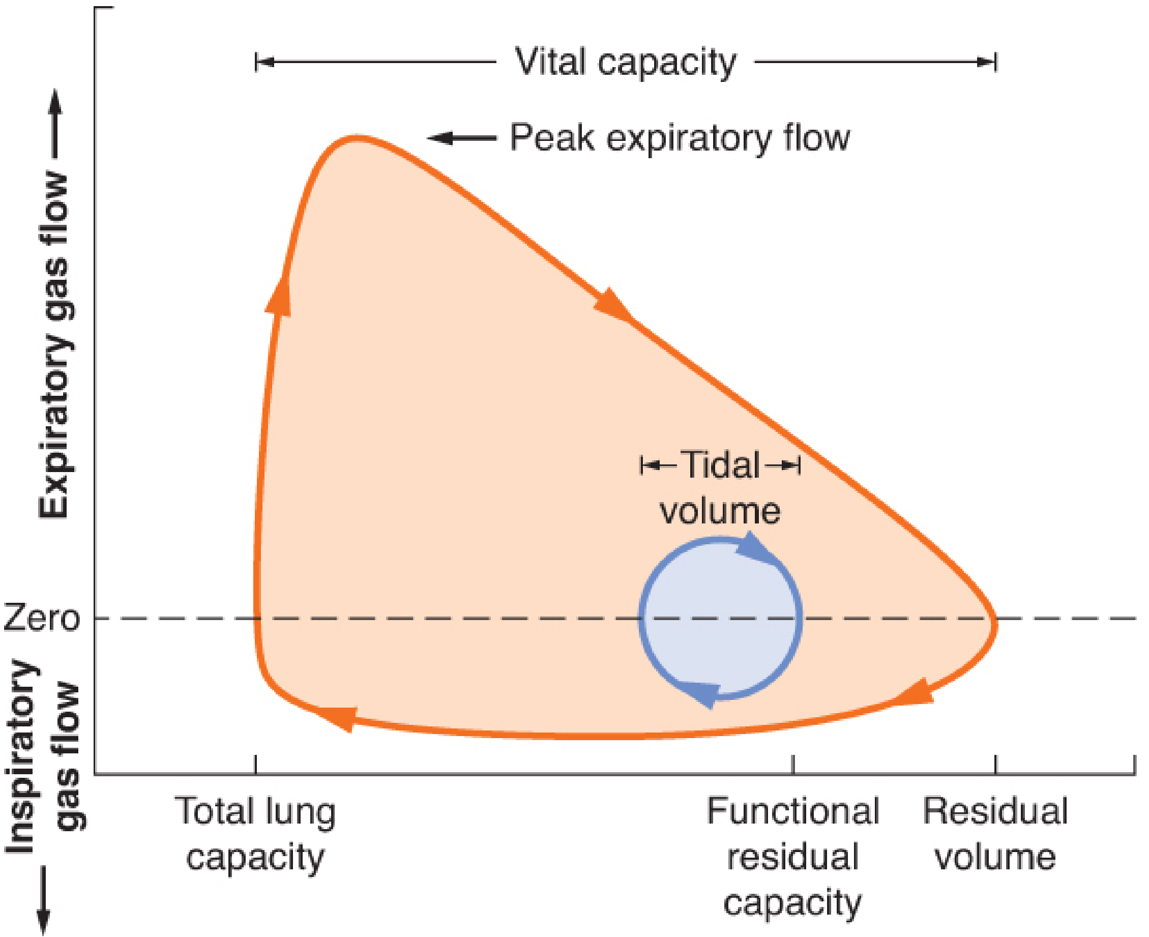
Figure: Normal adult flow-volume loop. Expiratory flow is above the zero line; inspiratory flow is below. Tidal volume is the small circle near functional residual capacity (FRC). - Barash Clinical Anesthesia, p. 1137
Axes and Orientation
| Axis | Represents | Direction |
|---|---|---|
| Y-axis | Airflow (L/s) | Above zero = expiration; below zero = inspiration |
| X-axis | Lung volume (L) | Left = TLC; right = RV |
- The expiratory limb (upper curve): starts at TLC, rises sharply to peak expiratory flow (PEF), then declines linearly toward RV
- The inspiratory limb (lower curve): begins at RV, peak inspiratory flow (PIF) occurs near the midpoint, and returns to TLC
Normal: PEF > PIF, because dynamic compression of intrathoracic airways limits expiratory flow more than inspiratory flow, while the inspiratory portion is entirely effort-dependent.
Effort Dependence
- First 25% of forced expiration (near TLC): effort-dependent - increased effort increases flow
- Remaining 75%: effort-independent - flow is determined by lung elastic recoil and airway mechanics (dynamic compression); more effort does not increase flow
- Entire inspiratory limb: effort-dependent throughout
Normal FVL Characteristics
- Rapid rise to PEF shortly after maximal exhalation begins
- Near-linear decline in expiratory flow from peak to RV
- Smooth, roughly semicircular inspiratory curve
- PIF slightly less than PEF
- FEF50% (mid-expiratory flow) is normally less than FIF50% (mid-inspiratory flow)
Pathological Patterns
1. Obstructive (COPD, Asthma, Emphysema)
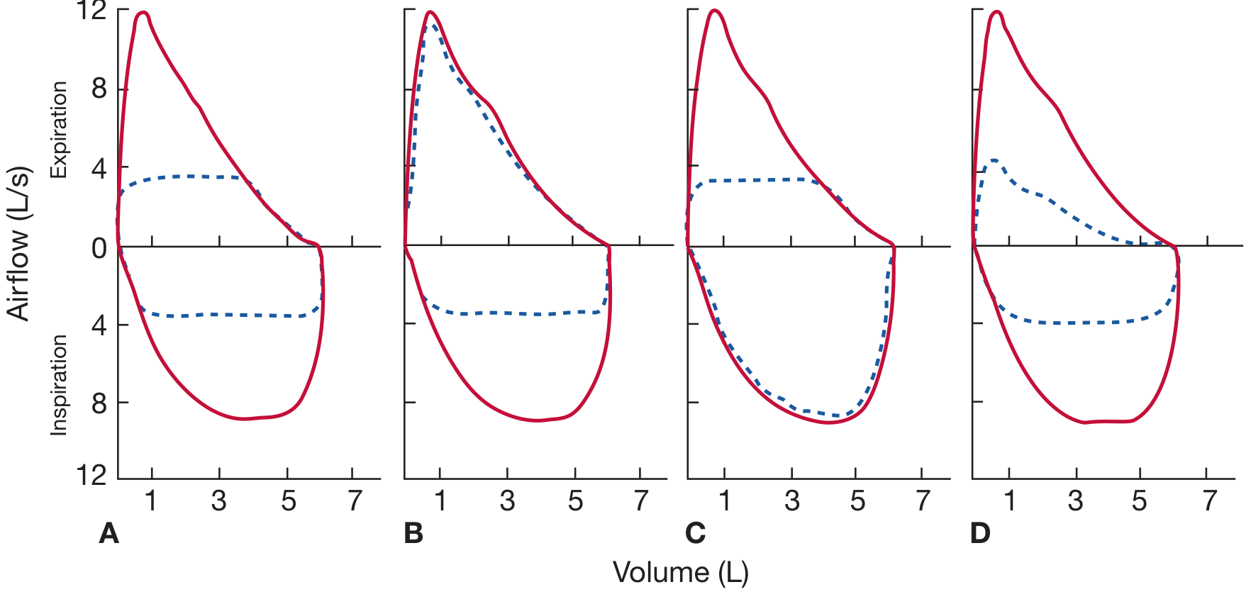
Figure: Schematic flow-volume loops in four pathologic conditions. Red = patient; blue dashed = normal. A: Fixed upper-airway obstruction (both limbs truncated). B: Variable extrathoracic obstruction (inspiratory limb flattened). C: Variable intrathoracic obstruction (expiratory limb flattened). D: COPD - reduced expiratory flow with preserved curve shape/taper. - Fishman's Pulmonary Diseases, p. 605
- Expiratory limb is concave upward (scooped/curvilinear) - this is the hallmark
- Reflects greater flow reduction at low lung volumes vs high lung volumes
- FVC and FEV1 are reduced; FEV1/FVC ratio is decreased
- The loop is shifted to the right (air trapping, increased RV)
- Asthma: loop may normalize post-bronchodilator
- Emphysema: severe scooping, DLCO reduced, TLC increased
2. Restrictive (Interstitial Lung Disease, Chest Wall)
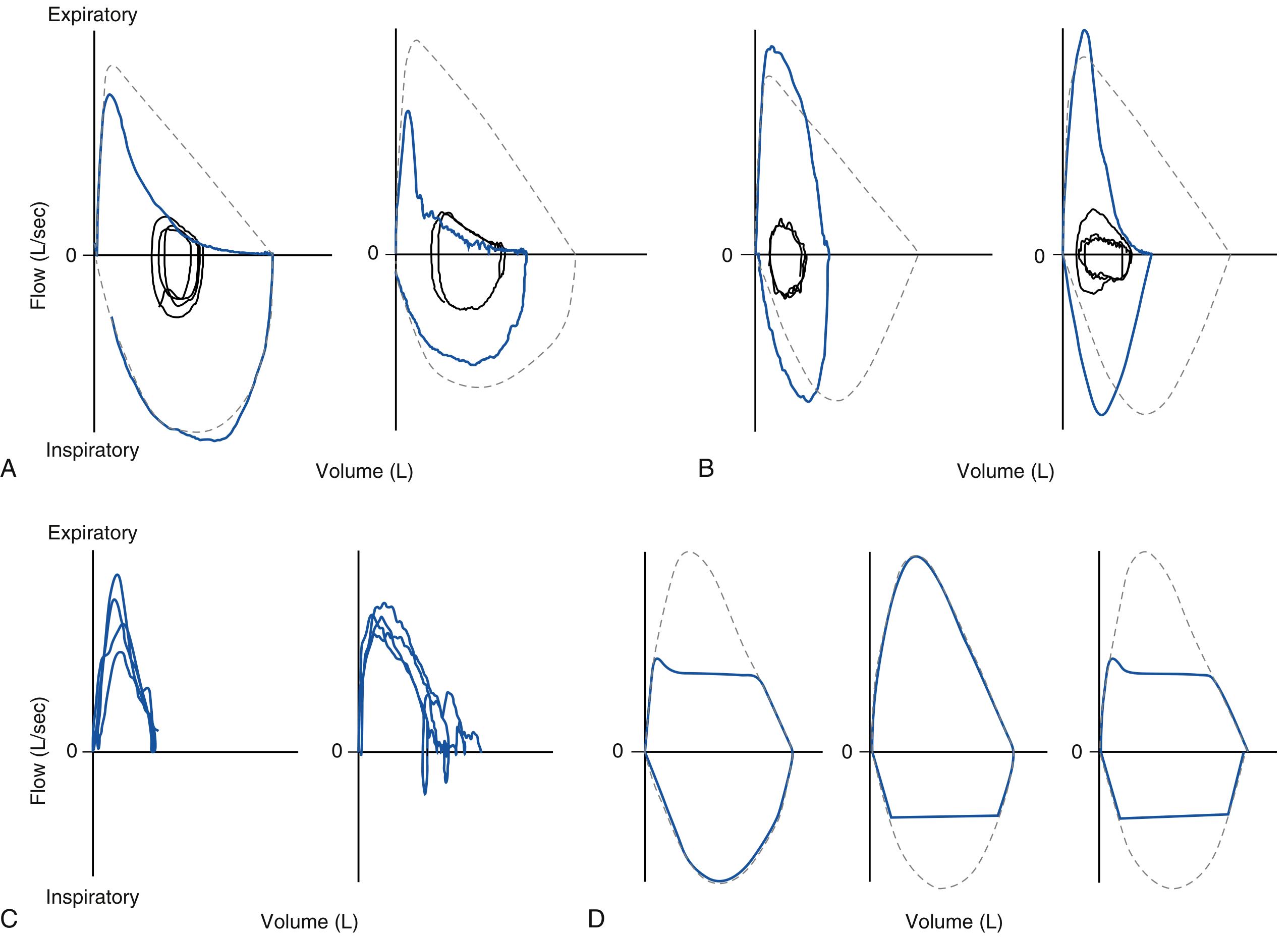
Figure: Real flow-volume curve patterns. A: COPD (concave expiratory limb). B: ILD/restriction (steep, vertically-oriented expiratory limb, reduced VC). C: Poor effort (irregular, non-reproducible). D: Large airway obstruction subtypes. - Murray & Nadel's Respiratory Medicine, p. 763
- Expiratory limb is convex upward (steep, vertically oriented) - increased elastic recoil drives higher peak flows at high volumes
- FVC and FEV1 both reduced proportionally; FEV1/FVC ratio is normal or increased
- Total lung capacity (TLC) is reduced - confirmation requires plethysmography
- Important caveat: spirometry alone identifies restrictive pattern with only ~50% sensitivity; TLC measurement is required for confirmation
3. Upper Airway Obstruction (UAO)
Three subtypes, each with a characteristic loop shape:
| Type | Location | Mechanism | Loop Pattern |
|---|---|---|---|
| Fixed | Either (tracheal scar, stenosis) | Cross-section fixed regardless of breathing phase | Both inspiratory AND expiratory limbs truncated/flattened equally |
| Variable extrathoracic (e.g., vocal cord paralysis, subglottic stenosis) | Above thoracic inlet | Forced inspiration reduces intratracheal pressure below atmospheric → airway collapses | Inspiratory limb flattened; expiratory limb normal |
| Variable intrathoracic (e.g., tracheal tumor, tracheomalacia) | Below thoracic inlet | Forced expiration increases pleural pressure above intratracheal → airway collapses | Expiratory limb flattened; inspiratory limb normal |
Key ratio: FEV1/PEFR > 8 mL/L/min on routine spirometry should prompt careful review of the FVL for UAO.
Distinguishing UAO from COPD: In COPD the expiratory limb is reduced but retains its normal tapering shape (gradual deceleration). In UAO the expiratory limb is flat (uniform flow plateau) throughout expiration - a qualitatively distinct pattern.
FVL in Specific Conditions
| Condition | FEV1 | FVC | FEV1/FVC | DLCO | FVL finding |
|---|---|---|---|---|---|
| Chronic bronchitis | ↓ | ↓ | ↓ | Normal | Abnormal (scooped expiratory) |
| Emphysema | ↓ | ↓ | ↓ | ↓ | Abnormal (scooped, large VC) |
| Asthma | ↓ | ↓ | ↓ | Normal | Abnormal; reverses post-BD |
| ILD (restrictive) | ↓ | ↓ | Normal/↑ | ↓ | Steep/convex expiratory |
| Fixed UAO | ↓ | ↓ | ↓ | Normal | Both limbs truncated |
| Variable extrathoracic | ↓ | ↓ | ↓ | Normal | Inspiratory flattening |
| Variable intrathoracic | ↓ | ↓ | ↓ | Normal | Expiratory flattening |
| Small airway disease | Normal | Normal | Normal | Normal | Normal |
- Fishman's Pulmonary Diseases, Table 31-19
FVL During Exercise
During cardiopulmonary exercise testing (CPET), tidal flow-volume loops (tidal FVLs) are plotted within the maximal flow-volume loop (MFVL) envelope:
- Expiratory flow limitation: tidal loop encroaches on the expiratory limb of the MFVL
- Dynamic hyperinflation: end-expiratory lung volume rises above resting FRC (seen as leftward shift of tidal loop within MFVL)
- Inspiratory muscle limitation: tidal inspiratory flows approach the inspiratory MFVL - indicates near-maximal inspiratory effort
This analysis is particularly useful in COPD, where both flow limitation and dynamic hyperinflation contribute to exertional dyspnea. - Fishman's Pulmonary Diseases, p. 619
Other FVL Findings
- Sawtooth pattern: oscillatory notching on the inspiratory limb (and sometimes expiratory) - associated with obstructive sleep apnea and upper airway instability, or extrapyramidal disease (e.g., Parkinson's)
- Tracheal plateau / vocal cord dysfunction: mid-inspiratory flow plateau suggesting episodic vocal cord adduction
- Suboptimal effort: irregular, slurred curves without repeatability; no consistent peak; diagnose as poor quality
- Negative effort dependence (in severe COPD): tidal expiratory flow at low lung volumes actually exceeds maximal forced flow - the airway collapses more with greater effort, a sign of severe dynamic airway collapse
Clinical Uses
- Diagnosis and classification of obstructive vs. restrictive ventilatory defects
- Detection and characterization of upper airway obstruction (fixed vs. variable, extra vs. intrathoracic)
- Monitoring response to bronchodilators in asthma/COPD
- Preoperative assessment (historically) - though CT/MRI has largely replaced FVL for structural UAO
- Exercise physiology - detection of expiratory flow limitation and dynamic hyperinflation during CPET
- Identifying poor patient effort and technical artifact
Sources: Fishman's Pulmonary Diseases and Disorders (pp. 605, 619) | Murray & Nadel's Textbook of Respiratory Medicine (pp. 707, 761, 763) | Barash Clinical Anesthesia (p. 1137) | Goldman-Cecil Medicine | KJ Lee's Essential Otolaryngology
What is dynamic compression
Dynamic compression of airways occurs during forced expiration when pleural pressure exceeds the pressure inside intrathoracic airways, causing them to narrow or collapse. This creates a flow-limiting segment and explains why expiratory flow becomes effort-independent beyond the first 25% of the FVC maneuver.
Searching sources...
Finding Sources
Finding Sources
Reading File
Finding Sources
Reading File
Reading File
Reading File
Loading Image
Loading Image
Dynamic Compression of Airways
Definition
Dynamic compression is the narrowing or collapse of intrathoracic airways during forced expiration, caused by the surrounding pleural pressure rising to exceed the pressure inside the airway lumen. This creates a flow-limiting segment and is the reason why expiratory flow beyond the first ~25% of the FVC maneuver becomes effort-independent - no matter how hard you try, you cannot increase flow further. - Morgan & Mikhail's Clinical Anesthesiology, p. 931
The Pressure Mechanics - Step by Step
The following diagram shows the pressure distribution across the airways at rest, during quiet expiration, and during forced expiration:
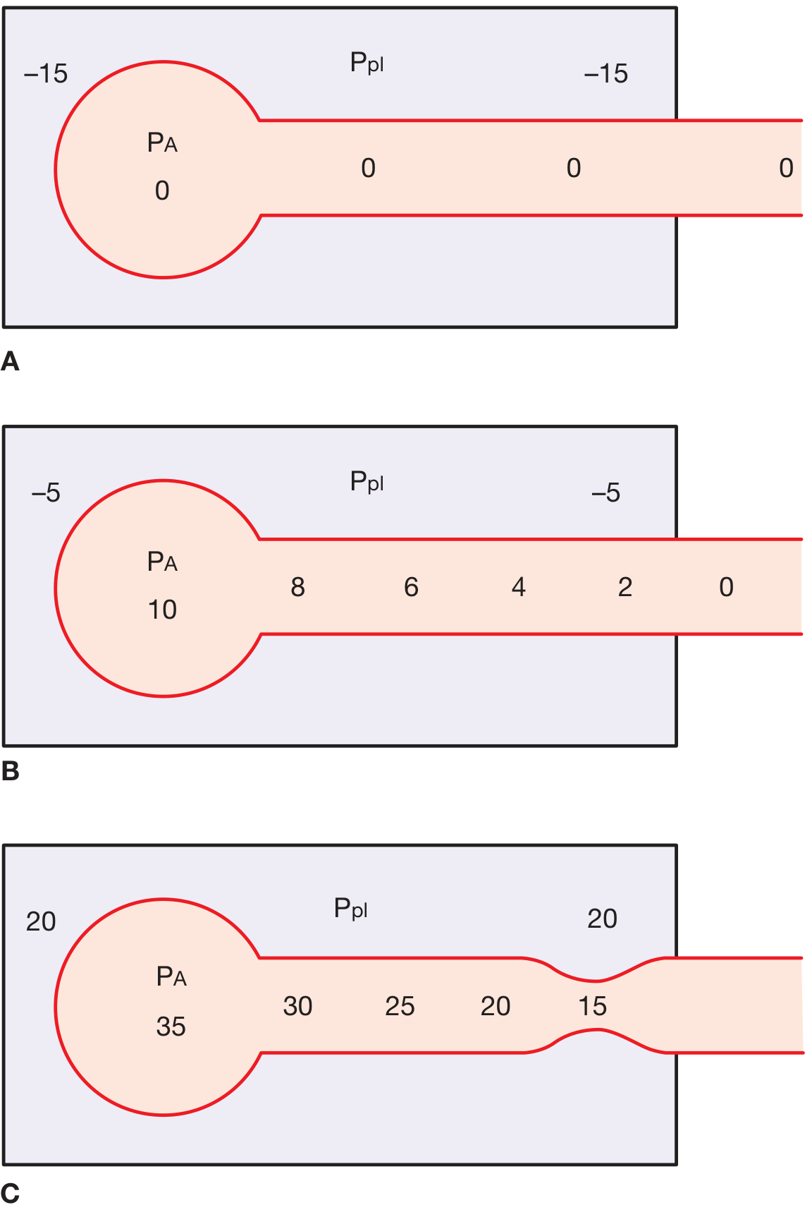
Figure: Equal pressure point concept. A: At rest - Ppl = -15, PA = 0, no flow. B: Quiet expiration - Ppl = -5, PA = +10, airway pressure drops from 8 → 0 toward mouth. C: Forced expiration - Ppl = +20, PA = +35; airway pressure falls from 30 → 15 along the tube. At the point where airway intraluminal pressure = +20 = Ppl, the transmural pressure is zero, and the airway collapses/narrows (pinch point visible). - Fishman's Pulmonary Diseases, p. 200
What happens in each phase:
At rest (A): Ppl is negative (-15 cmH₂O). PA = 0 (no airflow). The airway is held open because intraluminal pressure exceeds pleural pressure at every point.
Quiet expiration (B): Ppl rises to -5. PA becomes +10 (due to elastic recoil). Pressure drops from 10 → 0 along the airway toward the mouth, but intraluminal pressure still exceeds Ppl everywhere - no collapse.
Forced expiration (C): Ppl rises sharply to +20 cmH₂O. PA = +35 (elastic recoil + pleural pressure). As air flows toward the mouth, intraluminal pressure drops progressively (30 → 25 → 20...). At the point where intraluminal pressure = +20 = Ppl, the transmural pressure = 0 and the airway wall experiences no distending force. Beyond that point (toward the mouth), Ppl exceeds intraluminal pressure, and the airway is compressed from outside - this is dynamic compression.
The Equal Pressure Point (EPP)
The point where intraluminal airway pressure exactly equals pleural pressure is the equal pressure point (EPP).
- Airways upstream (alveolar side) of the EPP: intraluminal > pleural - airway held open
- Airways downstream (mouth side) of the EPP: pleural > intraluminal - airway compressed
The EPP divides the airways into two series segments:
| Segment | Location | Behavior |
|---|---|---|
| Upstream segment | Alveoli → EPP | Driving pressure = lung elastic recoil (P_L); determines maximum flow |
| Downstream segment | EPP → mouth | Compressed; increasing effort only compresses it more, does NOT increase flow |
Key formula:
V_max = P_L / R_upstream
where P_L = elastic recoil pressure and R_upstream = resistance of the upstream segment. This is why maximum expiratory flow depends on lung elastic recoil and upstream airway resistance, NOT on how hard you exhale.
Effort Independence - Explained
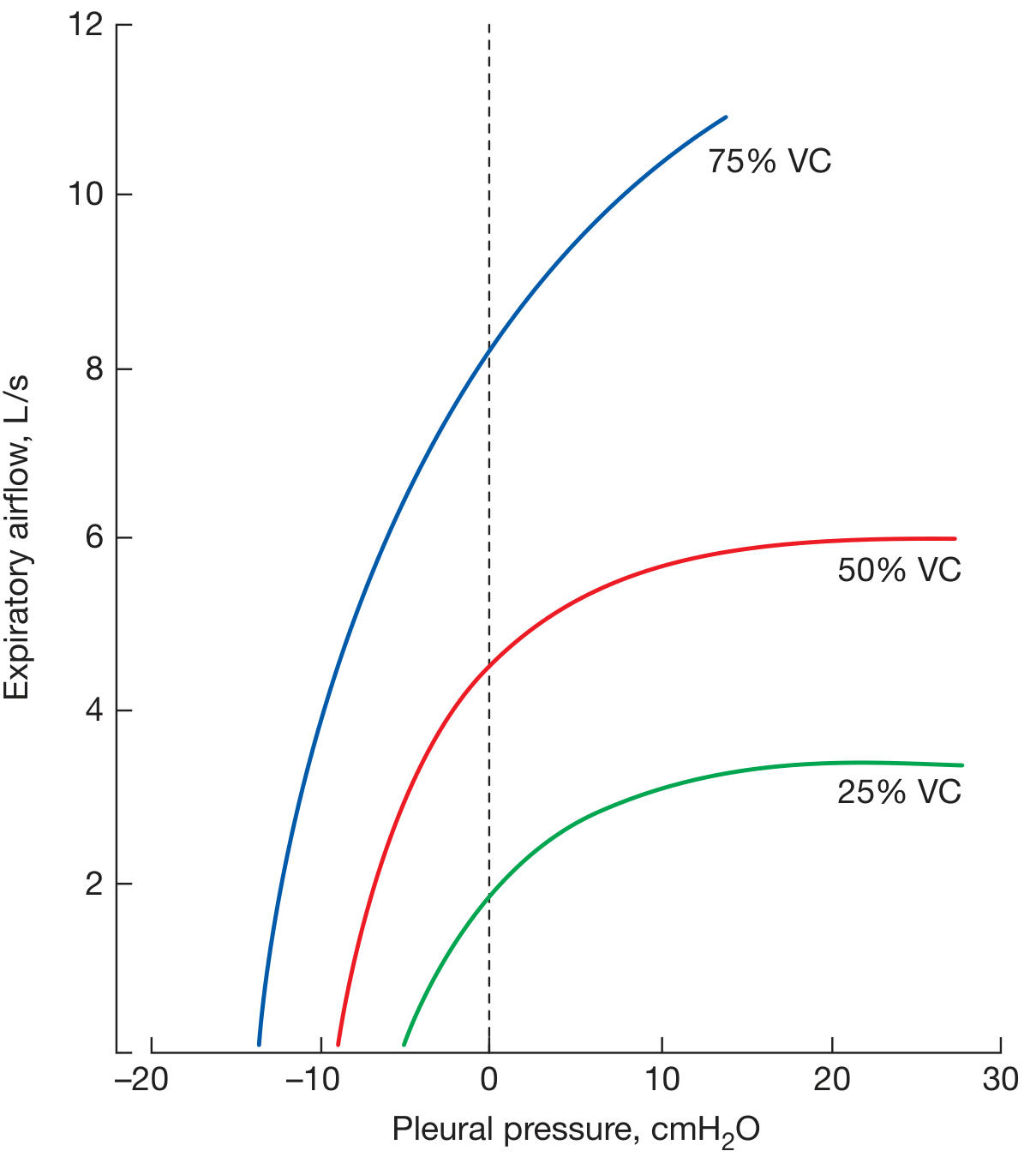
Figure: Isovolume pressure-flow curves. At 75% VC, flow is effort dependent (no plateau). At 50% and 25% VC, flow reaches a ceiling regardless of increasing pleural pressure - the hallmark of dynamic compression. - Fishman's Pulmonary Diseases, p. 199
- At high lung volumes (>75% VC): elastic recoil is high, airways are wide and stiff - the EPP has not been reached even at atmospheric Ppl, so increasing effort still increases flow = effort dependent
- At lower lung volumes (<75% VC): elastic recoil is lower, airway caliber is smaller - even modest expiratory effort creates an EPP, and further effort just compresses the downstream airway = effort independent
As lung volume falls during expiration, the EPP moves progressively upstream (toward smaller airways).
Where Does Dynamic Compression Normally Occur?
The equal pressure point is normally located at the 11th to 13th generation bronchioles, where cartilaginous support is absent (making them susceptible to collapse). - Morgan & Mikhail, p. 932
At high lung volumes, the choke point sits near the lower trachea/lobar bronchi. As lung volume decreases, it migrates toward smaller, more peripheral airways.
Two Theories of Flow Limitation
1. Equal Pressure Point (EPP) Theory
As described above - flow limited when pleural pressure exceeds intraluminal pressure, creating dynamic compression.
2. Wave Speed Theory
An alternative model: flow is limited when the linear velocity of gas molecules equals the velocity of propagation of pressure waves along the airway wall (the "wave speed"). A choke point develops where gas cannot travel faster than the pressure wave can propagate. Flow at wave speed depends on:
- Cross-sectional area of the airway at the choke point
- Stiffness of the airway wall (dP/dA)
Both theories give the same practical result: maximum expiratory flow is determined by elastic recoil and upstream airway properties, not by expiratory effort. - Fishman's Pulmonary Diseases, p. 200
Clinical Relevance
| Condition | Effect on Dynamic Compression | Consequence |
|---|---|---|
| COPD / Emphysema | Destruction of elastic tissue supporting small airways + reduced elastic recoil | EPP moves peripherally even at rest; severe dynamic compression; air trapping |
| Asthma | Bronchoconstriction and mucosal edema narrow airways, intensify collapse | More dynamic compression; patients may purse lips or terminate exhalation early |
| Both | Auto-PEEP / intrinsic PEEP from premature airway closure | FRC rises above normal; air trapping |
| Coughing | Deliberate use of dynamic compression | High intrathoracic pressure collapses central airways, accelerating airflow and clearing secretions |
In cough, dynamic compression is actually therapeutic - it dramatically increases air velocity in central airways (kinetic energy and turbulence increase), shearing secretions from the airway wall. As successive coughs occur at progressively lower lung volumes, the EPP migrates peripherally, progressively clearing more distal airways. - Murray & Nadel's Respiratory Medicine, p. 868
Sources: Fishman's Pulmonary Diseases and Disorders (pp. 199-200) | Morgan & Mikhail's Clinical Anesthesiology (pp. 931-932) | Murray & Nadel's Textbook of Respiratory Medicine (p. 868)
This is a shared conversation. Sign in to Orris to start your own chat.