
In clinical trails explain detail about hypokalemiametabolic alkolsis
"metabolic alkalosis" AND hypokalemia
hypokalemia metabolic alkalosis mechanism causes treatment
Hypokalemia and Metabolic Alkalosis
1. The Bidirectional Relationship
- K+ depletion causes H+ to shift intracellularly, producing intracellular acidosis in renal tubular cells
- This intracellular acidosis enhances H+ secretion and HCO3- reabsorption in the collecting duct
- Hypokalemia also stimulates renal ammoniagenesis and impairs Cl- reabsorption
- "Severe hypokalemia alone can cause alkalosis" - Morgan and Mikhail's Clinical Anesthesiology, 7e
- Alkalemia drives H+ out of cells in exchange for K+ moving in, lowering plasma K+
- Volume contraction (from vomiting, diuretics) activates the renin-angiotensin-aldosterone system; aldosterone increases distal K+ and H+ secretion by principal cells, worsening both the hypokalemia and the alkalosis
- Costanzo Physiology, 7th Edition summarizes this as a vicious cycle: "loss of gastric H+ generated the metabolic alkalosis, and volume contraction maintained it... [while] secondary hyperaldosteronism causes increased K+ secretion... which leads to further hypokalemia" (p. 337-338)
2. Classification by Urine Chloride (key diagnostic step)
| Type | Urine Cl- | Volume status | Response to saline |
|---|---|---|---|
| Chloride-sensitive | < 10-20 mEq/L | Volume contracted | Corrects with 0.9% saline |
| Chloride-resistant | > 20 mEq/L | Normal/expanded | Does NOT correct with saline |
- Vomiting / nasogastric suction (loss of gastric H+, Cl-, K+, and Na+)
- Diuretics (loop, thiazide) - increase Na+, Cl-, and K+ excretion
- Post-hypercapnic alkalosis, chloride-poor infant formula
- Primary hyperaldosteronism, Cushing syndrome, exogenous steroids
- Renal artery stenosis, renin-secreting tumors
- Liddle syndrome, glucocorticoid-remediable hyperaldosteronism, apparent mineralocorticoid excess, 11β/17α-hydroxylase deficiency
- Bartter syndrome and Gitelman syndrome (genetic tubulopathies mimicking loop/thiazide diuretic effect respectively) - Comprehensive Clinical Nephrology, 7th Edition
3. Why Alkalosis Persists (Maintenance Mechanisms)
- Increased proximal tubular HCO3- reabsorption (Starling forces)
- Angiotensin II-stimulated Na+-H+ exchange (more HCO3- reabsorption)
- Aldosterone-stimulated H+ secretion and "new" HCO3- generation
4. Treatment
- Chloride-sensitive: IV 0.9% normal saline + KCl repletion. Volume expansion suppresses aldosterone release, allowing renal excretion of the excess HCO3-.
- Chloride-resistant (mineralocorticoid excess): treat the underlying cause (adrenal/pituitary tumor resection); spironolactone or other mineralocorticoid receptor antagonists; potassium-sparing diuretics for severe hypokalemia.
- Refractory/severe cases: acetazolamide (promotes renal HCO3- excretion), rarely dilute HCl via central line, or dialysis against high-chloride/low-bicarbonate dialysate in renal failure.
- Correcting potassium is essential in all cases, since ongoing hypokalemia will itself perpetuate the alkalosis regardless of volume status.
Current Literature Check
Explain detail human bones and eachone bone with detailed explanation
Overview
Topic Breakdown
- Skull - cranial bones (frontal, parietal x2, temporal x2, occipital, sphenoid, ethmoid) and facial bones (maxilla, mandible, zygomatic, nasal, lacrimal, palatine, inferior nasal concha, vomer)
- Vertebral column - cervical (C1-C7, including atlas/axis), thoracic (T1-T12), lumbar (L1-L5), sacrum, coccyx
- Thoracic cage - sternum (manubrium, body, xiphoid process) and 12 pairs of ribs (true, false, floating)
- Upper limb and shoulder girdle - clavicle, scapula, humerus, radius, ulna, carpals (8), metacarpals (5), phalanges (14)
- Lower limb and pelvic girdle - hip bone (ilium, ischium, pubis), femur, patella, tibia, fibula, tarsals (7), metatarsals (5), phalanges (14)
- Bone tissue fundamentals - classification by shape, gross structure (diaphysis/epiphysis, compact vs spongy bone, periosteum, marrow), ossification types
Which section of the skeleton do you want a detailed, bone-by-bone explanation of first?
Hypokalemia and Metabolic Alkalosis
1. The Bidirectional Relationship
- K+ depletion causes H+ to shift intracellularly, producing intracellular acidosis in renal tubular cells
- This intracellular acidosis enhances H+ secretion and HCO3- reabsorption in the collecting duct
- Hypokalemia also stimulates renal ammoniagenesis and impairs Cl- reabsorption
- "Severe hypokalemia alone can cause alkalosis" - Morgan and Mikhail's Clinical Anesthesiology, 7e
- Alkalemia drives H+ out of cells in exchange for K+ moving in, lowering plasma K+
- Volume contraction (from vomiting, diuretics) activates the renin-angiotensin-aldosterone system; aldosterone increases distal K+ and H+ secretion by principal cells, worsening both the hypokalemia and the alkalosis
- Costanzo Physiology, 7th Edition summarizes this as a vicious cycle: "loss of gastric H+ generated the metabolic alkalosis, and volume contraction maintained it... [while] secondary hyperaldosteronism causes increased K+ secretion... which leads to further hypokalemia" (p. 337-338)
2. Classification by Urine Chloride (key diagnostic step)
| Type | Urine Cl- | Volume status | Response to saline |
|---|---|---|---|
| Chloride-sensitive | < 10-20 mEq/L | Volume contracted | Corrects with 0.9% saline |
| Chloride-resistant | > 20 mEq/L | Normal/expanded | Does NOT correct with saline |
- Vomiting / nasogastric suction (loss of gastric H+, Cl-, K+, and Na+)
- Diuretics (loop, thiazide) - increase Na+, Cl-, and K+ excretion
- Post-hypercapnic alkalosis, chloride-poor infant formula
- Primary hyperaldosteronism, Cushing syndrome, exogenous steroids
- Renal artery stenosis, renin-secreting tumors
- Liddle syndrome, glucocorticoid-remediable hyperaldosteronism, apparent mineralocorticoid excess, 11β/17α-hydroxylase deficiency
- Bartter syndrome and Gitelman syndrome (genetic tubulopathies mimicking loop/thiazide diuretic effect respectively) - Comprehensive Clinical Nephrology, 7th Edition
3. Why Alkalosis Persists (Maintenance Mechanisms)
- Increased proximal tubular HCO3- reabsorption (Starling forces)
- Angiotensin II-stimulated Na+-H+ exchange (more HCO3- reabsorption)
- Aldosterone-stimulated H+ secretion and "new" HCO3- generation
4. Treatment
- Chloride-sensitive: IV 0.9% normal saline + KCl repletion. Volume expansion suppresses aldosterone release, allowing renal excretion of the excess HCO3-.
- Chloride-resistant (mineralocorticoid excess): treat the underlying cause (adrenal/pituitary tumor resection); spironolactone or other mineralocorticoid receptor antagonists; potassium-sparing diuretics for severe hypokalemia.
- Refractory/severe cases: acetazolamide (promotes renal HCO3- excretion), rarely dilute HCl via central line, or dialysis against high-chloride/low-bicarbonate dialysate in renal failure.
- Correcting potassium is essential in all cases, since ongoing hypokalemia will itself perpetuate the alkalosis regardless of volume status.
Current Literature Check
Overview
Topic Breakdown
- Skull - cranial bones (frontal, parietal x2, temporal x2, occipital, sphenoid, ethmoid) and facial bones (maxilla, mandible, zygomatic, nasal, lacrimal, palatine, inferior nasal concha, vomer)
- Vertebral column - cervical (C1-C7, including atlas/axis), thoracic (T1-T12), lumbar (L1-L5), sacrum, coccyx
- Thoracic cage - sternum (manubrium, body, xiphoid process) and 12 pairs of ribs (true, false, floating)
- Upper limb and shoulder girdle - clavicle, scapula, humerus, radius, ulna, carpals (8), metacarpals (5), phalanges (14)
- Lower limb and pelvic girdle - hip bone (ilium, ischium, pubis), femur, patella, tibia, fibula, tarsals (7), metatarsals (5), phalanges (14)
- Bone tissue fundamentals - classification by shape, gross structure (diaphysis/epiphysis, compact vs spongy bone, periosteum, marrow), ossification types
Cranial bones expect position in the human body
skull anterior lateral view showing cranial bones frontal parietal temporal occipital sphenoid
lateral view of skull cranial bones diagram
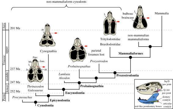
An evolutionary diagram and cladogram illustrating the cranial and mandibular morphology of Cynodontia leading to Mammalia. The main figure presents a phylogenetic timeline from the Permian to the Jurassic, showing dorsal views of skulls from taxa including Thrinaxodon, Cynognathia, Probainognathus, Tritylodontidae, and non-mammalian mammaliaforms. Key anatomical trends highlighted include the simplification of the circumorbital region (loss of prefrontal and postorbital bones) and the posterior migration of the internal choanae (indicated by red arrows), reflecting the development of the secondary palate. Labels identify cranial bones such as the Maxilla (Mx), Nasal (N), Frontal (F), and Parietal (P). An inset diagram focuses on the 'dual jaw joint' condition, showing a lateral view of the skull and a medial view of the mandible. It differentiates the ancestral quadrate-articular (Q-Art) joint from the derived squamosal-dentary (Sq-D) joint. The mandible illustration highlights the expansion of the dentary bone (D) and the reduction of postdentary bones into a rod-like structure, foundational to the evolution of the mammalian middle ear.
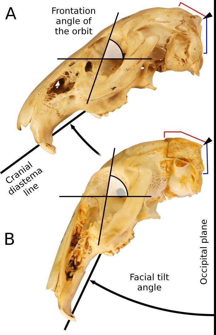
This comparative anatomical diagram illustrates the cranial morphology and facial tilt in two species of the Leporidae family, serving as an educational resource for comparative osteology and biomechanics. The image displays two crania, Caprolagus hispidus (A) and Pronolagus crassicaudatus (B), in a left lateral view to demonstrate the relationship between skull structure and functional ecology.
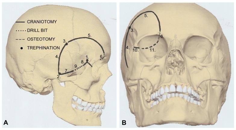
Two-panel anatomical diagram illustrating the bone work sequence for a modified orbitozygomatic (mOZ) one-piece craniotomy on a human skull. Panel A shows a lateral view, while Panel B shows an anteroposterior view. The illustration employs a numbered, coded system to detail surgical steps: trephinations (points 1, 2, 3) at the keyhole, posterior zygomatic arch, and superior temporal line; craniotomy cuts (lines 4, 5, 7, 8, 10) across the temporal, frontal, and sphenoid bones; drill bit maneuvers (dotted lines 6, 8, 9) at the orbital rim and zygomatic area; and osteotomy (dashed line 11) of the orbital roof and superior orbital fissure. This systematic approach facilitates access to the anterior and middle cranial fossae and the superior orbit. The diagram serves as a technical surgical guide for neurosurgical residents and surgeons focusing on skull base approaches for tumors, such as epidermoid cysts, in the superolateral orbital region.
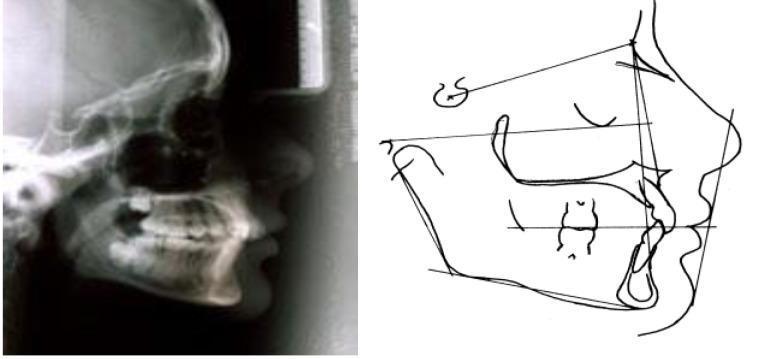
This diagnostic and comparison image consists of a lateral cephalometric radiograph (left) and a corresponding anatomical tracing (right) illustrating craniofacial morphology. The radiograph shows a lateral view of the human skull, detailing the cranial vault, sella turcica, facial bones, maxilla, mandible, and dental occlusion. The accompanying cephalometric diagram outlines key landmarks and planes used in orthodontic and maxillofacial surgery analysis, including the SN plane, mandibular plane, and soft tissue profile (nose, lips, and chin). The image specifically demonstrates an 'anterior rotation type' growth pattern, highlighting the relationship between skeletal bases and dental alignment. This material is designed for intermediate to advanced dental education, focusing on orthodontics and cephalometry to assess jaw discrepancies and facial growth vectors.
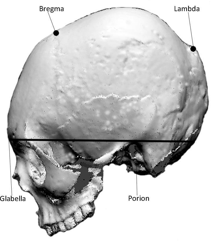
This diagnostic image shows a lateral view of a human skull, reconstructed from a 3D scan, serving as an anatomical diagram for craniometric analysis. Key osteological landmarks are labeled to define the morphology of the cranial vault and base. On the superior aspect of the cranium, the 'Bregma' (the junction of the coronal and sagittal sutures) is identified anteriorly, and the 'Lambda' (the junction of the lambdoid and sagittal sutures) is identified posteriorly. These points help characterize the curvature of the frontal, parietal, and occipital bones. At the base, a straight horizontal reference line is drawn connecting the 'Glabella' (the most prominent point between the eyebrows on the frontal bone) and the 'Porion' (the uppermost point on the margin of the external auditory meatus), extending toward the occipital region. This line serves to demarcate the inferior margin of the cranial vault for quantitative shape analysis. The image is used in clinical imaging and osteological research to assess cranial morphology and variations such as intentional cranial modification.
Cranial Bones - Position and Detailed Anatomy
1. Frontal Bone
- Sits at the front of the skull, articulating posteriorly with both parietal bones at the coronal suture.
- Contains the frontal sinuses (air-filled cavities) internally.
- Key landmarks: glabella (smooth area between the eyebrows), supraorbital ridge/margin (superior rim of the orbit), superciliary arches (brow ridges).
- "The frontal bone forms the contour of the forehead... the junction between the cranium and the face" - Cummings Otolaryngology Head and Neck Surgery
2. Parietal Bones (paired - left and right)
- Meet each other at the top in the midline at the sagittal suture.
- Meet the frontal bone anteriorly at the coronal suture.
- Meet the occipital bone posteriorly at the lambdoid suture.
- Meet the temporal bones laterally/inferiorly at the squamous suture.
- The junction of the coronal and sagittal sutures is the bregma; the junction of the sagittal and lambdoid sutures is the lambda - both are important craniometric landmarks.
3. Temporal Bones (paired - left and right)
- Composed of four parts: squamous (flat, anterior-superior part), petrous (dense, pyramid-shaped part housing the inner ear), mastoid (posterior part with the mastoid process, the bony bump behind the ear), and tympanic part (surrounds the ear canal).
- "The mastoid part is the most posterior part of the temporal bone" - Gray's Anatomy for Students
- Houses the structures of hearing and balance (cochlea, semicircular canals) within the petrous portion.
- Contains the external acoustic meatus (ear canal opening), styloid process (attachment for neck muscles/ligaments), and zygomatic process (articulates forward with the zygomatic bone to help form the cheekbone arch).
4. Occipital Bone
- Articulates with both parietal bones superiorly (lambdoid suture) and both temporal bones laterally.
- Contains the foramen magnum, the large opening through which the spinal cord passes to connect with the brainstem.
- Has the occipital condyles, which articulate with the first cervical vertebra (C1, the atlas), forming the joint that allows nodding of the head.
- The external occipital protuberance is a palpable bump at the base of the skull, a landmark for locating the confluence of venous sinuses inside.
5. Sphenoid Bone
- Shaped like a butterfly, with a body in the center and greater wings and lesser wings extending laterally.
- The body contains the sella turcica ("Turkish saddle"), a saddle-shaped depression that houses the pituitary gland.
- The greater wing forms part of the lateral orbital wall and part of the base of the skull (visible from below); the lesser wing forms part of the floor of the anterior cranial fossa.
- Inferiorly, the pterygoid processes project down and are important attachment sites for chewing (pterygoid) muscles.
- Contains key foramina for cranial nerves and vessels: optic canal, superior orbital fissure, foramen ovale, foramen rotundum.
- "The greater wing also forms part of the infratemporal fossa" - Gray's Anatomy for Students
6. Ethmoid Bone
- Lightweight, sponge-like bone forming part of the anterior cranial fossa floor, the medial orbital walls, the nasal septum, and the lateral walls of the nasal cavity.
- Contains the cribriform plate (perforated horizontally, allowing olfactory nerve fibers to pass from the nasal cavity to the brain), the perpendicular plate (forms the upper nasal septum), and the superior/middle nasal conchae (turbinate structures inside the nose).
- Its air cells form the ethmoid sinuses.
How They Fit Together (Summary of Position)
- Facial bones next (maxilla, mandible, zygomatic, nasal, etc.) to complete the skull, or
- Move to a different region (vertebral column, limbs, etc.)