

Explain pathological anatomy
infected heel pressure ulcer with slough necrosis and erythema
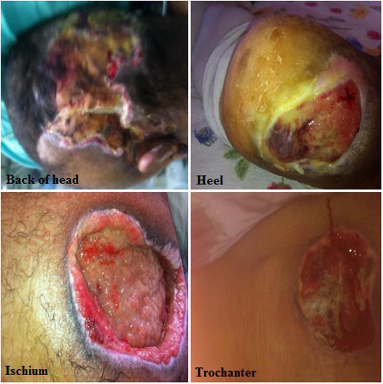
This composite clinical photograph displays four examples of pressure ulcers (PUs) at common anatomical locations: the back of the head, heel, ischium, and trochanter. The images illustrate varying stages of tissue damage. The 'Back of head' photograph shows a large, irregular ulcer with a mix of red granulation tissue, yellow slough, and dark necrotic areas. The 'Heel' ulcer demonstrates a thick, yellowish layer of slough or eschar with peripheral erythema. The 'Ischium' ulcer is a deep, cavitated wound featuring prominent beefy red granulation tissue and well-defined, raised inflammatory borders. The 'Trochanter' image shows a shallower ulceration with central slough and surrounding skin maceration and erythema. Collectively, these images serve as an educational resource for identifying decubitus ulcers over bony prominences, emphasizing variability in wound morphology, tissue composition (slough, granulation, necrosis), and wound depth (Stages II-IV) in patients with compromised mobility.
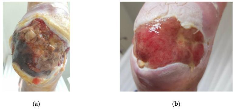
Clinical photograph of a diabetic foot ulcer on the heel, showing a comparison of wound healing over 33 days. Panel (a) illustrates the baseline (Day 0) state, characterized by a large, deep ulcer with significant necrotic tissue, dark eschar in the lower wound bed, and greenish-yellow slough indicative of Pseudomonas aeruginosa infection. The wound margins are irregular with surrounding erythema. Panel (b) shows the same wound on Day 33 after topical medical grade honey treatment. There is a marked reduction in total wound surface area. The wound bed has transitioned to healthy, beefy red granulation tissue with no visible necrosis or slough. Evidence of epithelialization is visible at the wound edges, and the surrounding skin appears less inflamed and macerated. The image sequence demonstrates the progression from a chronic, infected Stage B, Grade 3 diabetic ulcer to a vital, granulating wound bed suitable for continued secondary intention healing.
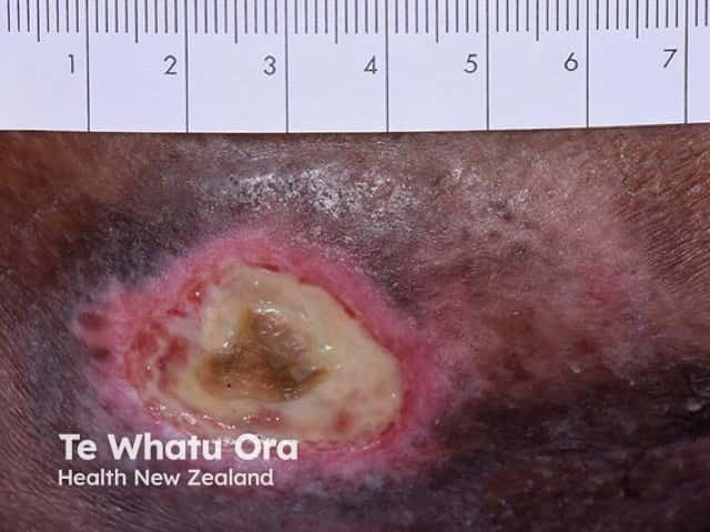
Clinical photography of a well-circumscribed pressure ulcer on the skin with a central necrotic slough overlying a granulating wound bed. The lesion shows a defined, circular/oval outline with surrounding erythema and a thick layer of slough occupying the wound center. A ruler is included in the frame for scale, indicating a lesion diameter of several centimeters. The wound bed demonstrates mixed tissue composition with necrotic slough (yellow-white) atop a pink-to-red granulating base; periwound skin is inflamed. The clinical impression is a chronic decubitus ulcer with suspected underlying osteomyelitis, given the depth and probable bone involvement. No radiographic image is provided; radiology notes would seek MRI or X-ray to confirm osteomyelitis and assess bone marrow edema or cortical disruption. Management implications include aggressive wound debridement, infection control, appropriate antibiotic therapy, and pressure offloading to prevent progression. This image is suitable for wound-care education, clinical case repositories, and decision-support datasets focusing on chronic wounds with osseous involvement. Keywords: pressure ulcer, decubitus ulcer, necrotic slough, osteomyelitis, wound bed, periwound inflammation, granulation tissue, offloading, debridement, antibiotics, MRI, X-ray, infection, chronic wound, dermatology imaging, clinical photography. Clinical relevance includes staging, mobility planning, and multidisciplinary care; potential for necrosis, tunneling, malodor, and systemic sepsis risk if untreated.
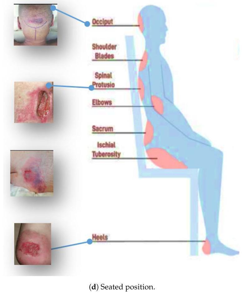
This medical infographic illustrates common anatomical sites for pressure ulcer development in a seated individual. The primary element is a lateral-view silhouette of a person seated in a chair, with red-highlighted zones identifying pressure-prone areas over bony prominences. These include the occiput, shoulder blades, spinal protrusions, elbows, sacrum, ischial tuberosity (buttocks), and heels. To the left, four clinical photographs provide real-world examples of decubitus ulcers at varying stages: 1) A closed, deep-tissue injury on the occiput with dark purple bruising; 2) A severe, full-thickness open wound (Stage III/IV) with visible slough and surrounding erythema at a spinal protrusion; 3) A reddish-purple, non-blanchable lesion at the ischial tuberosity indicative of Stage I/II damage; and 4) An open, erosive lesion on the heel. This educational graphic is intended for nursing and geriatric training to demonstrate the correlation between patient positioning and localized tissue necrosis caused by prolonged mechanical stress and friction.
Pathological Anatomy of the Wound in the Image
What the Image Shows
1. Wound Location and Type
- Site: Posterior calcaneal (heel) region - one of the highest-risk bony prominences for pressure injury
- Type: Deep open ulcer - full-thickness skin loss
- Likely etiology: Pressure ulcer (Stage III or unstageable), or diabetic/neuropathic foot ulcer given the calcaneal location
2. Layers Involved - Anatomical Depth
| Layer | Status |
|---|---|
| Epidermis | Completely absent |
| Dermis (papillary + reticular) | Destroyed |
| Subcutaneous fat | Exposed and largely necrotic |
| Deep fascia/tendon | Possibly involved but obscured by devitalized tissue |
- Stage III = full-thickness skin loss with damage to subcutaneous tissue, extending down to (but not including) the underlying fascia; presents as a crater-like ulcer
- Stage IV = full-thickness skin loss and extensive tissue necrosis with destruction extending to muscle, bone, or tendons; undermining or sinus tracts may be present
3. Wound Bed Components - Pathological Anatomy
a. Slough (Yellowish-Green Material)
b. Moist Necrosis (Dark Central Area)
c. Granulation Tissue
d. Exudate
4. Surrounding Skin (Periwound) Changes
| Feature | Pathological Significance |
|---|---|
| Bright red erythema halo | Active cellulitis / inflammatory vasodilation extending into periwound tissue |
| Swelling/edema | Local inflammatory exudate accumulation; suggests infection spreading into surrounding tissue |
| White macerated skin at wound edges | Prolonged moisture exposure causing epidermal softening and breakdown |
| Epidermal peeling | Friction damage, blister resolution, or maceration effect |
5. Pathogenesis - Why This Happens
- External pressure - interstitial pressure >32 mmHg (above normal capillary pressure of 12-32 mmHg) compromises microcirculation and tissue oxygenation
- Shearing forces - sliding displacement between tissue planes causes vessel angulation and thrombosis, leading to undermining
- Friction - abrades the stratum corneum, removing the protective skin barrier
- Moisture - increases ulcer risk fivefold by macerating the skin
6. Signs of Active Infection
- Greenish slough - suggests Pseudomonas aeruginosa or other gram-negative organisms
- Perilesional cellulitis (spreading erythema beyond wound margins)
- Heavy purulent exudate
- Periwound edema
7. Reference Image - Pressure Ulcer Staging
8. Clinical Urgency
- Wound debridement - surgical, enzymatic, or autolytic removal of slough and necrotic tissue to expose the true wound depth and allow healing
- Wound culture - to identify the causative organism(s) and guide antibiotic therapy
- Assess for osteomyelitis - calcaneal osteomyelitis is a serious complication at this depth; MRI is the most sensitive imaging modality
- Offloading - complete pressure relief from the heel using foam wedges, total contact cast, or specialized offloading footwear
- Appropriate wound dressing - an absorptive dressing (e.g., alginate or foam) for heavy exudate management, transitioning to hydrocolloid/hydrogel as the wound improves
- Systemic evaluation - assess for diabetes, peripheral vascular disease, malnutrition, and any condition impairing wound healing
Sources: Dermatology 2-Volume Set 5e, pp. 2210-2213; Andrews' Diseases of the Skin, p. 1854-1856