
Pres syndrome as a short note for md Anaesthesia exam
"posterior reversible encephalopathy syndrome" PRES
posterior reversible encephalopathy syndrome PRES MRI FLAIR occipital
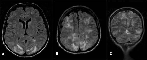
This diagnostic image set consists of three brain MRI sequences (two axial FLAIR images, A and B, and one coronal T2W image, C) demonstrating classic findings of Posterior Reversible Encephalopathy Syndrome (PRES). The images reveal bilateral, largely symmetric, patchy hyperintense lesions within the cortical and subcortical regions. In the axial FLAIR views (A, B), there is prominent involvement of the occipital lobes and frontal-parietal watershed zones. The coronal T2W image (C) further highlights the predilection for the posterior circulation, showing extensive vasogenic edema in the parietal and occipital lobes, as well as the cerebellar hemispheres. These neuroimaging features are consistent with the clinical presentation of acute encephalopathy and seizures, often associated with systemic triggers like hypertension or chemotherapy. The visual data emphasizes the characteristically reversible vasogenic edema pattern that defines RPLS/PRES in a clinical neurology or neuroradiology context.
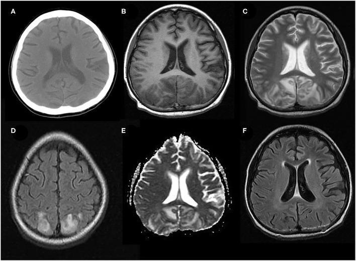
A composite diagnostic image set displaying typical neuroimaging findings of Posterior Reversible Encephalopathy Syndrome (PRES) in a pediatric patient. (A) Unenhanced axial brain CT shows subtle, symmetrical cortico-subcortical hypodensities in the bilateral parieto-occipital regions. (B-E) Acute stage axial MRIs further characterize these lesions: (B) T1-weighted MRI shows hypointensities; (C) T2-weighted and (D) T2-FLAIR MRI sequences reveal prominent, symmetrical hyperintensities consistent with vasogenic edema; and (E) the Apparent Diffusion Coefficient (ADC) map shows corresponding hyperintensity, indicating absence of diffusion restriction. (F) Follow-up axial FLAIR MRI at two months demonstrates complete resolution of the previous abnormal signal intensities, highlighting the reversible nature of the pathology. The primary educational focus is the recognition of classic parieto-occipital distribution and the signal characteristics (T2/FLAIR hyperintensity and high ADC) that distinguish vasogenic edema in PRES from cytotoxic edema in acute infarction.
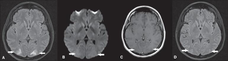
Diagnostic axial brain MRI series illustrating Posterior Reversible Encephalopathy Syndrome (PRES). Panel A (FLAIR) shows hallmark bilateral, symmetric hyperintensities in the white matter of the occipital lobes, indicative of vasogenic edema. Panel B (Diffusion-Weighted Imaging) demonstrates no restricted diffusion in these areas, confirming the absence of cytotoxic edema or acute infarction. Panel C (T1-weighted post-contrast) shows no pathological gadolinium enhancement, highlighting the lack of blood-brain barrier breakdown. Panel D (Follow-up FLAIR at 4 weeks) shows complete radiological resolution of the previous hyperintense lesions. This comparison demonstrates the classic reversible neuroimaging features of vasogenic edema associated with PRES, typically seen in clinical contexts such as hypertensive crisis, preeclampsia, or immunosuppressive therapy.
PRES (Posterior Reversible Encephalopathy Syndrome)
Definition
Aetiology / Precipitating Causes
- Hypertensive emergencies (most common) - systolic BP >195 mmHg, or a sudden rise even at lower levels
- Eclampsia / Pre-eclampsia / HELLP syndrome - most common obstetric scenario encountered by anaesthetists
- Immunosuppressive drugs - tacrolimus, cyclosporine/ciclosporin, FK-506 (calcineurin inhibitors)
- Chemotherapeutic agents - cyclophosphamide, interferons, bevacizumab, cisplatin
- Renal failure / acute glomerulonephritis
- Systemic lupus erythematosus, TTP/HUS
- Pheochromocytoma, cocaine, sympathomimetics (phenylephrine, aminophylline)
- Post-transplant (kidney, bone marrow)
Pathophysiology
1. Autoregulatory Failure (Breakthrough Hyperperfusion) - Dominant Theory
- Sudden severe hypertension overwhelms cerebral autoregulation
- Cerebral vessels passively dilate, blood-brain barrier (BBB) disrupts
- Vasogenic edema results from fluid extravasation through leaky capillary endothelial junctions
- Posterior cerebral areas are more susceptible due to relatively sparse sympathetic innervation of posterior circulation vessels (less vasoconstrictive tone)
2. Vasospasm / Endothelial Dysfunction Theory
- Endothelial injury (from toxins, immunosuppressants, cytokines) causes vasospasm
- Ischemia and cytotoxic edema can also occur
- Explains PRES in normotensive or mildly hypertensive patients (e.g., drug toxicity, eclampsia at moderate BP)
Clinical Features (The Classic Tetrad)
| Feature | Details |
|---|---|
| Headache | Sudden, severe, often the first symptom |
| Seizures | Often the presenting feature; may be focal or generalised |
| Visual disturbances | Visual field defects, cortical blindness, Balint syndrome, hallucinations |
| Altered consciousness | Confusion, encephalopathy, obtundation, coma in severe cases |
In eclampsia, all four features may appear rapidly - this is one of the most common perioperative presentations.
Imaging (Key for Exam)
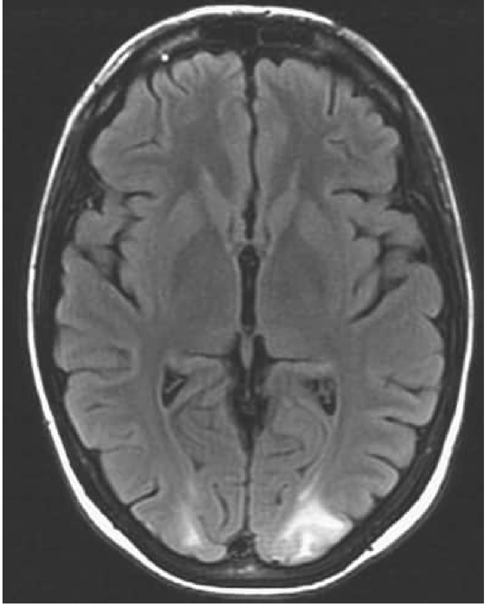
- T2/FLAIR: Bilateral symmetric hyperintensities in posterior parieto-occipital white matter (can involve cortex too)
- DWI (Diffusion-Weighted Imaging): Typically no diffusion restriction - distinguishes vasogenic edema from cytotoxic edema of infarction (high ADC values)
- T1: Hypointense lesions
- Gadolinium enhancement: Usually absent (BBB not fully broken down)
- Follow-up MRI: Complete resolution within 2-4 weeks with treatment
Distribution of Edema
- Parieto-occipital lobes - most common (posterior cerebral predominance)
- Frontal lobes, temporal lobes, cerebellum, brainstem - less frequent
- Can be atypical: frontal-predominant, asymmetric, or involve basal ganglia
- Not restricted to white matter - cortex also involved in many cases
Diagnosis
- Ischemic stroke (DWI restriction differentiates)
- Venous sinus thrombosis
- Encephalitis (fever, CSF pleocytosis)
- Reversible Cerebral Vasoconstriction Syndrome (RCVS) - can overlap with PRES
Management (Anaesthesia-Focused)
1. Treat the Underlying Cause
- Control hypertension - rapid, controlled BP reduction
- IV labetalol, hydralazine, nicardipine, sodium nitroprusside
- Target: reduce MAP by ~20-25% in the first hour; avoid overcorrection
- In eclampsia: MgSO4 + antihypertensives + delivery
- Withdraw offending drug (tacrolimus, cyclosporine) immediately if drug-induced
- Treat renal failure, autoimmune cause
2. Seizure Management
- IV benzodiazepines (lorazepam, diazepam) acutely
- Levetiracetam or phenytoin/fosphenytoin for ongoing seizures
- Long-term antiepileptics usually not required if underlying cause is treated (PRES seizures tend to be self-limiting once etiology resolved)
3. Airway / ICU
- Protect airway in encephalopathic/seizing patients
- Anaesthetist's role: RSI if needed, haemodynamic monitoring, vasopressor choice in hypotensive patients (avoid agents that aggravate hypertension)
- Avoid hypoxia (worsens edema)
4. MgSO4 in Eclampsia
- Both anticonvulsant and mild vasodilatory - first-line in eclampsia-related PRES
- Monitor for toxicity (respiratory depression, loss of patellar reflex)
Prognosis
- Generally favorable with early diagnosis and treatment
- Neuroimaging normalises within days to weeks
- Neurological recovery is usually complete
- Poor outcome predictors: delayed diagnosis, intracerebral hemorrhage, diffusion restriction on MRI (cytotoxic component), prolonged seizures, recurrence
Anaesthesia-Specific Points (Exam High-Yield)
- Eclampsia and preeclampsia are the most common obstetric causes - anaesthetist must recognise PRES perioperatively
- PRES can occur intraoperatively with sudden BP spikes or postoperatively
- In organ transplant patients on calcineurin inhibitors presenting with new neurological symptoms - think PRES
- DWI non-restriction is the key MRI finding to differentiate PRES (vasogenic) from stroke (cytotoxic)
- Seizures in PRES context - treat the cause, not just the seizure; routine anticonvulsants are not started long-term
- BP overcorrection is dangerous - aim for controlled, gradual reduction
- MgSO4 level monitoring is essential intraoperatively in eclampsia patients
Can you make it more towards Anaesthesia and take reference from standard anesthesia textbook like millers
PRES (Posterior Reversible Encephalopathy Syndrome)
Short Note - MD Anaesthesia Exam
Definition
Relevance to Anaesthesia
- Obstetric anaesthesia - eclampsia/pre-eclampsia (most common perioperative trigger)
- Spine surgery - listed by Miller's Anesthesia 10e as a cause of perioperative visual loss after spinal procedures
- Transplant anaesthesia - calcineurin inhibitor toxicity (tacrolimus, cyclosporine)
- Postoperative hypertensive surges - untreated emergence hypertension
- Oncology/ICU patients - chemotherapy-associated PRES
Aetiology
| Category | Specific Causes |
|---|---|
| Hypertensive emergencies | Accelerated/malignant hypertension, renal artery stenosis, pheochromocytoma, Cushing syndrome |
| Obstetric | Pre-eclampsia, eclampsia, HELLP syndrome |
| Immunosuppressants | Tacrolimus, cyclosporine/ciclosporin (calcineurin inhibitors), FK-506 |
| Chemotherapy | Cyclophosphamide, bevacizumab, cisplatin, interferons |
| Renal | Acute glomerulonephritis, CKD, hemodialysis |
| Drugs/Toxins | Cocaine, aminophylline, sympathomimetics (phenylephrine) |
| Autoimmune | SLE, TTP/HUS |
Absolute BP level is less important than the rate of rise. Eclampsia can cause PRES at relatively modest BP levels due to endothelial dysfunction overwhelming autoregulatory reserve. - Adams & Victor's Principles of Neurology, 12th Ed.
Pathophysiology
Theory 1: Autoregulatory Breakthrough (Primary Mechanism)
- Sudden BP surge overwhelms cerebral autoregulatory capacity
- Passive forced vasodilatation of cerebral vessels
- Disruption of capillary endothelial tight junctions
- Fluid extravasation → vasogenic edema
- Posterior hemispheres are preferentially affected due to relatively sparse sympathetic adrenergic innervation of posterior circulation, reducing their vasoconstrictive reserve
Theory 2: Endothelial Dysfunction / Vasospasm
- Toxins (calcineurin inhibitors), cytokines (eclampsia), immune complex deposition directly injure the endothelium
- Vasoconstriction → ischemia → BBB disruption
- Explains PRES in normotensive patients
Clinical Features - "The Tetrad"
| Symptom | Features |
|---|---|
| Headache | Sudden, severe; often the first symptom |
| Seizures | Most dramatic; generalised or focal; may be status epilepticus |
| Visual disturbances | Cortical blindness, homonymous hemianopia, visual hallucinations, Balint syndrome |
| Encephalopathy | Confusion → obtundation → coma in severe cases |
Imaging
| Sequence | Finding |
|---|---|
| T2 / FLAIR | Bilateral, largely symmetric hyperintensities in posterior parieto-occipital white matter ± cortex |
| DWI | No diffusion restriction - high ADC (vasogenic edema); this is the KEY differentiator from ischemic stroke |
| ADC map | Increased (confirms free water movement - vasogenic not cytotoxic) |
| T1 + Gadolinium | Usually no enhancement (intact BBB) |
| Follow-up MRI | Complete resolution within 2-4 weeks with treatment |
Diagnosis
- Clinical + radiological correlation is essential
- CSF: elevated protein (up to >100 mg/dL), normal cells; not required routinely
- LP done only to exclude subarachnoid haemorrhage if suspected
| Condition | Differentiating Feature |
|---|---|
| Ischemic stroke | DWI restriction (cytotoxic edema), arterial territory distribution |
| Venous sinus thrombosis | Thrombosis on MRV, haemorrhagic infarction |
| Encephalitis | Fever, CSF pleocytosis, temporal lobe involvement |
| RCVS | "Thunderclap" headache, vasoconstriction on angiography; can coexist with PRES |
Anaesthesia Management
A. Control the Precipitating Cause
- IV labetalol (alpha + beta blockade) - drug of choice, especially in eclampsia
- IV nicardipine (calcium channel blocker) - titratable infusion
- IV hydralazine - arteriolar vasodilator; useful in obstetrics
- IV sodium nitroprusside - avoid prolonged use (cyanide toxicity); use only in refractory cases
- Target: Reduce MAP by 20-25% over the first hour; avoid aggressive overcorrection (risk of watershed infarction)
- Avoid: nifedipine sublingual (unpredictable BP drop)
- MgSO4 is first-line (both anticonvulsant and vasodilatory)
- Loading: 4-6 g IV over 15-20 min; Maintenance: 1-2 g/hr infusion
- Monitor for toxicity: loss of patellar reflex (Mg 7-10 mEq/L), respiratory depression (>12 mEq/L)
- Antidote: 10 mL of 10% calcium gluconate IV
- Antihypertensives as above
- Delivery is definitive treatment
- Anaesthetic implications: MgSO4 potentiates both depolarising and non-depolarising NMBAs - reduce NDMBA doses, use neuromuscular monitoring
- Immediate withdrawal or dose reduction of tacrolimus/cyclosporine
- Neurological recovery typically follows drug cessation
B. Seizure Management (Perioperative)
- Acute: IV benzodiazepines (lorazepam 0.1 mg/kg or diazepam 0.15 mg/kg)
- Second line: Levetiracetam, phenytoin/fosphenytoin, valproate
- Status epilepticus: anaesthetic doses of propofol or thiopentone + airway protection
- Long-term antiepileptics are not routinely indicated - once the underlying cause is treated, seizures resolve
C. Airway and Haemodynamic Management
- Encephalopathic/seizing patient: RSI with cricoid pressure - protect airway
- Induction agents: Propofol (reduces ICP and seizure threshold) preferred
- Ketamine: traditionally avoided in PRES (raises BP), but increasingly used at low doses in eclampsia; avoid in frank hypertensive crisis
- Intraoperative hypertensive surges - adequate depth of anaesthesia, opioids, vasodilators
- Invasive arterial line: mandatory for continuous BP monitoring
- Vasopressors: phenylephrine (pure alpha) preferred over ephedrine (sympathomimetic - raises BP further) in eclampsia-associated hypotension from spinal anaesthesia
- Avoid hypoxia and hypercapnia (both worsen cerebral edema and seizure threshold)
D. PRES in Spine Surgery (Miller's Anesthesia 10e)
- Avoid sustained hypotension (especially with controlled hypotension techniques)
- Head position: neutral, at or above heart level - no prolonged neck flexion/rotation
- No direct pressure on the eyes
- Staged procedures in high-risk patients
- Colloids alongside crystalloids for volume replacement
- Periodic haemoglobin monitoring in major blood loss cases
- Case-by-case decision for deliberate hypotension in high-risk patients
Prognosis
- Excellent with early recognition and treatment
- MRI normalises within days to weeks
- Complete neurological recovery in most cases
- Poor prognostic indicators:
- Delayed diagnosis
- DWI restriction on MRI (cytotoxic component - irreversible infarction)
- Intracerebral haemorrhage
- Status epilepticus
- Recurrence (possible with ongoing or recurrent triggers)
Summary - High-Yield Points for MD Anaesthesia
| Point | Key Fact |
|---|---|
| Most common anaesthesia trigger | Eclampsia / pre-eclampsia |
| Mechanism | Vasogenic edema from autoregulatory failure |
| Why posterior? | Sparse sympathetic innervation of posterior circulation |
| MRI key finding | T2/FLAIR hyperintensity + DWI non-restriction (high ADC) |
| Drug of choice for seizures | MgSO4 in eclampsia; BDZ acutely otherwise |
| BP target | Reduce MAP by 20-25% over first hour; avoid overcorrection |
| MgSO4 interactions | Potentiates all NMBAs - reduce doses, monitor with nerve stimulator |
| Miller's specific context | Perioperative visual loss after spine surgery (PRES is one of 4 causes) |
| Long-term anticonvulsants | Not routinely required |
| Calcineurin inhibitor PRES | Stop the drug immediately |
- Miller's Anesthesia, 10th Ed. (2-Volume Set) - Perioperative Visual Loss, Spine Surgery Chapter
- Barash, Cullen & Stoelting's Clinical Anesthesia, 9th Ed. - Neurological complications, Diagnosis
- Adams & Victor's Principles of Neurology, 12th Ed. - PRES/Hypertensive Encephalopathy
- Bradley & Daroff's Neurology in Clinical Practice, 9th Ed. - MRI characteristics of PRES
- Fugate JE, Hawkes MA, Rabinstein AA. Posterior reversible encephalopathy syndrome: evolving insights. Lancet Neurology, 2025.