
I need a to z about Cataract Pterygium Aphakia Pseudophakia Chronic dacryocystitis Chalazion Corneal opacity Stye ( hordeolum externum)
Reading File
Now let me search for all 8 conditions simultaneously:
Finding Sources
Finding Sources
Finding Sources
Finding Sources
Finding Sources
Finding Sources
Good, I have file paths. Now let me read the most comprehensive ophthalmology textbook (Kanski's) sections for cataract, pterygium, and dacryocystitis, plus the Goldman-Cecil sections for hordeolum, and do a broader search for aphakia/pseudophakia and corneal opacity:
Reading File
Reading File
Reading File
Finding Sources
Now let me read the cataract section from Kanski's (main block), the chalazion/stye section, and dacryocystitis section:
Reading File
Reading File
Finding Sources
Now let me read the main Kanski's cataract section (it's in a different block) and look for corneal opacity:
Finding Sources
Finding Sources
Now let me read the Kanski's cataract section (block2) and pterygium treatment section:
Reading File
Reading File
Now let me also read the cataract management/surgery section and the Goldman-Cecil section on cataract:
Reading File
Reading File
I now have comprehensive data from the textbooks. Let me also fetch a couple of key images to include visually:
Loading Image
Loading Image
Here is a comprehensive A-Z guide on all eight conditions, sourced from Kanski's Clinical Ophthalmology, Goldman-Cecil Medicine, Tintinalli's Emergency Medicine, and The Wills Eye Manual.
Comprehensive Ophthalmology Guide
1. CATARACT
Definition
Any opacification of the crystalline lens. The lens doubles in volume between birth and age 70 as new lens fiber cells are laid down externally, compressing older fibers into the center. - Goldman-Cecil Medicine
Classification
A. Age-Related (Acquired) Cataract - most common
| Type | Location | Key Feature |
|---|---|---|
| Posterior Subcapsular (PSC) | Just in front of posterior capsule | Granular/plaque appearance on slit lamp; black and vacuolated on retroillumination; Wedl (bladder) cells; profound glare effect |
| Nuclear Sclerotic | Central nucleus | Yellowish hue (urochrome pigment); associated myopic shift ("second sight of the aged"); brown or even black when advanced (brunescent); Morgagnian cataract = liquefaction of cortex with inferior sinking of nucleus |
| Cortical | Anterior, posterior, or equatorial cortex | Starts as clefts/vacuoles due to cortical hydration; cuneiform (wedge-shaped) or radial spoke-like opacities; commonly begins inferonasal quadrant |
B. Congenital Cataract
- Lamellar (zonular): Most common type - white opacities surrounding nucleus with alternating clear and white cortical lamella ("onion skin")
- Polar: Small anterior or posterior polar opacities; anterior polar usually small and visually insignificant
- Causes: Rubella, Down syndrome, galactosaemia, Lowe syndrome, trauma
C. Secondary/Complicated Cataract
- Metabolic: Diabetes (osmotic swelling; "snowflake" cortical in juvenile DM), galactosaemia, hypocalcaemia (polychromatic lamellar)
- Drug-induced: Systemic corticosteroids (posterior subcapsular), chlorpromazine, amiodarone, busulphan, gold (anterior capsular)
- Radiation: Posterior subcapsular; posterior pole accumulates non-translucent damaged cells
- Ocular disease: Uveitis (PSC), angle-closure glaucoma, high myopia, retinitis pigmentosa
- Trauma: "Rosette" cataract after blunt trauma; Vossius ring (iris pigment on lens after impact)
- Post-vitrectomy: Nuclear sclerosis, especially in patients >50 years
- Silicone oil: Feathering of posterior subcapsular lens; virtually all phakic eyes with silicone oil eventually develop cataract
- Myotonic dystrophy: Fine iridescent "Christmas tree" cortical opacities in 3rd decade → visually disabling by 5th decade
Symptoms
- Reduced visual acuity (initially correctable with spectacles)
- Loss of contrast sensitivity
- Change in colour perception (blue colours particularly affected)
- Glare - difficulty with night driving
- Monocular diplopia
- Difficulty in low-light situations
Investigations
- Slit-lamp biomicroscopy (oblique beam for nuclear, retroillumination for PSC and cortical)
- Red reflex assessment
- Visual acuity and contrast sensitivity testing
- B-scan ultrasound if fundus not visible
Management
Conservative: Refractive correction while cataract is mild; adequate lighting.
Surgical - indicated when vision impairs daily activities:
- Phacoemulsification (standard): Ultrasound probe fragments and aspirates nucleus through a small incision (~2.4 mm); IOL inserted into capsular bag. Results equivalent to femtosecond laser-assisted surgery.
- ECCE (extracapsular cataract extraction): Used when nucleus too hard for phaco; larger incision
- ICCE (intracapsular): Entire lens including capsule removed; now rarely performed
- IOL (intraocular lens) implantation: Synthetic lens replaces natural lens; standard = posterior chamber IOL in capsular bag
- Femtosecond laser-assisted: Capsulotomy and lens fragmentation prior to phaco; outcomes similar to standard phaco
Post-operative complications: Posterior capsular opacification (PCO - "after-cataract"), endophthalmitis, cystoid macular oedema, retinal detachment
Prognosis: Excellent for restoration of vision if retina is healthy.
2. PTERYGIUM
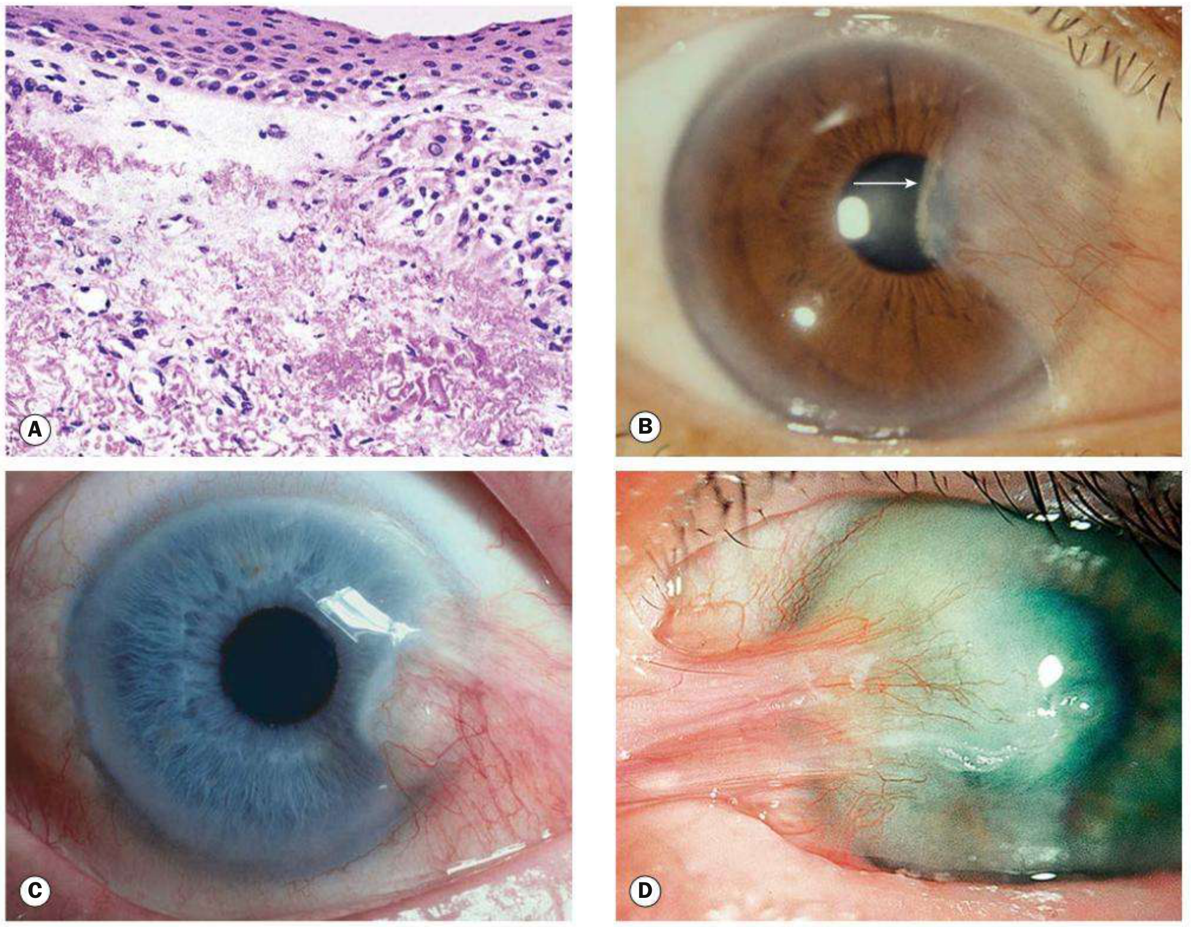
Definition
A triangular fibrovascular sub-epithelial ingrowth of degenerative bulbar conjunctival tissue over the limbus onto the cornea. Histologically shows elastotic degenerative changes in vascularized subepithelial stromal collagen. - Kanski's Clinical Ophthalmology
Pathogenesis
- UV exposure (main risk factor; occurs in sunny countries)
- Chronic surface dryness
- Familial tendency
- Invades Bowman's layer of the cornea (distinguishing it from pinguicula which does not cross the limbus)
Anatomy of a Pterygium
Three parts:
- Cap - avascular halo-like zone at the advancing edge
- Head - apex on the cornea
- Body - vascularized conjunctival part on the sclera
- Stocker line: linear iron deposition in corneal epithelium anterior to the head
- Fuchs islets: small discrete whitish flecks of pterygial epithelial cells at the advancing edge
Symptoms
- Small lesions: asymptomatic
- Irritation and grittiness (dellen effect at advancing edge - localized drying due to disruption of precorneal tear film)
- Contact lens intolerance
- Visual disturbance (obscuring visual axis, inducing astigmatism)
- Intermittent inflammation (pterygitis)
- Cosmetic concern
- Extensive/recurrent lesions: subconjunctival fibrosis extending to fornices → restricted ocular excursion
Pseudopterygium vs True Pterygium
| Feature | True Pterygium | Pseudopterygium |
|---|---|---|
| Location | Horizontal meridian (nasal most common) | Any position |
| Attachment to cornea | Along full length | Only at apex |
| Cause | UV degeneration | Chemical burn, corneal ulcer, trauma, cicatrizing conjunctivitis |
| Probe test | Cannot pass probe under it | Probe can pass under body |
Treatment
Medical (symptomatic):
- Lubricating drops/artificial tears
- Topical NSAIDs or mild steroids for inflammation
- UV-blocking sunglasses (advice for all patients)
Surgical indications: Encroachment toward visual axis, significant astigmatism, recurrent inflammation, cosmesis
Surgical options:
- Bare sclera excision: Simple excision - high recurrence rate (~80%); not recommended alone
- Conjunctival autografting: Donor conjunctiva from superior/upper-temporal para-limbal region; sutured or glued (fibrin glue, e.g., TISSEEL) into defect - significantly reduces recurrence
- Adjuvant treatment: Mitomycin C (anti-metabolite) or beta-irradiation to reduce recurrence
- Peripheral lamellar keratoplasty: For deep lesions involving stroma
TIP: Recurrence risk is reduced by using conjunctival autograft or applying mitomycin C to the operation site. - Kanski's Clinical Ophthalmology
3. APHAKIA
Definition
Absence of the crystalline lens from its normal position, with the lens capsule also absent (or predominantly absent). The term derives from Greek: "a" (without) + "phakos" (lens).
- Primary aphakia: Failure of lens development during embryogenesis (failure of lens placode to form in week 4, or failure of lens induction by optic vesicle). Usually associated with microphthalmia and severe intraocular dysgenesis (retinal dysplasia, corneal opacity). High risk of glaucoma. - The Wills Eye Manual
- Acquired aphakia: Most commonly results from cataract surgery (ICCE - intracapsular cataract extraction) or from traumatic lens dislocation/expulsion
Optical Consequences
- Loss of approximately +10 to +12 dioptres of refracting power
- Severe hypermetropia
- Loss of accommodation (no lens)
- Increased depth of field
- Enlarged retinal image
- Altered colour perception (increased UV transmission without lens filtering)
- Aphakic bullous keratopathy: corneal oedema and blistering
Visual Rehabilitation in Aphakia
- Aphakic spectacles: +10 to +12D lenses; magnify image ~30% → ring scotoma, jack-in-the-box phenomenon; useful for older children with bilateral aphakia
- Contact lenses: Superior optical solution for unilateral or bilateral aphakia; preferred in children (compliance worsens after ~age 2)
- IOL implantation: Now standard; secondary IOL can be implanted even years later if aphakia results from ICCE or trauma
Aphakia as a Risk Factor
- Predisposing condition for retinal detachment (along with high myopia, pseudophakia, lattice degeneration) - The Wills Eye Manual
4. PSEUDOPHAKIA
Definition
A state in which the natural crystalline lens has been replaced by an artificial intraocular lens (IOL) implant. The prefix "pseudo" (false) + "phakos" (lens). This is now the standard outcome of modern cataract surgery.
IOL Types
| Type | Position | Notes |
|---|---|---|
| Posterior chamber IOL (PCIOL) | In capsular bag or ciliary sulcus | Standard; most physiological position |
| Anterior chamber IOL (ACIOL) | In front of iris/angle | Used when posterior capsule is absent |
| Iris-fixated IOL | Clipped to iris | Used in select cases |
Special IOL Designs
- Monofocal: Corrects for one focal distance; most common
- Multifocal/trifocal: Splits light to correct near, intermediate, and distance
- Extended depth of focus (EDOF): Elongated focal point
- Toric: Corrects astigmatism
- Phakic IOL: Implanted without removing the natural lens (for high refractive errors)
Complications of Pseudophakia
- Posterior capsular opacification (PCO): Most common; "after-cataract"; treated with Nd:YAG laser posterior capsulotomy
- Cystoid macular oedema (Irvine-Gass syndrome)
- Pseudophakic bullous keratopathy: corneal decompensation
- Glare and halos: Particularly with multifocal IOLs; also a cause of glare/halos in the Wills Eye differential
- IOL dislocation/decentration
- Endophthalmitis (infection)
- Retinal detachment: Pseudophakia is a predisposing factor alongside aphakia
Pseudophakia vs Aphakia vs Phakia
| Term | Lens Status |
|---|---|
| Phakia | Natural crystalline lens present |
| Aphakia | No lens at all (no capsule) |
| Pseudophakia | Artificial IOL in place of natural lens |
5. CHRONIC DACRYOCYSTITIS
Anatomy Recap
The lacrimal drainage system: puncta → canaliculi (2 mm vertical + 8 mm horizontal) → common canaliculus → lacrimal sac (10-12 mm; in lacrimal fossa) → nasolacrimal duct (12-18 mm) → inferior nasal meatus (valve of Hasner). - Kanski's Clinical Ophthalmology
Definition
Chronic inflammation/infection of the lacrimal sac, most commonly secondary to obstruction of the nasolacrimal duct. Causative organisms are usually Staphylococcus or Streptococcus species.
Pathogenesis
Obstruction of the nasolacrimal duct → stasis of tears in lacrimal sac → bacterial colonization and chronic infection → mucocoele or mucopurulent discharge.
Clinical Features
- Chronic epiphora (watering eye) - the cardinal symptom
- Chronic or recurrent unilateral conjunctivitis
- Mucocoele: painless swelling at the inner canthus (below medial canthal tendon) - the hallmark sign
- If swelling is absent, pressure over the sac produces mucopurulent canalicular reflux (regurgitation test positive)
- Usually painless (contrast with acute dacryocystitis which is painful)
Diagnosis
- Regurgitation test (syringing): reflux of mucopurulent material confirms obstruction
- Dacryocystography (DCG) or dacryoscintigraphy to locate the obstruction
- CT/MRI if neoplasm or dacryolithiasis suspected
Treatment
- Definitive: Dacryocystorhinostomy (DCR) - creation of an anastomosis between the lacrimal sac and nasal mucosa to bypass the obstruction. Can be performed externally (standard) or endonasally (endoscopic).
- Topical/systemic antibiotics: temporary suppression only; do not cure
- Important warning: Postpone any intraocular surgery if there are signs of lacrimal drainage infection - risk of endophthalmitis - Kanski's Clinical Ophthalmology
Complications
- Mucocoele (distended, mucus-filled sac)
- Acute on chronic dacryocystitis
- Sac-skin fistula
- Orbital cellulitis (rare)
6. CHALAZION
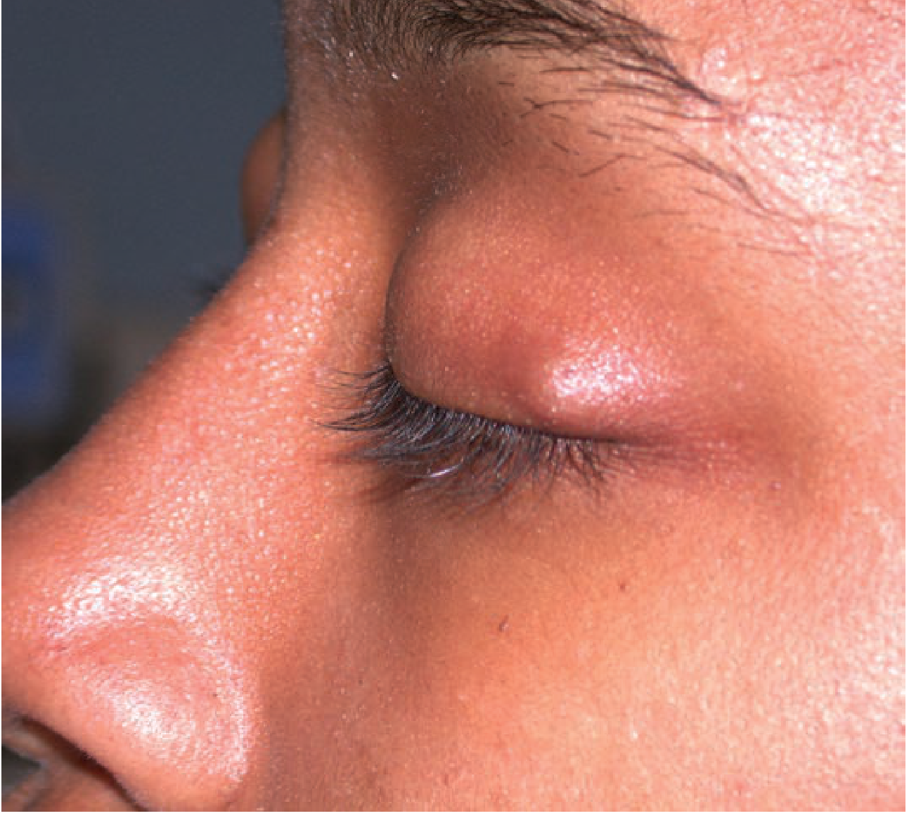
Definition
An acute or chronic lipogranulomatous inflammation of the eyelid caused by obstruction and subsequent rupture of a meibomian gland (or Zeis gland) in the tarsal plate, with leakage of lipid secretions into surrounding tissue. This provokes a foreign body giant cell granulomatous reaction. - Tintinalli's Emergency Medicine
Pathogenesis
Meibomian gland ostia obstruction → lipid retention → gland rupture → lipid into surrounding tissue → chronic granulomatous inflammation
Clinical Features
- Usually painless or mildly tender lid lump
- Located within the tarsal plate (deep to skin), felt as a firm nodule
- May point on the inner (conjunctival) surface of the eyelid
- Subacute to chronic onset (distinguishes from hordeolum)
- Occasionally mild erythema overlying
- Can grow large enough to indent the cornea and cause astigmatism
- Recurrent chalazia in an elderly patient: consider sebaceous gland carcinoma (masquerade lesion)
Chalazion vs Internal Hordeolum
Both can look identical clinically. An internal hordeolum is an acute purulent infection of a meibomian gland, whereas a chalazion is a chronic sterile lipogranuloma. Treatment is the same.
Treatment
- Warm compresses + gentle lid massage: first-line; promotes drainage
- Topical antibiotics (e.g., erythromycin): for secondary infection
- Intralesional corticosteroid injection: triamcinolone; effective for persistent chalazia (caution near medial canthus due to depigmentation risk)
- Incision and curettage (I&C): Under local anaesthetic; lid everted; vertical incision on conjunctival surface; contents curetted; preferred for large or chronic lesions
- Referral: To ophthalmologist at 1-2 weeks if not resolving
7. CORNEAL OPACITY
Definition
Any loss of transparency of the corneal stroma or surface due to scarring, deposition, oedema, or infiltration. Can be congenital or acquired.
Grades of Corneal Opacity (by density)
| Grade | Opacity | Vision |
|---|---|---|
| Nebula | Faint, hazy; can only be seen with slit lamp in oblique illumination | Minimal or no effect |
| Macula | Moderate, visible to naked eye | Some visual impairment |
| Leucoma | Dense white opacity | Significant visual impairment |
| Leucoma adherens | Dense opacity with iris adherent behind | Iris tissue incorporated into scar (after perforating injury) |
Causes
Congenital:
- Sclerocornea (cornea resembles sclera)
- Peter's anomaly (central corneal leucoma with posterior defects)
- Birth trauma (forceps injury)
- Primary aphakia with corneal dysgenesis
Acquired:
- Infective: Bacterial keratitis (Pseudomonas, Staphylococcus, Streptococcus), fungal keratitis, herpetic keratitis (HSV, HZV), Acanthamoeba
- Trachoma: Most common infectious cause of corneal blindness worldwide (Chlamydia trachomatis)
- Vitamin A deficiency: Xerophthalmia → keratomalacia → corneal melting → opacity
- Trauma: Chemical burns, perforating injury
- Exposure keratopathy: Lagophthalmos → drying → secondary infection → scarring
- Interstitial keratitis: Stromal vascularization and scarring (congenital syphilis - "salmon patches" with ghost vessels)
- Corneal dystrophies: Granular, lattice, macular, Fuchs endothelial
- Bullous keratopathy: Aphakic or pseudophakic; repeated epithelial bullae → scarring
- Band keratopathy: Calcium deposition in Bowman's layer (horizontal band); seen with hypercalcaemia, chronic uveitis, extended silicone oil tamponade
Clinical Assessment
- Slit-lamp biomicroscopy
- Corneal topography (assess regularity and induced astigmatism)
- Specular microscopy (endothelial cell count in bullous keratopathy)
- Anterior segment OCT
Treatment
Optical rehabilitation:
- Spectacles or contact lenses for irregular astigmatism
- Low vision aids
Surgical:
- Penetrating keratoplasty (PK): Full-thickness corneal transplant; for deep/total stromal opacities
- Lamellar keratoplasty (DALK): For stromal opacities with healthy endothelium; preserves host endothelium
- DSAEK/DMEK: For endothelial failure (bullous keratopathy)
- PTK (phototherapeutic keratectomy): Excimer laser ablation; for superficial opacities/anterior stromal scars
- EDTA chelation: For band keratopathy (removes calcium)
- Amniotic membrane transplant: For acute chemical burns to reduce scarring
Complications of Corneal Opacity
- Amblyopia (especially in children with opacities involving visual axis)
- Irregular astigmatism
- Secondary glaucoma (if adhesions involve the angle)
- Nystagmus (if bilateral and early onset)
8. STYE (HORDEOLUM EXTERNUM)
Definition
An acute bacterial infection (usually Staphylococcus aureus) of the follicle of an eyelash and the adjacent sebaceous glands of Zeis or sweat glands of Moll, located at the eyelid margin. - Tintinalli's Emergency Medicine
The term "stye" or "external hordeolum" specifically refers to this external infection, as distinct from an internal hordeolum (infection of the meibomian glands, deeper in the tarsal plate).
Pathogenesis
- Staphylococcal (usually S. aureus) infection of lash follicle
- Spreads to adjacent Zeis (sebaceous) or Moll (sweat) glands
- Forms a localized abscess at the lash line
Clinical Features
- Small pustule at the eyelid margin (at the lash line)
- Pain, swelling, and erythema of the eyelid - usually localized
- Tender nodule at lid margin
- May discharge spontaneously with relief
- Associated with blepharitis (chronic staphylococcal lid margin disease)
- Recurrent styes → investigate for diabetes mellitus
Comparison: External vs Internal Hordeolum vs Chalazion
| Feature | External Hordeolum (Stye) | Internal Hordeolum | Chalazion |
|---|---|---|---|
| Structure involved | Lash follicle + Zeis/Moll glands | Meibomian gland | Meibomian/Zeis gland |
| Location | Lid margin (lash line) | Inner surface of tarsal plate | Within tarsal plate |
| Nature | Acute purulent infection | Acute purulent infection | Chronic lipogranuloma |
| Pain | Yes - tender | Yes - tender | Usually painless |
| Organism | S. aureus | S. aureus | Sterile (lipid reaction) |
| Pointing | External (skin side) | Internal (conjunctival side) | Rarely points |
Treatment
- Warm compresses: 4-5 times/day, 10-15 minutes each; promotes localization and spontaneous drainage
- Topical antibiotic ointment: Erythromycin ophthalmic ointment twice daily for 7-10 days
- Removal of offending eyelash: Can be considered to aid drainage
- Systemic antibiotics: If significant surrounding cellulitis (preseptal or orbital)
- Incision and drainage: Refer to ophthalmologist if spontaneous drainage does not occur or if abscess is large
- Avoid squeezing: May spread infection
Quick Reference Summary Table
| Condition | Key Pathology | Hallmark Sign | Treatment |
|---|---|---|---|
| Cataract | Lens opacification | Reduced red reflex; visual loss | Phacoemulsification + IOL |
| Pterygium | Fibrovascular conjunctival ingrowth onto cornea | Triangular nasal lesion with Stocker line | Excision + conjunctival autograft ± MMC |
| Aphakia | Absent lens | Deep anterior chamber; iridodonesis; no red reflex in capsular bag area | Spectacles, contact lens, or secondary IOL |
| Pseudophakia | Artificial IOL in place of natural lens | IOL visible on slit lamp | Monitor for PCO (YAG capsulotomy if needed) |
| Chronic Dacryocystitis | Lacrimal sac infection secondary to NLD obstruction | Painless medial canthal swelling; regurgitation test positive | DCR (dacryocystorhinostomy) |
| Chalazion | Lipogranuloma from blocked meibomian gland | Painless firm tarsal nodule | Warm compresses; I&C if persistent; steroid injection |
| Corneal Opacity | Stromal scarring/deposition | White/grey corneal haze | PTK (superficial); keratoplasty (deep) |
| Stye (Hordeolum Externum) | Staph infection of lash follicle + Zeis/Moll glands | Painful pustule at lash margin | Warm compresses; topical erythromycin |
Sources: Kanski's Clinical Ophthalmology: A Systematic Approach, 10th Ed. | Goldman-Cecil Medicine, International Edition | Tintinalli's Emergency Medicine | The Wills Eye Manual, 8th Ed. | The Developing Human: Clinically Oriented Embryology
This is a shared conversation. Sign in to Orris to start your own chat.