


Explain in form of flowchart in form of image use pastel colours
A detailed medical flowchart explaining Leishmaniasis using soft pastel colours (light pink, lavender, mint green, peach, light blue, pale yellow). The flowchart starts at the top with a main box "LEISHMANIASIS" and branches downward as follows: Top box: LEISHMANIASIS - Caused by obligatory intracellular protozoa, genus Leishmania. Affects reticulo-endothelial system. Below that, three main branches side by side: 1. VECTOR (light blue box): Female sandfly bite 2. SUBGENERA (lavender box): L. Leishmania (L.L.) and L. Viannia (L.V.) - ~20 species each 3. CLINICAL FORMS (peach box) The CLINICAL FORMS branch splits into three sub-boxes: - Visceral Leishmaniasis VL (mint green) - Post Kala-Azar Dermal Leishmaniasis PKDL (pale yellow): Occurs months to years after VL - Cutaneous Forms (light pink): CL, DCL (Diffuse CL), LR (Leishmania Recidivans), MCL (Mucocutaneous CL) Below SUBGENERA, a box: GEOGRAPHICAL DISTRIBUTION splits into two columns: OLD WORLD (light orange box): - Asia, Africa, Europe - Vector: Phlebotomus sandfly - L.L. donovani → VL + PKDL - L.L. infantum → VL + PKDL - L.L. tropica complex → CL NEW WORLD (light green box): - Central and South America - Vector: Lutzomyia sandfly - L.L. chagasi → VL + CL - L.L. mexicana complex → CL - L. Viannia braziliensis complex → MCL + CL At the bottom, a note box (pale yellow): Indian Subcontinent = Anthropophilic (humans only). Rest of world = Zoonotic (from animals). Clean, well-organized flowchart with arrows connecting all boxes, soft pastel background, clear readable fonts, medical textbook style.
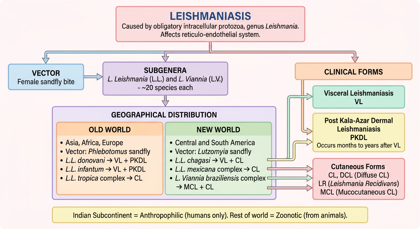
- Visceral Leishmaniasis (VL) - Kala-azar
- Post-Kala-Azar Dermal Leishmaniasis (PKDL) - occurs months to years after VL
- Cutaneous forms - CL, DCL, LR, MCL
| Old World | New World |
|---|---|
| Asia, Africa, Europe | Central & South America |
| Vector: Phlebotomus | Vector: Lutzomyia |
| L.L. donovani → VL + PKDL | L.L. chagasi → VL + CL |
| L.L. infantum → VL + PKDL | L.L. mexicana → CL |
| L.L. tropica complex → CL | L. Viannia braziliensis → MCL + CL |


Explain all given data in form of flowchart in form of image include the lufe cycle under headinfg of life cycle use pastel colours
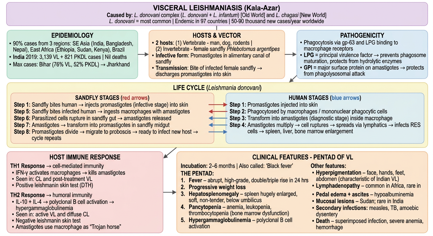
A large, detailed, well-organized medical flowchart about Visceral Leishmaniasis (Kala-Azar) using soft pastel colours (mint green, lavender, peach, light blue, pale yellow, light pink, pale orange). White background. Clear readable text. Professional medical textbook style. Layout (top to bottom, left to right): === TOP HEADER BOX (wide, lavender) === VISCERAL LEISHMANIASIS (Kala-Azar) Caused by: L. donovani complex (L. donovani + L. infantum) [Old World] and L. chagasi [New World] L. donovani = most common | Endemic in 97 countries | 50-90 thousand new cases/year worldwide === ROW 1: Three boxes side by side === Box 1 (mint green): EPIDEMIOLOGY • 90% cases from 3 regions: SE Asia (India, Bangladesh, Nepal), East Africa (Ethiopia, Sudan, Kenya), Brazil • India 2019: 3,139 VL + 821 PKDL cases | Nil deaths • Max cases: Bihar (76% VL, 52% PKDL) → Jharkhand Box 2 (peach): HOSTS & VECTOR • 2 hosts: (1) Vertebrate - man, dog, rodents | (2) Invertebrate - female sandfly Phlebotomus argentipes • Infective form: Promastigotes in alimentary canal of sandfly • Transmission: Bite of infected female sandfly → discharges promastigotes into skin Box 3 (light blue): PATHOGENICITY • Phagocytosis via gp-63 and LPG binding to macrophage receptors • LPG = principal virulence factor → prevents phagosome maturation, protects from hydrolytic enzymes • GPI = major surface protein on amastigotes → protects from phagolysosomal attack === ROW 2: LIFE CYCLE (wide box, pale yellow, titled in bold) === LIFE CYCLE (Leishmania donovani) Two columns inside: SANDFLY STAGES (red arrows): Step 1: Sandfly bites human → injects promastigotes (infective stage) into skin Step 5: Sandfly bites infected human → ingests macrophages with amastigotes Step 6: Parasitized cells rupture in sandfly gut → amastigotes released Step 7: Amastigotes → transform into promastigotes in sandfly midgut Step 8: Promastigotes divide → migrate to proboscis → ready to infect new host → cycle repeats HUMAN STAGES (blue arrows): Step 1: Promastigotes injected into skin Step 2: Phagocytosed by macrophages / mononuclear phagocytic cells Step 3: Transform into amastigotes (diagnostic stage) inside macrophage Step 4: Amastigotes multiply → cell ruptures → spreads via lymphatics → infects RES cells → spleen, liver, bone marrow enlargement === ROW 3: Two boxes side by side === Box 1 (light pink): HOST IMMUNE RESPONSE TH1 Response → cell-mediated immunity • IFN-γ activates macrophages → kills amastigotes • Seen in: CL and post-treatment VL • Positive leishmanin skin test (DTH) TH2 Response → humoral immunity • IL-10 + IL-4 → polyclonal B cell activation → hypergammaglobulinemia • Seen in: active VL and diffuse CL • Negative leishmanin skin test • Amastigotes use macrophage as "Trojan horse" Box 2 (pale orange): CLINICAL FEATURES - PENTAD OF VL Incubation: 2–6 months | Also called: "Black fever" THE PENTAD: 1. Fever – abrupt, high-grade, double/triple rise in 24 hrs 2. Progressive weight loss 3. Hepatosplenomegaly – spleen hugely enlarged, soft, non-tender, below umbilicus 4. Pancytopenia – anemia, leukopenia, thrombocytopenia (bone marrow dysfunction) 5. Hypergammaglobulinemia – polyclonal B cell activation Other features: • Hyperpigmentation – face, hands, feet, abdomen (characteristic of Indian VL) • Lymphadenopathy – common in Africa, rare in India • Pedal edema + ascites – hypoalbuminemia • Mucosal lesions – Sudan; rare in India • Secondary infections: measles, TB, amoebic dysentery • Death – superimposed infection, severe anemia, hemorrhage Arrows connecting all sections with flow direction indicators. Clean professional layout.
| Section | Key Points |
|---|---|
| Causative agents | L. donovani + L. infantum (Old World), L. chagasi (New World) |
| Epidemiology | 50-90k cases/year; India 2019: 3,139 VL cases; Bihar = max burden |
| Vector | Female sandfly (Phlebotomus); infective form = promastigotes |
| Life Cycle | 8-step cycle between human and sandfly; promastigote ↔ amastigote conversion |
| Pathogenicity | LPG (principal virulence factor), gp-63, GPI protect parasite inside macrophage |
| Immune Response | TH1 = protective (IFN-γ, kills parasite); TH2 = disease progression (IL-10, IL-4) |
| Pentad of VL | Fever + Weight loss + Hepatosplenomegaly + Pancytopenia + Hypergammaglobulinemia |
| Hallmark sign | Hyperpigmentation (face, hands, feet, abdomen) = classic Indian VL = "black fever" |
| Death | Superimposed infection, severe anemia, hemorrhage |


Explain in form of flowchart in form of image use pastel colours