
write about PACS in detail from basic to advanced level in very detail; how does it happen; why does it happen; pathogenesis; everything; clinical trilas and recent advances in chat
"post-acute COVID-19 syndrome" OR "long COVID" pathogenesis
PACS post-acute COVID syndrome pathogenesis clinical features management 2025 2026
"post-acute sequelae" AND "SARS-CoV-2" AND pathogenesis
https://recovercovid.org/news/recover-research-update-march-…
PMID: 39326415
PMID: 40474772
"long COVID" OR "PASC" treatment clinical trial
PMID: 37667052
PMID: 39603702
PACS - Post-Acute COVID-19 Syndrome (Long COVID / PASC)
A Complete Guide: Basic to Advanced
1. TERMINOLOGY AND DEFINITIONS
- PASC - Post-Acute Sequelae of SARS-CoV-2
- Long COVID (the lay/common term)
- Post-COVID Condition (PCC) - WHO preferred term
- Post-COVID Syndrome (PCS)
- Long Haulers syndrome
Formal Definitions
| Organization | Definition |
|---|---|
| WHO | Symptoms persisting for at least 2 months after resolution of acute infection (usually 3 months from initial onset), not explained by an alternative diagnosis |
| CDC | New, returning, or ongoing health problems 4 or more weeks after first SARS-CoV-2 infection |
| NICE (UK) | Health problems from 4 weeks or more after acute infection |
| German AWMF | Persistent or new-onset symptoms attributed to SARS-CoV-2 beyond the acute phase, including exacerbation of pre-existing conditions |
- Goldman-Cecil Medicine, PASC section; Robbins Pathologic Basis of Disease, p. 331; RKI Journal of Health Monitoring 2026
2. EPIDEMIOLOGY
Global Burden
- Estimated 6% overall incidence of long COVID among people who have had acute COVID-19 (Robbins Pathologic Basis of Disease), though some early studies suggested up to 10-30%
- Wide variation in reported prevalence due to differing case definitions, study populations, variant periods, and vaccination status
- A 2025 systematic review and meta-analysis (PMID: 40073162) estimated global prevalence from prospective evidence
- A 2025 meta-analysis of 3-year data (PMID: 40476637) documented persistent symptoms up to 3 years post-infection
Who Gets PACS?
| Risk Factor | Evidence |
|---|---|
| Female sex | Consistently reported |
| Age >20 years | Spectrum of risk |
| Severe acute COVID-19 | Strongest single predictor |
| Lack of vaccination | Vaccination reduces PACS risk |
| Type 2 diabetes | Independent risk factor |
| Obesity / high BMI | Risk for long COVID |
| 5+ symptoms in first week of illness | OR 3.5 (95% CI 2.75-4.50) for Long COVID |
| SARS-CoV-2 RNAemia during acute phase | Predicts PASC development |
| Epstein-Barr virus reactivation | Associated, not causative |
- Braunwald's Heart Disease, p. 2551; Rheumatology 2-Vol Set, p. 1656
Variants and Vaccination Impact
- Omicron-associated PACS appears to have lower incidence than Delta/ancestral strain
- Vaccination before infection reduces but does not eliminate PACS risk
- Reinfections carry risk of PACS with each episode
3. PATHOGENESIS - HOW AND WHY DOES PACS HAPPEN?
Mechanism 1: Viral Persistence (Viral Reservoir Theory)
- SARS-CoV-2 RNA, viral proteins, and even replication-competent virus have been detected in tissues - gut, lymph nodes, lung, brain, heart - weeks to months after apparent clinical recovery
- These viral reservoirs continue to trigger immune responses and release viral proteins into circulation
- Evidence from Proal et al., Nature Immunology 2023 (PMID: 37667052): documented SARS-CoV-2 RNA/protein in PASC tissue samples, with evidence these reservoirs drive coagulation abnormalities, microbiome dysbiosis, and neuroimmune dysfunction
- The gut is a particularly significant reservoir - viral RNA persists in gut epithelium for months
- This explains why antivirals (e.g., Paxlovid) are being tested in PACS - to clear the reservoir
Mechanism 2: Immune Dysregulation and Chronic Inflammation
-
Persistent immune activation with cytokine imbalances - elevated TNF, IL-6, IFN-alpha
-
T-cell exhaustion: CD8+ T cells lose effector function, impairing viral clearance
-
NK cell dysfunction
-
Activated mast cells (mast cell activation syndrome overlap)
-
Elevated complement activation
-
January 2026 Nature Immunology study (from RECOVER Initiative): "Long COVID involves activation of proinflammatory and immune exhaustion pathways"
-
This mirrors patterns seen in ME/CFS (Myalgic Encephalomyelitis/Chronic Fatigue Syndrome)
Mechanism 3: De Novo Autoimmunity
-
SARS-CoV-2 triggers formation of new autoantibodies - against nuclear antigens, immunomodulatory proteins, G-protein coupled receptors, and coagulation factors
-
Population data (TriNetX U.S. Network): COVID-19 survivors had significantly higher rates of:
- Rheumatoid arthritis (aHR 2.98)
- SLE (aHR 2.99)
- Ankylosing spondylitis (aHR 3.21)
-
German insurance data (640,000 patients): Autoimmune disease incidence 15.05 per 1000 person-years post-COVID vs. 10.55 in unexposed controls
-
Autoantibodies documented include: anti-nuclear, anti-phospholipid, anti-IFN, anti-ACE2 antibodies
-
Mechanism: Molecular mimicry (viral proteins resemble self-antigens), bystander activation, and direct immune dysregulation
-
Firestein & Kelley's Textbook of Rheumatology, pp. 2760-2782
Mechanism 4: Endothelial Dysfunction and Microthrombi
-
SARS-CoV-2 infects ACE2-expressing endothelial cells directly
-
Triggers endothelialitis - endothelial inflammation
-
Results in:
- Microclot formation (fibrin amyloid microclots resistant to fibrinolysis)
- Capillary rarefaction
- Platelet hyperactivation
- Impaired microvascular flow
-
Explains symptoms: brain fog (cerebral microvascular disease), exercise intolerance, fatigue, dyspnea
-
D-dimer, fibrin degradation products often elevated
-
Gupta et al. 2025; Braunwald's Heart Disease
Mechanism 5: Neuroinflammation and Autonomic Dysfunction
-
SARS-CoV-2 enters the CNS via: olfactory nerve, disruption of blood-brain barrier, or via infected monocytes
-
Neuroinflammation: activated microglia, astrocyte dysfunction, elevated neuroinflammatory markers (GFAP, NfL)
-
Autonomic dysfunction: SARS-CoV-2 infects autonomic ganglia; antibodies against autonomic receptors (adrenergic, muscarinic)
-
POTS (Postural Orthostatic Tachycardia Syndrome) is one of the most well-documented PACS manifestations - results from autonomic dysregulation, hypovolemia, and small fiber neuropathy
-
Serotonin dysregulation: vagal serotonin signaling impaired by low platelet serotonin (trapped in microthrombi)
-
Neuroscience: Exploring the Brain 5th ed.; Harrison's Principles of Internal Medicine 22E (2025)
Mechanism 6: Reactivation of Latent Viruses
- SARS-CoV-2-induced immune suppression allows reactivation of:
- Epstein-Barr virus (EBV): EBV reactivation detected in PASC patients; associated with fatigue
- Herpes simplex virus (HSV)
- Human herpesvirus 6 (HHV-6)
- EBV reactivation is a specific risk factor for PASC development (Goldman-Cecil Medicine)
- Similar to EBV-driven post-infectious fatigue syndromes
Mechanism 7: Gut Microbiome Dysbiosis
- SARS-CoV-2 extensively infects the GI tract (ACE2 highly expressed in enterocytes)
- Results in dysbiosis: decreased Lactobacillus and Bifidobacterium, increased pathobionts
- Disrupted gut-brain axis contributes to fatigue, cognitive symptoms, mood changes
- Altered microbiome persists months after acute infection
- Harrison's (2025): "Among the mechanisms for long COVID-associated fatigue and exercise intolerance are oxidative stress, altered energy metabolism, and dysbiosis of the gut microbiome."
Mechanism 8: Mitochondrial Dysfunction and Metabolic Disturbances
- SARS-CoV-2 impairs mitochondrial function, reducing ATP production
- Results in post-exertional malaise (PEM) - signature symptom shared with ME/CFS
- Iron dysregulation, persistent oxidative stress
- Altered glycolysis and fatty acid metabolism
- This explains why exercise worsens rather than helps many PACS patients (unlike most fatigue conditions)
4. CLINICAL FEATURES - SYMPTOMS BY ORGAN SYSTEM
Most Common Symptoms
| Symptom | Approximate Frequency |
|---|---|
| Fatigue | Most common (>50%) |
| Post-exertional malaise (PEM) | Very common |
| Cognitive impairment / "Brain fog" | ~20-30% |
| Dyspnea / exercise intolerance | Common |
| Palpitations | Common |
| Headache | Common |
| Sleep disturbance | Common |
| Chest pain | Common |
| Myalgia / arthralgia | Common |
By Organ System
- Brain fog: poor concentration, memory lapses, word-finding difficulties
- Anxiety, depression, PTSD
- Headache (often new-onset or different character)
- Anosmia / parosmia (smell disturbance) - often persisting
- Dysgeusia (taste disturbance)
- Peripheral neuropathy / paresthesias
- Cognitive decline measurable on neuropsychometric testing
- Sleep disorders (insomnia, hypersomnia)
- Palpitations - highly reported
- POTS - among the most common PACS presentations, especially in younger women
- Chest pain
- Myocarditis (subacute)
- Cardiac arrhythmias
- Tachycardia at rest / orthostatic tachycardia
- Exertional dyspnea without structural cause
- Persistent dyspnea
- Chronic cough
- Reduced exercise capacity (6-minute walk test abnormal)
- Pulmonary fibrosis sequelae (post-severe disease)
- Abnormal PFTs (restrictive or diffusion defects)
- Myalgia
- Arthralgia (migratory, often non-inflammatory on examination)
- New-onset inflammatory arthritis
- Post-exertional malaise after minimal activity
- Nausea, abdominal pain, diarrhea
- Altered bowel habits
- New food intolerances
- New-onset type 1 and type 2 diabetes
- Thyroid dysfunction (subacute thyroiditis)
- Adrenal insufficiency (rare)
- Persistent proteinuria
- Reduced eGFR - COVAN (coronavirus-associated nephropathy)
-
Hair loss (telogen effluvium)
-
"COVID toes" (pernio-like lesions)
-
Rashes
-
Miller's Anesthesia 10th ed., Table 29.23 (citing Nalbandian et al., Nat Med 2021); Braunwald's Heart Disease, p. 2553
PACS Phenotypes / Subtypes
- Fatigue-predominant (overlaps with ME/CFS)
- Cardiorespiratory (dyspnea, exercise intolerance, POTS)
- Neurocognitive (brain fog, memory, mood)
- Multi-system / Complex (multiple domain involvement)
- Musculoskeletal (myalgia, arthralgia)
5. DIAGNOSIS
Diagnostic Criteria (WHO-based)
- History of confirmed or probable SARS-CoV-2 infection
- Symptoms persisting or newly arising at least 3 months from infection onset
- Symptoms lasting at least 2 months
- Symptoms not explained by an alternative diagnosis
Workup
- CBC with differential (often lymphopenia may persist)
- CMP / metabolic panel
- CRP, ESR (usually normal or mildly elevated in PACS - unlike acute COVID)
- D-dimer, fibrinogen
- Thyroid function (TSH, fT4)
- Ferritin
- Cardiac troponin (if cardiac symptoms)
- ECG (POTS workup: standing/lying HR, tilt table test)
- Pulmonary function tests / 6-minute walk test
- Brain MRI (if cognitive symptoms - may show white matter changes)
- Echocardiogram (if cardiorespiratory symptoms)
- Autonomic function testing
- SARS-CoV-2 antigen persistence in plasma
- Microclot detection (fibrin amyloid)
- T-cell exhaustion markers (PD-1, TIM-3 on CD8+)
- Reactivated EBV titers
- GFAP, NfL (neuroinflammation markers)
- Gut microbiome profiling
6. DIFFERENTIAL DIAGNOSIS
- Myalgic Encephalomyelitis / Chronic Fatigue Syndrome (ME/CFS) - significant overlap
- Fibromyalgia
- PTSD / anxiety / depression (often comorbid, not exclusive)
- Autoimmune diseases (newly triggered)
- Thyroid disorders
- Anemia
- Cardiac disease
- Sleep apnea
- Post-ICU/post-intensive care syndrome (PICS)
- MIS-A (Multisystem Inflammatory Syndrome in Adults) - distinct: more acute, severe cardiovascular compromise, within 2-6 weeks of COVID
7. MANAGEMENT
Evidence-Based Interventions
| Intervention | Evidence Level | Effect |
|---|---|---|
| Online CBT programme | Moderate certainty | Probably reduces fatigue and improves concentration |
| Combined physical + mental health rehabilitation | Moderate certainty | Probably improves overall health, reduces depression, improves QoL; 161 more per 1000 experiencing meaningful improvement |
| Intermittent aerobic exercise (3-5x/week, 4-6 weeks) | Moderate certainty | Probably improves physical function vs. continuous exercise |
| Pacing (energy management) | Low certainty / expert consensus | Essential for PEM - prevents "crash-and-burn" cycles |
Post-Hospitalisation COVID Rehabilitation
- PHOSP-R trial (Daynes et al., ERJ 2025, PMID: 39978856): RCT of exercise-based rehabilitation post-hospitalisation for COVID-19 - important evidence for structured pulmonary rehabilitation
Symptom-Specific Management
- Energy pacing ("envelope theory")
- Heart rate-limited activity (keep HR <110 bpm during activity to prevent PEM)
- Treat sleep disorders
- Methylphenidate / amantadine (low-quality evidence)
- Treat coexisting anemia, thyroid dysfunction
- Cognitive rehabilitation
- CBT (Gorenshtein et al. systematic review, PMID: 38695969)
- JAMA Neurology RCT (Nov 2025, RECOVER Initiative): tested interventions for cognitive symptoms - did not find an effective treatment, but informed future trial design
- Treat sleep, mood disorders first
- Increased salt and fluid intake
- Compression stockings
- Beta-blockers (low-dose propranolol, ivabradine)
- Fludrocortisone (if hypovolemic component)
- Supervised recumbent exercise program (rowing, swimming initially - horizontal)
- Pulmonary rehabilitation
- Breathing exercises, diaphragmatic training
- Spirometry-guided management
- CBT - best evidence
- SSRIs for depression and anxiety
- Screen and treat PTSD
Emerging and Investigational Therapies
| Drug/Intervention | Mechanism Targeted | Status |
|---|---|---|
| Nirmatrelvir-ritonavir (Paxlovid) | Clear viral reservoir | Phase 3 RCT (RECOVER-VITAL) |
| Anticoagulants / antiplatelet | Microclots, hypercoagulability | Ongoing trials |
| Low-dose naltrexone | Immune modulation, neuroinflammation | Pilot studies |
| Metformin | Antiviral, anti-inflammatory (mTOR) | COVID-OUT trial (PMID: 37302406) showed ~41% reduction in PACS incidence |
| Probiotics | Gut microbiome restoration | Trials ongoing; no compelling evidence yet (BMJ SR) |
| SSRIs (fluvoxamine) | Anti-inflammatory, sigma-1 receptor | Clinical trials |
| Baricitinib | JAK inhibitor, immune modulation | Studies ongoing |
| Hyperbaric oxygen | Neuroinflammation, endothelial function | No compelling evidence (BMJ SR) |
| IV immunoglobulin (IVIG) | Autoimmune / dysregulated immune | Case series; trials ongoing |
| Fecal microbiota transplantation | Gut dysbiosis | Early trials |
- Metformin reduced PACS incidence by ~41% when given during acute COVID-19
- Mechanism: mTOR inhibition reducing viral replication + anti-inflammatory effects
- This is one of the most exciting prevention strategies
- November 2025 Nature Communications: Long COVID trajectories in prospective RECOVER-Adult cohort
- January 2026 Nature Immunology: Long COVID involves proinflammatory and T-cell exhaustion pathways
- November 2025 eLife: Role of co-infections in PASC pathogenesis
- October 2025 JAMA Network Open: Schizophrenia, bipolar disorder linked to increased PASC risk
Physical Exercise - Important Nuances
"Focused exercise prescriptions guided by patient-specific pathophysiology have been shown to relieve symptoms in many individuals."
- PEM-positive patients (ME/CFS-like): pacing, heart rate-controlled, avoid triggering PEM
- PEM-negative, deconditioning-predominant patients: gradually structured aerobic exercise can help
8. SPECIAL POPULATIONS
Pediatric PACS
- Less common than in adults but documented
- JAMA Pediatrics 2026 (RECOVER): Social determinants of health linked to pediatric Long COVID
- Post-acute dyslipidemia and BMI abnormalities documented in children (Journal of Pediatrics 2026)
- Must be distinguished from MIS-C
PACS vs. ME/CFS
- PEM is the diagnostic hallmark of ME/CFS and is present in many PACS patients
- Overlapping immune dysfunction (T-cell exhaustion, cytokine abnormalities)
- Overlapping autonomic dysfunction
- PACS may "unmask" or trigger ME/CFS in genetically predisposed individuals
- Neuroscience: Exploring the Brain 5th ed.: "Long COVID is dominated by neurological symptoms" paralleling ME/CFS
Pregnancy
- Limited data; ongoing surveillance needed
9. PREVENTION
- Vaccination is the most effective prevention strategy - reduces risk of severe acute illness and subsequent PACS
- Antiviral treatment during acute illness (Paxlovid, remdesivir): some data suggest reduced PACS incidence
- Metformin during acute COVID: COVID-OUT trial showed ~41% reduction in PACS
- Avoiding reinfection: each reinfection carries independent PACS risk
- Early identification of high-risk patients for closer follow-up
10. PROGNOSIS
- Most people recover within 1 year of symptom onset (Robbins)
- A relapsing-remitting pattern is the most common trajectory, but progressive and persistent courses occur
- Omicron-era prognosis appears better than earlier variants
- Severe long COVID (especially ME/CFS-like) can cause significant disability lasting years
- 3-year follow-up data (PMID: 40476637) shows some patients have symptoms persisting beyond 3 years
11. SUMMARY TABLE: PACS AT A GLANCE
| Feature | Detail |
|---|---|
| Definition | Symptoms >3 months from initial COVID-19, lasting >2 months, not otherwise explained |
| Prevalence | ~6-10% of COVID-19 survivors |
| Core mechanisms | Viral reservoir, immune dysregulation, autoimmunity, microthrombi, neuroinflammation, dysbiosis |
| Hallmark symptoms | Fatigue, brain fog, PEM, dyspnea, POTS, palpitations |
| Diagnosis | Clinical; exclude alternatives; no single test |
| Best-evidence treatment | CBT, combined physical-mental rehab, paced exercise |
| Active drug trials | Paxlovid (RECOVER-VITAL), metformin, anticoagulants |
| Prevention | Vaccination, metformin during acute illness, antiviral therapy |
| Prognosis | Most recover within 1 year; some persistent/relapsing cases |
Key References
- Peluso & Deeks, Cell 2024 - Mechanisms of long COVID and therapeutics (PMID: 39326415)
- Gupta et al., Comprehensive Physiology 2025 - Viral persistence, immune dysregulation, multi-organ dysfunction (PMID: 40474772)
- Proal et al., Nature Immunology 2023 - SARS-CoV-2 reservoir in PASC (PMID: 37667052)
- Zeraatkar et al., BMJ 2024 - Living systematic review of interventions for long COVID (PMID: 39603702)
- Bramante et al., Lancet Infect Dis 2023 - Metformin and PACS prevention (COVID-OUT) (PMID: 37302406)
- Rahmati et al., J Med Virol 2025 - Long-term sequelae 3 years post-infection (PMID: 40476637)
- Goldman-Cecil Medicine, PASC section
- Braunwald's Heart Disease, Postacute Sequelae section
- Robbins, Cotran & Kumar Pathologic Basis of Disease, COVID-19 chapter
- Firestein & Kelley's Textbook of Rheumatology, Autoantibodies post-COVID
- Harrison's Principles of Internal Medicine 22E (2025), Exercise intolerance and long COVID
wait I said PRIMARY ANGLE CLOSURE GLAUCOMA
"primary angle closure glaucoma" pathogenesis mechanisms treatment
primary angle closure glaucoma PACG PACS pathogenesis management 2025 guidelines
"angle closure glaucoma" laser iridotomy lens extraction EAGLE
https://www.aao.org/education/preferred-practice-pattern/pri…
PMID: 42035831
PMID: 35387778
Primary Angle Closure Glaucoma (PACG) - Complete Guide
1. THE DISEASE SPECTRUM - TERMINOLOGY
| Stage | Abbreviation | Definition |
|---|---|---|
| Primary Angle Closure Suspect | PACS | ≥180° of iridotrabecular contact (ITC); no elevated IOP, no PAS, no optic nerve damage |
| Primary Angle Closure | PAC | ITC + peripheral anterior synechiae (PAS) and/or elevated IOP; still no glaucomatous optic neuropathy |
| Primary Angle-Closure Glaucoma | PACG | ITC + PAS/elevated IOP + glaucomatous optic neuropathy (structural or functional damage) |
- Kanski's Clinical Ophthalmology 10th ed., p. 374; AAO PPP 2025 guidelines
2. GLOBAL BURDEN AND EPIDEMIOLOGY
- PACG may be responsible for up to half of all glaucoma cases globally (Kanski)
- Particularly prevalent in Asia (Far Eastern and Indian populations)
- More likely to result in visual loss than primary open-angle glaucoma (POAG) - it progresses more rapidly
- Accounts for a disproportionate share of bilateral blindness worldwide
- Highest burden in Asian populations, especially Chinese and Inuit descent (AAO PPP 2025)
Risk Factors
| Risk Factor | Detail |
|---|---|
| Age | Average presentation ~62 years for pupillary block form |
| Sex | Females more affected than males |
| Race | Far Eastern and Indian Asians; Inuit |
| Hyperopia | Shortened axial length, shallow AC; up to 1 in 6 patients with >1D hyperopia are PACS |
| Short axial length | Short eyes have anteriorly positioned lens; nanophthalmos (axial length <20 mm) at extreme risk |
| Shallow anterior chamber depth | Primary anatomical predisposing factor |
| Thick crystalline lens | Large lens vault pushes iris-lens diaphragm forward |
| Family history | Genetic factors important but poorly defined |
| Smaller corneal diameter | Reduced anterior chamber volume |
- Kanski's Clinical Ophthalmology 10th ed., p. 390
3. ANATOMY OF THE ANTERIOR CHAMBER ANGLE
- Schwalbe line (anterior limit of TM)
- Trabecular meshwork (TM) - the drainage site
- Scleral spur
- Ciliary body band
- Iris root
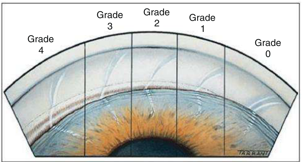
| Shaffer Grade | Degrees | What is Visible | Clinical Significance |
|---|---|---|---|
| Grade 4 | 35-45° | Ciliary body visible | Widest; characteristic of myopia |
| Grade 3 | 25-35° | Scleral spur visible | Open; cannot close |
| Grade 2 | 20° | Trabeculum, not scleral spur | Gonioscopy recommended |
| Grade 1 | 10° | Schwalbe line ± top of TM | Dangerously narrow |
| Slit | ~0° | No angle structures, no contact | Near-closure |
| Grade 0 | 0° | Iridocorneal contact present | Closed |
| AC depth vs corneal thickness | Grade | Comment |
|---|---|---|
| ≥1x corneal thickness | 4 | Wide open |
| 1/4 - 1/2 | 3 | Cannot close |
| 1/4 | 2 | Should undergo gonioscopy |
| <1/4 | 1 | Dangerously narrow on gonioscopy |
- Kanski's Clinical Ophthalmology 10th ed., p. 389
4. PATHOGENESIS - HOW AND WHY DOES ANGLE CLOSURE HAPPEN?
Mechanism 1: Relative Pupillary Block (Most Common - ~70-80%)
- In a predisposed eye (short axial length, large lens, shallow AC), the posterior surface of the iris rests in close apposition to the anterior lens surface
- This contact impedes aqueous flow from the posterior chamber (where it is produced by the ciliary body) through the pupil to the anterior chamber
- A pressure differential develops: posterior chamber pressure > anterior chamber pressure
- This causes the peripheral iris to bow forward (iris bombé configuration)
- The bowed iris then comes into contact with the peripheral cornea/trabecular meshwork - iridotrabecular contact
- Aqueous outflow is blocked → IOP rises
- At mid-dilation (3-5 mm), the iris is thickest and the contact with the lens is maximum - this is the most dangerous pupil size for triggering an acute attack
- Full constriction (pilocarpine) thins the iris
- Full dilation (dilated by drops) pulls the iris root away from the angle
- Peripheral iridotomy (PI/LPI) creates a bypass channel between posterior and anterior chambers, equalizing pressure and eliminating the bowing
Mechanism 2: Plateau Iris Configuration / Syndrome
- The ciliary processes are anteriorly rotated, pushing the peripheral iris into the angle even without significant pupillary block
- The iris root is inserted anteriorly and the peripheral iris "bunches up" in the angle during dilation
- The anterior chamber may appear deceptively normal centrally, but the angle is narrow peripherally
- Seen as younger patients, often non-hyperopic (occasionally myopic)
- Plateau iris configuration: anatomical finding (UBM/AS-OCT shows anteriorly rotated ciliary processes)
- Plateau iris syndrome: angle closure persists despite a patent iridotomy in a patient with plateau iris configuration
- An element of pupillary block is often also present; pure plateau iris is less common
- Treatment: LPI does not fully relieve it; laser iridoplasty (peripheral laser burns to contract and flatten peripheral iris) or pilocarpine long-term
Mechanism 3: Lens-Related (Phacomorphic) Component
- As the crystalline lens grows with age, it increases in thickness and vault (the "lens vault" = the distance of the anterior lens pole anterior to the scleral spur plane)
- A large lens vault is independently associated with angle closure
- Pushes the iris-lens diaphragm anteriorly, shallowing the AC and worsening both pupillary block and angle crowding
- This is why cataract/lens extraction is so effective at treating PACG - it removes the primary anatomical driver
- Explains the age-related onset
Mechanism 4: Choroidal Expansion (Dynamic)
- Transient posterior pressure from choroidal expansion (choroidal effusion, suprachoroidal fluid) can push the lens-iris diaphragm forward
- Contributes to the dynamic nature of angle closure
- Important in understanding why attacks can be precipitated by certain medications or positions
Downstream Consequences of Closure
- Appositional obstruction: iris physically covers the TM - reversible if caught early
- TM degeneration: chronic or intermittent iris-TM contact causes trabecular damage - partially reversible
- Peripheral anterior synechiae (PAS): permanent adhesion of the peripheral iris to the TM/angle - irreversible; IOP control correlates inversely with PAS extent
- Kanski's Clinical Ophthalmology 10th ed., p. 390; Li et al., Prog Retin Eye Res 2026 (PMID: 42035831)
5. CLINICAL PRESENTATIONS
A. Acute Angle Closure Crisis (AACC) / Acute Congestive Attack
- Watching TV / being in a dark room (mild dilation, pupil in "danger zone")
- Pharmacological mydriasis (dilating drops)
- Certain systemic medications: anticholinergics, sympathomimetics, motion sickness patches, cold remedies
- Semi-prone position (reading in bed)
- Acute emotional stress
- Sudden, severe, unilateral eye pain
- Headache (often frontal - can be confused with migraine or cluster headache)
- Blurred vision with colored halos around lights (corneal edema)
- Nausea and vomiting - can be so severe it simulates acute abdomen
- ~5% of cases occur simultaneously in both eyes
- Markedly elevated IOP (can exceed 60 mmHg - approaching diastolic arterial pressure)
- Corneal epithelial edema (steamy/cloudy cornea)
- Conjunctival and episcleral vascular congestion (red eye)
- Mid-dilated, non-reactive pupil (ischemic iris sphincter)
- Shallow anterior chamber
- Glaukomflecken: small anterior subcapsular lens opacities from ischemic necrosis of lens epithelium - permanent marker of prior acute attack
- Optic disc pallor/cupping (if chronic or after acute)
B. Subacute / Intermittent Angle Closure
- Recurrent mild episodes, usually resolving spontaneously (pupil re-constricts)
- Symptoms: intermittent eye ache, headache, colored halos, blurred vision
- Often misdiagnosed as migraine
- Each episode causes some PAS formation - cumulative damage
C. Chronic Angle Closure Glaucoma
- Gradual, insidious IOP elevation from progressive PAS formation
- Often asymptomatic until advanced field loss
- Mimics POAG in presentation
- Gonioscopy is essential for differentiation
6. DIAGNOSIS
Gonioscopy (Gold Standard)
- Mandatory for any angle closure diagnosis
- Performed with a gonioscopic lens (Goldmann, Zeiss 4-mirror)
- Dark-room dynamic gonioscopy recommended (AAO PPP 2025) to assess functional closure vs structural
- Indentation gonioscopy: differentiates appositional (opens with indentation - reversible) from synechial closure (does not open - PAS, irreversible)
- Angle is typically narrowest superiorly
- Diagnosis of PACS: ≥180° (≥3 quadrants) of ITC on gonioscopy
Slit Lamp Examination
- Shallow anterior chamber
- Iris bombé configuration
- Corneal edema (in acute attack)
- Glaukomflecken
- Posterior synechiae
Imaging
| Modality | Use |
|---|---|
| Anterior Segment OCT (AS-OCT) | Non-contact; measures angle opening distance, anterior chamber depth, lens vault; useful for screening |
| Ultrasound Biomicroscopy (UBM) | Gold standard for plateau iris (visualizes ciliary processes); contact procedure |
| Optical coherence tomography (RNFL/ONH) | Detects glaucomatous structural damage to RNFL |
| Visual field testing (perimetry) | Detects functional glaucomatous damage |
Differentiating PAC from POAG
7. MANAGEMENT
Decision Framework by Stage
PACS ──► Observe vs prophylactic LPI (risk stratification)
PAC  ──► LPI → if inadequate: CLE or MIGS
PACG ──► LPI → CLE (preferred) or trabeculectomy + medical therapy
AACC ──► Emergency medical IOP lowering → LPI → treat fellow eye
A. PACS Management
- LPI has a small prophylactic effect over 6 years in PACS
- Therefore, LPI should be offered only to those at highest risk of developing acute closure or PACG, not all PACS patients
- Kanski: Factors favoring prophylactic LPI in PACS:
- Symptoms suggestive of prior intermittent closure
- Systemic medication with atropine-like structure (high risk of dilation-induced attack)
- Need for frequent dilated examination (diabetes, AMD)
- Difficulty accessing ophthalmic care urgently
- Very narrow angle with significant ITC
- Observation (most cases)
- Laser iridoplasty
- Long-term pilocarpine 1% twice daily
- If symptomatic cataract: lens extraction usually opens the angle
B. PAC and PACG Management
- Creates a bypass channel in the peripheral iris
- Equalizes posterior and anterior chamber pressures
- Eliminates the pupillary block component
- Does NOT help plateau iris syndrome (requires additional therapy)
- Does NOT reverse PAS already formed
- After LPI, gonioscopy repeated to confirm angle opening
The EAGLE Trial (Effectiveness of Early Lens Extraction for the Treatment of Primary Angle-Closure Glaucoma)
| Outcome | CLE arm | LPI arm |
|---|---|---|
| "Good responders" (IOP <21 mmHg, no further surgery) | 90% | 67% |
| "Optimal responders" (good response + medication-free) | 66% | 18% |
| Drops/surgery-free survival | Significantly longer (p<0.05) |
- Patients randomised to CLE were 10x more likely to maintain drop-free IOP control (OR=10.1, 95% CI 6.1-16.8)
- CLE also corrects hypermetropia, deepens the anterior chamber, and opens the filtration angle
- Same as POAG: prostaglandin analogues, beta-blockers, carbonic anhydrase inhibitors, alpha-2 agonists
- Required when substantial synechial closure is present or IOP remains elevated despite open angle post-iridotomy
- Option for persistent IOP elevation despite above measures
- Risk of malignant glaucoma (aqueous misdirection) in angle closure eyes - important complication
C. Acute Angle Closure Crisis (AACC) - Emergency Management
| Drug | Dose/Route | Mechanism |
|---|---|---|
| Timolol 0.5% | 1 drop topical | Beta-blocker - reduces aqueous production |
| Apraclonidine 1% | 3x daily topical | Alpha-2 agonist - reduces aqueous production |
| Acetazolamide | 250-500 mg IV or 500 mg oral | Carbonic anhydrase inhibitor - reduces aqueous production |
| Pilocarpine 2-4% | 1 drop topical (once IOP partially reduced - ineffective at very high IOP as iris sphincter is ischemic) | Miotic - opens angle |
| IV mannitol 20% | 1-2 g/kg over 45 min | Osmotic agent - reduces vitreous volume |
- Goldman-Cecil Medicine: "Initial treatment is with topical (e.g., timolol 0.5%) and systemic pressure-lowering agents (e.g., acetazolamide 250-500 mg IV), followed by creation of a fistula in the peripheral iris with laser."
- Anterior chamber paracentesis (immediate IOP reduction)
- Laser peripheral iridoplasty
- Lens extraction (more definitive)
- Trabeculectomy may be needed
8. RECENT ADVANCES AND CLINICAL TRIALS
1. EAGLE Trial (landmark) - Already covered above
2. ZAP Trial (Zhongshan Angle Closure Prevention)
- Tested whether prophylactic LPI in PACS reduces progression
- Result: LPI has only a small protective effect in PACS over 6 years
- Changed practice: not all PACS patients need LPI; individualized risk stratification required
- Referenced directly in Kanski 10th edition
3. MIGS in PACG (Minimally Invasive Glaucoma Surgery)
"MIGS expands treatment options for PACG once angle access is restored... Techniques range from trabecular meshwork and Schlemm's canal-based procedures to subconjunctival stents and ciliary body approaches. Combined with lens extraction, these methods deepen the anterior chamber, release PAS, and restore physiological outflow, with clinical evidence showing 20-50% IOP reductions and reduced medication burden."
- KDB (Kahook Dual Blade) goniotomy - excisional TM removal
- iStent inject - Schlemm's canal microstent
- Hydrus Microstent - Schlemm's canal scaffold
- Goniosynechialysis (GSL) - physically breaking PAS (can be combined with phaco)
- Endocyclophotocoagulation (ECP) - ciliary body
- Xen gel stent - subconjunctival drainage
4. Imaging Advances
- Swept-source OCT for anterior segment provides better angle visualization
- AI-based AS-OCT analysis for PACS/PAC risk stratification - under development
- OCTA (optical coherence tomography angiography) for optic nerve head vessel density in PACG: Sener et al., Photodiagnosis 2023 (PMID: 36493693) - systematic review showing reduced ONH vessel density in angle closure disease
5. Rho-Kinase (ROCK) Inhibitors
- Netarsudil (Rhopressa) and ripasudil act on TM and Schlemm's canal
- Work by increasing trabecular outflow and reducing episcleral venous pressure
- Potentially valuable in PACG where TM function is impaired by PAS
9. OPTIC NERVE AND GLAUCOMATOUS DAMAGE IN PACG
- Loss of retinal ganglion cells and their axons (RNFL thinning)
- Optic disc cupping progresses
- Visual field loss - initially peripheral (arcuate scotomas, nasal steps) → if untreated → tunnel vision → blindness
- Important: Optic nerve damage from an acute angle closure episode may not appear as typical glaucomatous cupping - ischemic pallor may dominate
10. SUMMARY TABLE
| Feature | PACS | PAC | PACG |
|---|---|---|---|
| ITC (≥3 quadrants) | ✓ | ✓ | ✓ |
| Elevated IOP or PAS | ✗ | ✓ | ✓ |
| Glaucomatous ON damage | ✗ | ✗ | ✓ |
| First-line treatment | Observe ± LPI (risk-stratified) | LPI → CLE | CLE > LPI; ± medical/surgery |
| Key trial | ZAP | EAGLE | EAGLE |
Key References
- Kanski's Clinical Ophthalmology: A Systematic Approach, 10th Edition, Chapter 11 - Primary Angle-Closure Glaucoma (the primary textbook source)
- Goldman-Cecil Medicine, Angle-Closure Glaucoma section
- AAO Preferred Practice Pattern: Primary Angle-Closure Disease 2025
- Mitchell et al., Br J Ophthalmol 2023 - EAGLE trial predictors (PMID: 35387778)
- Li et al., Prog Retin Eye Res 2026 - MIGS in PACG (PMID: 42035831)
- Sener et al., Photodiagnosis 2023 - OCTA in angle closure disease (PMID: 36493693)
- Naravane & Liu, Glaucoma Physician March 2025 - Management of PACS