
Type of bone, joint, fracture and general examination
Reading File
Now let me search the medical library for all four topics simultaneously.
Finding Sources
Finding Sources
Finding Sources
Finding Sources
Finding Sources
Finding Sources
Finding Sources
Reading File
Reading File
Reading File
Finding Sources
Reading File
Finding Sources
Reading File
Reading File
Reading File
Reading File
Now I have rich material from the textbooks. Let me also grab the general examination section from an orthopaedic/surgical context and fetch key images.
Finding Sources
Loading Image
Loading Image
I now have comprehensive textbook content from Gray's Anatomy for Students, Rosen's Emergency Medicine, THIEME Atlas, and Bailey & Love. Let me compile the full answer.
Types of Bone, Joint, Fracture, and General Examination
1. TYPES OF BONE
Based on shape and function, bones are classified into 7 types:
| Type | Description | Examples |
|---|---|---|
| Long bones | Tubular shaft with two expanded ends | Femur, humerus, tibia, fibula, radius, ulna |
| Short bones | Roughly equal dimensions; spongy bone covered by compact | Carpal and tarsal bones |
| Flat bones | Thin, flat plates; two layers of compact bone sandwiching spongy bone | Scapula, ilium, calvaria (skull vault) |
| Irregular bones | Complex shape that doesn't fit other categories | Vertebrae, many facial bones |
| Pneumatic bones | Contain air-filled (paranasal) sinuses; reduce skull weight | Frontal, maxilla, ethmoid, sphenoid |
| Sesamoid bones | Develop within tendons; reduce friction | Patella (largest), fabella |
| Accessory bones | Anomalous supernumerary bones from failure of fusion of ossification centers | Os trigonum (foot), sutural (Wormian) bones of skull |
General Anatomy and Musculoskeletal System, THIEME Atlas
Bone Tissue Types
- Compact (cortical) bone - Dense outer layer organized into osteons (Haversian systems). Haversian canals run longitudinally and connect via transverse Volkmann canals.
- Cancellous (spongy/trabecular) bone - Lattice of trabeculae; nourished by diffusion from the medullary cavity. Trabeculae ~200-300 µm thick.
2. TYPES OF JOINTS
Joints are classified into two broad categories:
A. Synovial Joints (Cavity-containing)
The articular surfaces are separated by a fluid-filled articular cavity. Key features:
- Hyaline cartilage covers articulating surfaces
- Joint capsule: inner synovial membrane (produces synovial fluid) + outer fibrous membrane
- Accessory structures: articular discs, fat pads, bursae, tendon sheaths
Subtypes by shape and movement:
| Subtype | Movement | Axes | Example |
|---|---|---|---|
| Plane (gliding) | Sliding/gliding | Multiaxial (limited) | Acromioclavicular joint |
| Hinge | Flexion/extension | Uniaxial | Elbow (humeroulnar), ankle |
| Pivot | Rotation | Uniaxial (longitudinal) | Atlanto-axial joint |
| Bicondylar | Mainly one axis + limited rotation | Biaxial | Knee joint |
| Condylar (Ellipsoid) | Flexion, extension, abduction, adduction, circumduction | Biaxial | Wrist joint |
| Saddle | Flexion, extension, abduction, adduction, circumduction | Biaxial | 1st carpometacarpal joint (thumb) |
| Ball-and-socket | All movements including rotation | Multiaxial | Hip, shoulder |
Gray's Anatomy for Students, Fig. 1.22
B. Solid Joints (No cavity)
Components are linked by connective tissue or cartilage. Movement is more restricted.
1. Fibrous joints - joined by fibrous connective tissue:
- Sutures - skull only; joined by sutural ligament
- Gomphoses - teeth in bony sockets via periodontal ligament
- Syndesmoses - two bones linked by a ligament (e.g., ligamentum flavum between vertebral laminae; interosseous membrane of forearm)
2. Cartilaginous joints - joined by cartilage:
- Synchondroses - two ossification centers separated by cartilage; e.g., growth plate (epiphyseal plate). Eventually ossify.
- Symphyses - two bones joined by fibrocartilage; most in midline. e.g., pubic symphysis, intervertebral discs
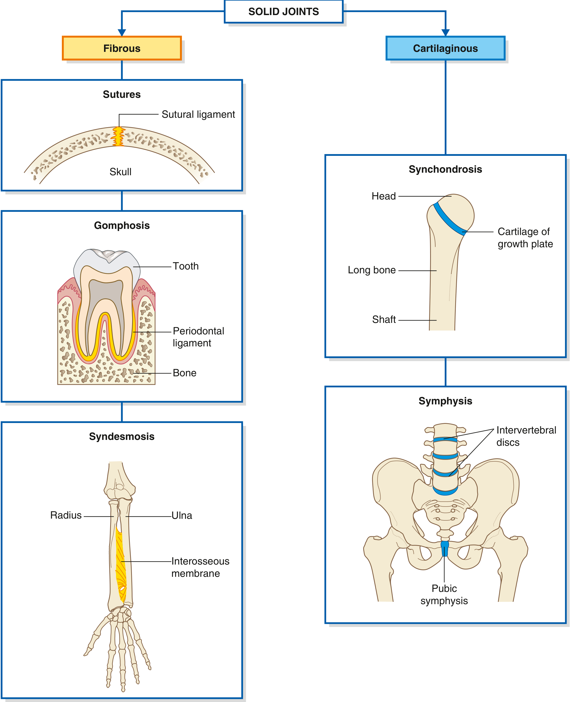
Gray's Anatomy for Students, Fig. 1.23
3. TYPES OF FRACTURES
A fracture is a break in the continuity of bone. Fractures are described systematically:
A. Skin Integrity
| Type | Definition |
|---|---|
| Closed (simple) | Skin/soft tissue overlying the fracture is intact |
| Open (compound) | Fracture exposed to the external environment (skin breached) |
B. Completeness
| Type | Definition |
|---|---|
| Complete | Both cortices disrupted |
| Incomplete | Only one cortex disrupted (common in children) |
- Greenstick fracture: incomplete - one side completely disrupted, other side buckled (children)
- Torus (buckle) fracture: cortex buckles without breaking through (children)
C. Direction of Fracture Line
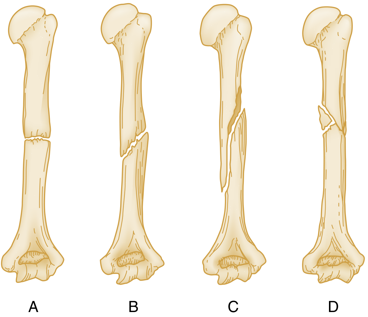
| Type | Mechanism | Direction |
|---|---|---|
| Transverse | Direct force | Right angle to long axis |
| Oblique | Angulation force | Diagonal to long axis |
| Spiral | Rotational/torsional force | Encircles shaft spirally |
| Comminuted | High-energy | More than 2 fragments |
Rosen's Emergency Medicine, Fig. 41.1
D. Fragment Number
- Simple (two-part) - one fracture line, two fragments
- Comminuted - multiple fragments
E. Position and Alignment of Fragments
- Displacement: deviation of distal fragment from normal position (described as none/minimal/moderate/severe, or in mm or %)
- Angulation (malalignment): axis deviation. Direction = apex of angle
- Valgus: apex toward midline
- Varus: apex away from midline
- Rotation: distal fragment rotated around the long axis
F. Special Types
| Type | Description |
|---|---|
| Pathological | Fracture through abnormal/diseased bone (tumor, osteoporosis, Paget's) |
| Stress (fatigue) | Repeated loading below single-fracture threshold; e.g., march fracture (2nd metatarsal) |
| Avulsion | Bony fragment pulled off by tendon/ligament |
| Impacted | One fragment driven into the other |
| Compression | Axial loading crushes bone (e.g., vertebral compression fracture) |
| Depression | Fragment pushed below normal surface level (e.g., skull, tibial plateau) |
G. Articular Involvement
- Extra-articular vs. intra-articular (articular surface involvement described as a %)
Rosen's Emergency Medicine, Box 41.1
4. GENERAL EXAMINATION
General examination is a systematic, head-to-toe clinical assessment performed before detailed regional or organ-specific examination.
Positioning
Patient lying flat, limbs extended (a pillow may be permitted). The region to be examined is adequately exposed. For abdominal examination, hips and knees are flexed during palpation to relax abdominal muscles.
Sequence of General Examination
1. Inspection (overall appearance)
- Look for signs of: weight loss, dehydration, pallor (anaemia), jaundice, abnormal pigmentation, oedema (pedal), distress/posture
2. Hands
- Clubbing, koilonychia, leukonychia (liver disease), palmar erythema, Dupuytren's contracture, peripheral cyanosis
3. Head and Neck
- Scleral jaundice, conjunctival pallor (anaemia)
- Lymphadenopathy - particularly left supraclavicular (Virchow's node = intra-abdominal malignancy)
- Parotid enlargement (liver disease, alcoholism)
4. Vital Signs
- Heart rate, blood pressure, respiratory rate, temperature
- In elective patients: weight, BMI
5. Special Inspection Signs
- Grey Turner's sign: flank discoloration (retroperitoneal haemorrhage - acute pancreatitis, leaking AAA)
- Cullen's sign: periumbilical discoloration (severe acute pancreatitis, ruptured ectopic pregnancy, hepatic trauma)
6. Abdominal Wall Movement
- In acute abdominal pain, observe whether wall moves with respiration (rigidity/peritonism restricts movement)
Bailey and Love's Short Practice of Surgery, 28th Edition
In Orthopaedic/Musculoskeletal General Examination
The classic sequence is:
- Look - inspection (swelling, deformity, muscle wasting, scars, skin changes, alignment)
- Feel - palpation (tenderness, warmth, effusion, crepitus)
- Move - active then passive range of motion; neurovascular assessment
- Measure - limb lengths and girth
- Special tests - specific to the joint/region
Sources:
- General Anatomy and Musculoskeletal System, THIEME Atlas of Anatomy
- Gray's Anatomy for Students
- Rosen's Emergency Medicine: Concepts and Clinical Practice
- Bailey and Love's Short Practice of Surgery, 28th Edition
- Junqueira's Basic Histology, 17th Edition
This is a shared conversation. Sign in to Orris to start your own chat.