35 years old lady presents with weakness of body for 3weeks. On examination diplopia & ptosis was found. Diagnosis of myesthinia gravis is likely A. There is urinary incontinence B. L/ pinpoint pupils C. Dry Mouth D. Ptosis improve with ice pack E. Global areflexia

Reading File
Finding Sources
Finding Sources
Reading File
Reading File
Finding Sources
Searching Images

ice pack test ptosis myasthenia gravis improvement
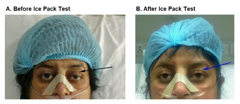
Two side-by-side clinical photographs labeled A and B demonstrate a bedside ice pack test in a patient with suspected myasthenia gravis. Image A (Before Ice Pack Test) shows significant left-sided blepharoptosis, with the upper eyelid drooping to partially cover the pupil (indicated by a black arrow). Image B (After Ice Pack Test) demonstrates a positive result, where the left eyelid shows temporary elevation and resolution of ptosis (indicated by a blue arrow) following local cooling. The patient is shown in a clinical setting wearing a blue surgical cap, a nasal cannula for oxygen delivery, and an adhesive nasal strip for stabilization. This comparative series illustrates the clinical utility of the ice pack test, which inhibits acetylcholinesterase activity at the neuromuscular junction to temporarily improve muscle strength in myasthenic patients. The images highlight key diagnostic physical exam findings in neuro-ophthalmology and neuromuscular medicine.
Two side-by-side clinical photographs labeled A and B demonstrate a bedside ice pack test in a patient with suspected myasthenia gravis. Image A (Before Ice Pack Test) shows significant left-sided blepharoptosis, with the upper eyelid drooping to partially cover the pupil (indicated by a black arrow). Image B (After Ice Pack Test) demonstrates a positive result, where the left eyelid shows temporary elevation and resolution of ptosis (indicated by a blue arrow) following local cooling. The patient is shown in a clinical setting wearing a blue surgical cap, a nasal cannula for oxygen delivery, and an adhesive nasal strip for stabilization. This comparative series illustrates the clinical utility of the ice pack test, which inhibits acetylcholinesterase activity at the neuromuscular junction to temporarily improve muscle strength in myasthenic patients. The images highlight key diagnostic physical exam findings in neuro-ophthalmology and neuromuscular medicine.

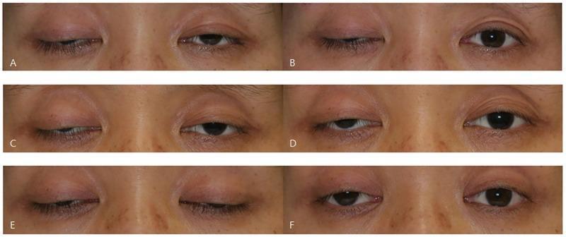
This set of six clinical photographs (A-F) illustrates the variability of the ice test in a patient with myasthenia gravis presenting with bilateral asymmetric ptosis. The images are arranged in pairs representing three different days, showing the patient's eyes before and after a 2-minute ice application. Frames A, C, and E show baseline bilateral blepharoptosis with varying degrees of upper eyelid drooping obscuring the pupil and iris. Frames B, D, and F show the post-intervention state. The series demonstrates three distinct clinical responses in the right eye: (B) a negative result with no significant change, (D) an equivocal result showing subtle 1.0 mm elevation, and (F) a positive result with a marked 2.0 mm increase in the palpebral fissure. This comparison serves as an educational tool for neurology and ophthalmology students to understand the fluctuating nature of neuromuscular junction disorders and the diagnostic utility (and inconsistency) of the ice pack test, which temporarily improves ptosis by inhibiting acetylcholinesterase at the motor endplate.

This set of six clinical photographs (A-F) illustrates the variability of the ice test in a patient with myasthenia gravis presenting with bilateral asymmetric ptosis. The images are arranged in pairs representing three different days, showing the patient's eyes before and after a 2-minute ice application. Frames A, C, and E show baseline bilateral blepharoptosis with varying degrees of upper eyelid drooping obscuring the pupil and iris. Frames B, D, and F show the post-intervention state. The series demonstrates three distinct clinical responses in the right eye: (B) a negative result with no significant change, (D) an equivocal result showing subtle 1.0 mm elevation, and (F) a positive result with a marked 2.0 mm increase in the palpebral fissure. This comparison serves as an educational tool for neurology and ophthalmology students to understand the fluctuating nature of neuromuscular junction disorders and the diagnostic utility (and inconsistency) of the ice pack test, which temporarily improves ptosis by inhibiting acetylcholinesterase at the motor endplate.

Excellent - comprehensive data gathered. Here is the full, detailed answer:

Answer: D. Ptosis improves with ice pack

This is the correct answer. The ice pack test is a highly specific bedside test for Myasthenia Gravis (MG), with a pooled sensitivity of 0.94 and specificity of 0.97 for ocular myasthenia.

The Ice Pack Test - Mechanism

Cooling the neuromuscular junction reduces the activity of acetylcholinesterase (the enzyme that breaks down acetylcholine). At lower temperatures, more ACh remains available in the synaptic cleft, partially compensating for the reduced number of functional AChRs. This transiently improves muscle function - the ptotic eyelid visibly lifts.
Procedure: An ice pack is applied over closed ptotic eyelids for 2 minutes. An improvement of ≥ 2 mm in palpebral fissure height is considered a positive result.
Ice pack test: before (left, ptosis present) and after (right, ptosis improved)

Why the Other Options Are Wrong (and What They Point To)

A. Urinary incontinence - Against MG

MG affects the neuromuscular junction of skeletal muscle only - it does not involve smooth muscle or the autonomic nervous system. Bladder (smooth muscle/autonomic) function is completely preserved in MG. Urinary incontinence would point toward an upper motor neuron lesion, spinal cord disease, or autonomic neuropathy.

B. Pinpoint pupils - Against MG

Miosis (pinpoint pupils) is a parasympathomimetic (muscarinic) sign, seen in:
  • Organophosphate/cholinesterase inhibitor toxicity
  • Pontine hemorrhage
  • Opioid overdose MG does not cause pupillary changes. In fact, pupils are classically spared in MG, which is one important distinguishing feature from other causes of external ophthalmoplegia (e.g., botulism, oculomotor nerve palsy from aneurysm which does affect the pupil).

C. Dry mouth - Against MG; points to Lambert-Eaton

Dry mouth is an autonomic (muscarinic) symptom. MG does not affect the autonomic nervous system. Dry mouth is a classic feature of Lambert-Eaton Myasthenic Syndrome (LEMS), where autoantibodies against presynaptic voltage-gated calcium channels (VGCC) disrupt both nicotinic (motor) and muscarinic (autonomic) transmission. LEMS also typically presents with proximal weakness that paradoxically improves with repeated use, and is associated with small cell lung carcinoma.

E. Global areflexia - Against MG; points to Lambert-Eaton or GBS

Deep tendon reflexes are preserved and normal in MG (as stated in multiple neurology texts: "reflexes and sensory exam are normal"). Global areflexia is a hallmark of:
  • Lambert-Eaton syndrome (hyporeflexia is classic - reflexes may transiently improve after repeated contraction)
  • Guillain-Barre syndrome
  • Severe peripheral neuropathies

Summary Table

OptionFavors MG?What It Actually Indicates
A. Urinary incontinenceNoUMN lesion / autonomic disorder
B. Pinpoint pupilsNoOrganophosphate toxicity / pontine lesion
C. Dry mouthNoLambert-Eaton syndrome
D. Ice pack improves ptosisYESClassic MG bedside test (Sp 97%)
E. Global areflexiaNoLambert-Eaton / GBS

Key Features of MG (for completeness)

  • Fatigable weakness - worsens with repeated use, improves with rest
  • Ocular involvement - ptosis + diplopia (often the presenting feature, especially in young women)
  • Bulbar symptoms - dysarthria, dysphagia, nasal voice
  • Normal reflexes, no sensory loss, no autonomic involvement
  • Anti-AChR antibodies positive in ~85% generalized, ~50% ocular MG
  • Thymoma association - CT chest is mandatory
  • Bimodal onset: young women (2nd-3rd decade) and older men (6th-7th decade)
Sources: Neuroanatomy through Clinical Cases 3rd Ed.; ROSEN's Emergency Medicine; Adams and Victor's Principles of Neurology 12th Ed.

48 years old sales representative, who is a chronic alcohol consumer, presents with confusion and fall due to unstable gait. He has double vision. Neurological examination can not be performed because he is confused. His vitals are stable. What is the next step of management? A. Intramuscular haloperidol B. IV cefrioxone and IV acyclovir C. IV Dectrose 50% D. IV diazepam E. IV Thiamine

Finding Sources
Finding Sources
Reading File
Finding Sources
Reading File
Searching Images

Wernicke encephalopathy MRI brain alcoholic thiamine deficiency
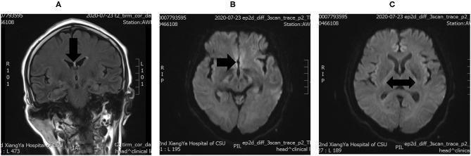
This diagnostic image set consists of three brain MRI panels (A, B, and C) demonstrating classic radiological findings of Wernicke's encephalopathy. Panel A is a coronal T2-weighted/FLAIR image showing symmetrical hyperintense signals in the periventricular region surrounding the third ventricle, highlighted by a vertical arrow. Panels B and C are axial Diffusion-Weighted Imaging (DWI) sequences demonstrating restricted diffusion characterized by high signal intensity within the bilateral basal ganglia and midline structures. Black arrows in the axial views indicate the symmetrical involvement of deep gray matter nuclei. These imaging findings are pathognomonic for thiamine deficiency-related neurotoxicity when correlated with clinical presentation. The modality demonstrates the importance of T2 and DWI sequences in identifying acute metabolic encephalopathy. This material is suitable for advanced medical education in neurology and neuroradiology, focusing on the identification of non-alcoholic Wernicke's encephalopathy manifestations.
This diagnostic image set consists of three brain MRI panels (A, B, and C) demonstrating classic radiological findings of Wernicke's encephalopathy. Panel A is a coronal T2-weighted/FLAIR image showing symmetrical hyperintense signals in the periventricular region surrounding the third ventricle, highlighted by a vertical arrow. Panels B and C are axial Diffusion-Weighted Imaging (DWI) sequences demonstrating restricted diffusion characterized by high signal intensity within the bilateral basal ganglia and midline structures. Black arrows in the axial views indicate the symmetrical involvement of deep gray matter nuclei. These imaging findings are pathognomonic for thiamine deficiency-related neurotoxicity when correlated with clinical presentation. The modality demonstrates the importance of T2 and DWI sequences in identifying acute metabolic encephalopathy. This material is suitable for advanced medical education in neurology and neuroradiology, focusing on the identification of non-alcoholic Wernicke's encephalopathy manifestations.

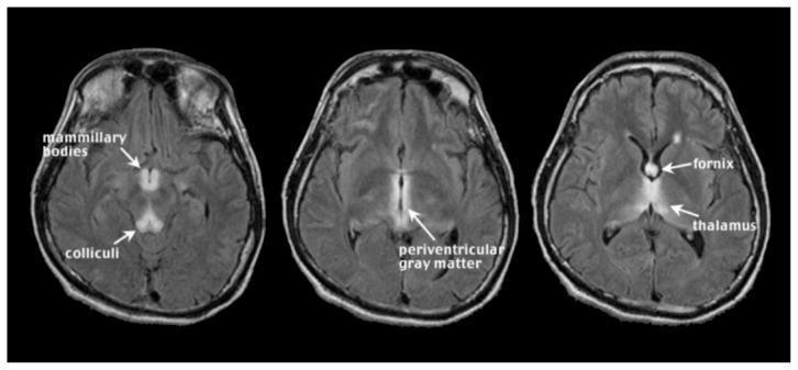
This diagnostic image consists of three axial MRI slices of the brain using Fluid Attenuated Inversion Recovery (FLAIR) sequences, illustrating the classic neuroimaging findings of acute Wernicke encephalopathy. The images demonstrate bilateral, symmetrical hyperintense (bright) signals in specific anatomical regions characteristic of thiamine deficiency. Key structures labeled include the mammillary bodies in the posterior hypothalamus and the colliculi in the midbrain. Higher slices show prominent hyperintensities within the periventricular gray matter surrounding the third ventricle, as well as involvement of the fornix and the medial aspects of the thalamus. These findings represent edema or glial changes associated with the acute clinical manifestation of the disease. The primary educational focus is to teach the radiological signature of Wernicke encephalopathy in the context of chronic alcoholism and nutritional deficiency, emphasizing the typical distribution of lesions in midline brain structures.

This diagnostic image consists of three axial MRI slices of the brain using Fluid Attenuated Inversion Recovery (FLAIR) sequences, illustrating the classic neuroimaging findings of acute Wernicke encephalopathy. The images demonstrate bilateral, symmetrical hyperintense (bright) signals in specific anatomical regions characteristic of thiamine deficiency. Key structures labeled include the mammillary bodies in the posterior hypothalamus and the colliculi in the midbrain. Higher slices show prominent hyperintensities within the periventricular gray matter surrounding the third ventricle, as well as involvement of the fornix and the medial aspects of the thalamus. These findings represent edema or glial changes associated with the acute clinical manifestation of the disease. The primary educational focus is to teach the radiological signature of Wernicke encephalopathy in the context of chronic alcoholism and nutritional deficiency, emphasizing the typical distribution of lesions in midline brain structures.

All the evidence needed is in. Here is the complete answer:

Answer: E. IV Thiamine

This patient has Wernicke's Encephalopathy (WE) - a neurological emergency caused by thiamine (Vitamin B1) deficiency, classically seen in chronic alcoholics.

Recognizing the Diagnosis

The clinical picture maps perfectly onto the classic triad of Wernicke's Encephalopathy:
FeatureThis Patient
Confusion / altered mental statusYes - "confused, exam not possible"
Ataxia (cerebellar/vestibular)Yes - "unstable gait, fall"
OphthalmoplegiaYes - "double vision (diplopia)"
The classic triad of ophthalmoplegia, ataxia, and confusion is present here. A presumptive diagnosis of Wernicke's encephalopathy should be made in any patient with alcohol dependence who has ANY ONE of these signs. - Maudsley Prescribing Guidelines, 15th Ed.
Importantly, the full triad is only present in a minority of cases (~16%), so this patient is actually a textbook presentation. Do not wait for all three to initiate treatment.

Why IV Thiamine is the IMMEDIATE Next Step

The Core Mechanism

Chronic alcohol use causes:
  1. Reduced dietary thiamine intake (poor nutrition)
  2. Reduced GI absorption of thiamine
  3. Impaired hepatic storage
Thiamine is an essential cofactor for key enzymes in glucose metabolism (pyruvate dehydrogenase, alpha-ketoglutarate dehydrogenase, transketolase). Without it, neurons - especially in the mammillary bodies, thalamus, periaqueductal gray, and cerebellar vermis - are selectively destroyed.

The Critical Rule: Thiamine BEFORE Glucose

As thiamine is required to utilise glucose, a glucose load in a thiamine-deficient patient can precipitate Wernicke's encephalopathy or worsen existing disease. - Maudsley Prescribing Guidelines, 15th Ed.
Hypoglycemia requiring glucose infusion must be supplemented with thiamine to prevent the emergence of Wernicke encephalopathy. - Adams & Victor's Principles of Neurology, 12th Ed.
This is why Option C (IV Dextrose 50%) alone is wrong - it would consume the last remaining thiamine stores and potentially worsen or precipitate WE. If hypoglycemia is also suspected (common in alcoholics), give thiamine first, then dextrose.

Dosing

  • Suspected WE: IV/IM Thiamine 200-500 mg three times daily for 3-5 days
  • Prophylaxis in at-risk patients: 100-300 mg IV/IM daily

Why the Other Options Are Wrong

A. IM Haloperidol - Wrong

Haloperidol is an antipsychotic used for agitation/psychosis. This patient's confusion has a clear organic, metabolic cause (thiamine deficiency). Treating it with an antipsychotic would be inappropriate and potentially dangerous (lowers seizure threshold). It does not address the underlying pathology.

B. IV Ceftriaxone + IV Acyclovir - Wrong

This combination is the empiric treatment for bacterial meningitis + viral encephalitis (HSV). While these must be considered in any confused patient with fever/neck stiffness, there is no mention of fever, headache, photophobia, or meningismus here. The clinical context strongly points to Wernicke's. However, if the diagnosis were uncertain, LP should be considered - but the alcohol history + classic triad makes WE the overwhelming priority.

C. IV Dextrose 50% - Wrong (and potentially harmful)

As explained above, giving glucose first in a thiamine-depleted state can precipitate or worsen WE by accelerating consumption of the remaining thiamine. Always give thiamine before (or simultaneously with) glucose. This is a classic exam trap.

D. IV Diazepam - Wrong

Benzodiazepines are the treatment of choice for alcohol withdrawal seizures / delirium tremens. There is no mention of seizures, tremor, autonomic instability (tachycardia, hypertension, sweating), or hallucinations suggestive of withdrawal. Giving a benzodiazepine here would not treat WE and would add CNS depression on top of existing encephalopathy.

What Happens If Untreated?

If WE goes untreated, it progresses to Korsakoff Syndrome (Korsakoff Psychosis) - an irreversible amnestic disorder characterized by:
  • Anterograde amnesia (inability to form new memories)
  • Confabulation (fabricating false memories without intent to deceive)
  • Relatively preserved consciousness and other cognition
Together, the acute + chronic stages are called Wernicke-Korsakoff Syndrome.

MRI Findings in Wernicke's (for completeness)

MRI showing classic Wernicke's encephalopathy: bilateral FLAIR hyperintensities in mammillary bodies, periaqueductal gray, and medial thalami
Classic MRI findings: symmetrical T2/FLAIR hyperintensities in the:
  • Mammillary bodies (most specific)
  • Medial thalami / dorsomedial thalamic nuclei
  • Periaqueductal gray matter
  • Cerebellar vermis

Quick Summary

OptionVerdictReason
A. IM HaloperidolWrongNo psychosis; misses metabolic cause
B. IV Ceftriaxone + AcyclovirWrongNo evidence of meningitis/encephalitis
C. IV Dextrose 50%Wrong (harmful)Worsens WE by depleting thiamine
D. IV DiazepamWrongNo alcohol withdrawal features
E. IV ThiamineCORRECTTreats underlying Wernicke's; must precede glucose
Sources: Maudsley Prescribing Guidelines 15th Ed.; Adams & Victor's Principles of Neurology 12th Ed.; ROSEN's Emergency Medicine; Tintinalli's Emergency Medicine
This is a shared conversation. Sign in to Orris to start your own chat.