
Acid fast bacilli staining
Reading File
Now let me search the medical library for acid fast bacilli staining content, in parallel with a heading search and paragraph search.
Finding Sources
Finding Sources
Good results. Let me now read the most relevant sections from Medical Microbiology 9e and Sherris & Ryan's for comprehensive content, in parallel.
Reading File
Reading File
Reading File
Let me read the full acid-fast stains table from Medical Microbiology 9e and the Sherris section on acid-fast stain variants, plus search for Ziehl-Neelsen mechanism details.
Reading File
Reading File
Finding Sources
Let me now read the detailed content from Harrison's, Quick Compendium, and Robbins for more clinical/pathological depth, plus fetch the key diagram image.
Reading File
Reading File
Loading Image
Let me also read the Kinyoun stain details and modified acid-fast info from Quick Compendium, plus the AFB smear photo from Harrison's.
Reading File
Loading Image
I now have comprehensive information to write a thorough answer. Let me compile everything.
Acid-Fast Bacilli (AFB) Staining
What is Acid-Fastness?
Acid-fast organisms stain very poorly with most dyes (including the Gram stain - they are typically neutral on Gram staining). However, once stained with a concentrated dye - aided by heat or a penetrating agent - they resist decolorization by acid-alcohol solutions that would strip the same dye from all other bacteria. This combination of difficult initial uptake followed by strong retention is the defining property.
The mechanism is the high lipid content of the mycobacterial cell wall, specifically mycolic acids - long-chain, cross-linked fatty acids (70-90 carbon atoms in mycobacteria) covalently linked to arabinogalactan and peptidoglycan. Carbolfuchsin (fuchsin dye) binds to mycolic acid in a way that resists acid-alcohol decolorization. - Harrison's Principles of Internal Medicine 22E, p. 1424; Quick Compendium of Clinical Pathology 5th ed.
The Three-Step Staining Process
The diagram below shows the principle clearly - compare it to the Gram stain side-by-side:
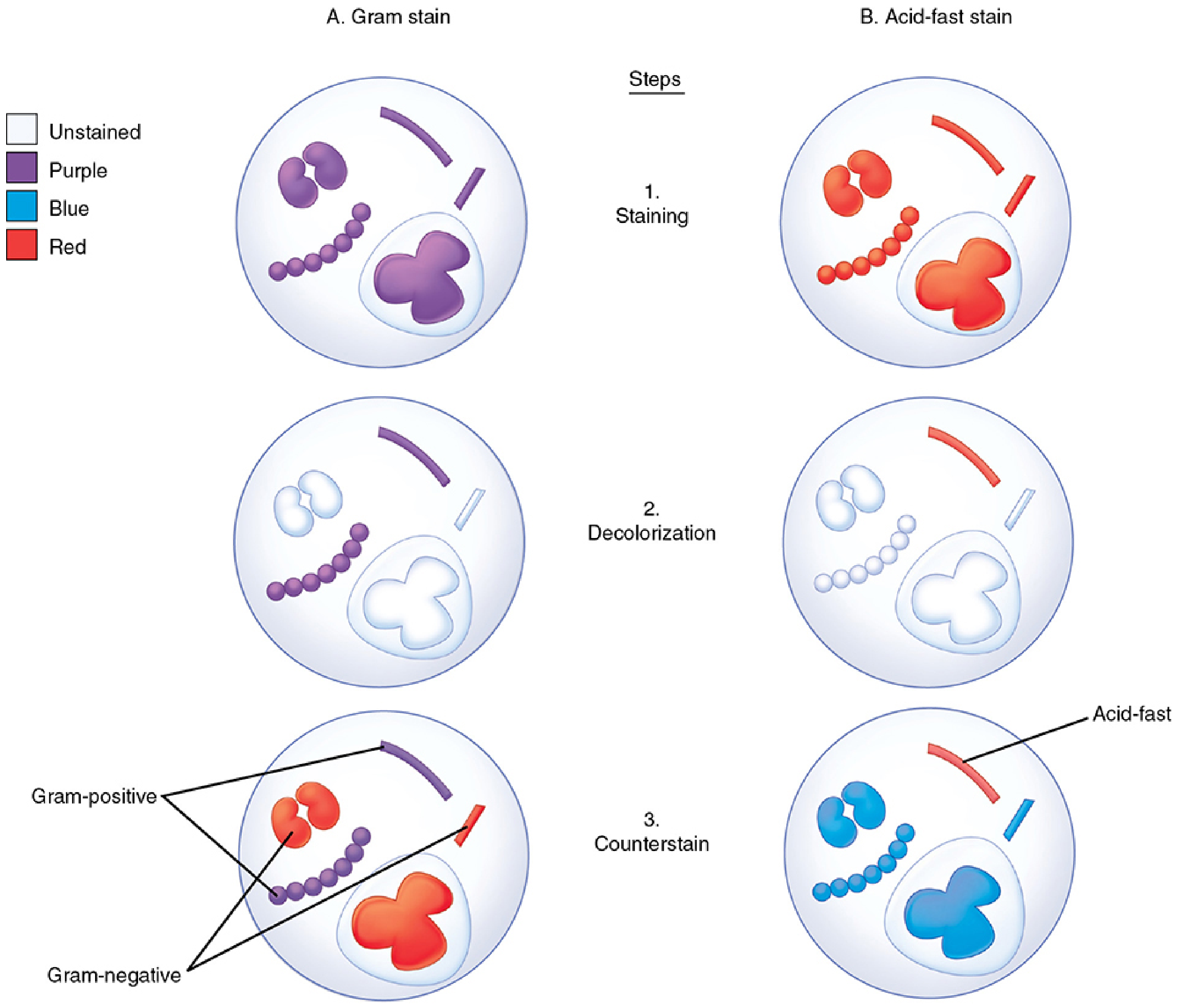
FIGURE: Gram and acid-fast stains compared. Step 1 (Staining): all organisms take up carbol fuchsin (red). Step 2 (Decolorization): acid-alcohol removes dye from non-acid-fast organisms. Step 3 (Counterstain): methylene blue stains the background/non-AFB blue, while acid-fast organisms remain red. - Sherris & Ryan's Medical Microbiology, 8th ed.
| Step | Agent | Effect |
|---|---|---|
| Primary stain | Carbol fuchsin (basic fuchsin + phenol/carbolic acid) | Stains all organisms red |
| Decolorization | Acid-alcohol (HCl in ethanol) | Removes dye from non-acid-fast organisms |
| Counterstain | Methylene blue | Turns background and non-AFB blue |
Result: AFB appear red (pink-red) rods against a light blue background.
Variants of the Acid-Fast Stain
1. Ziehl-Neelsen Stain (Hot Acid-Fast Stain)
- The classic method, most commonly used
- Carbolfuchsin penetration requires heating the slide (hot acid-fast stain) - heat drives the dye into the waxy cell wall
- Decolorized with acid-alcohol (HCl in ethanol)
- Counterstained with methylene blue
- Scanned at 100x oil-immersion lens
- Medical Microbiology 9e
2. Kinyoun Stain (Cold Acid-Fast Stain)
- Does NOT require heating
- Uses a higher concentration of carbolfuchsin plus a detergent (wetting agent) in the primary stain to enhance penetration without heat
- Same decolorization and counterstain principle as Ziehl-Neelsen
- Quick Compendium of Clinical Pathology 5th ed.; Medical Microbiology 9e
3. Fluorochrome Stain (Auramine-Rhodamine / Auramine-O)
- Uses fluorescent dyes (auramine alone, or auramine-rhodamine mixture) as the primary stain
- Decolorized with acid-alcohol
- Counterstained with potassium permanganate (a strong oxidizing agent that quenches unbound fluorochrome)
- AFB fluoresce golden yellow-green against a black background under fluorescence microscopy
- More sensitive than carbolfuchsin stains - bacteria detected at as few as 10⁴ AFB/mL; allows rapid screening at lower magnification (40x)
- Has become the method of choice in most high-volume laboratories
- Quick Compendium of Clinical Pathology 5th ed.; Medical Microbiology 9e; Sherris & Ryan's 8th ed.
4. Modified Acid-Fast Stain (Fite Stain / Modified Kinyoun)
- Uses a weaker decolorizing agent (e.g., sulfuric acid in ethanol instead of HCl-ethanol)
- Detects "weakly acid-fast" / "partially acid-fast" organisms that would be decolorized by the full-strength decolorizer used in ZN or Kinyoun
- Quick Compendium of Clinical Pathology 5th ed.
Microscopic Appearance of AFB
The actual appearance of M. tuberculosis bacilli on a Ziehl-Neelsen smear:
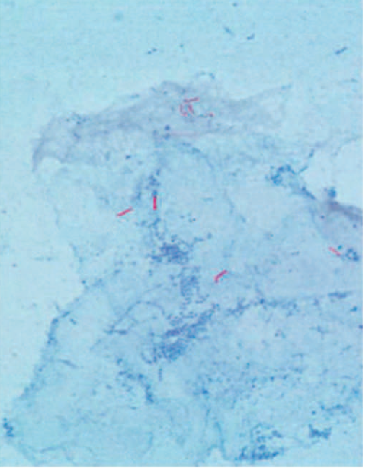
FIGURE 183-1: Acid-fast bacillus smear showing M. tuberculosis bacilli (Ziehl-Neelsen). Thin, red rods scattered on blue background. (CDC/Harrison's Principles of Internal Medicine 22E)
- Slender rods, may be slightly bent or beaded
- In sputum, organisms often lie parallel, or two organisms adhere at one end to form a "V" shape
- Quick Compendium of Clinical Pathology 5th ed.
Organisms That Stain Acid-Fast
| Category | Examples |
|---|---|
| Strongly acid-fast (true AFB) | Mycobacterium tuberculosis, M. leprae, other mycobacteria |
| Weakly / partially acid-fast | Nocardia spp., Rhodococcus equi, Tsukamurella, Gordonia |
| Other partially acid-fast | Legionella micdadei, Cryptosporidium, Cyclospora, Cystoisospora (formerly Isospora), Sarcocystis |
The difference in degree of acid-fastness relates to mycolic acid chain length - mycobacteria have 70-90 carbons vs. Nocardia's 50-62, explaining why Nocardia is only "weakly" acid-fast and requires modified (weaker) decolorizer. - Medical Microbiology 9e
Quick Comparison Table
| Feature | Ziehl-Neelsen | Kinyoun | Auramine-Rhodamine | Modified (Fite) |
|---|---|---|---|---|
| Heat | Yes (hot) | No (cold) | No | No |
| Primary dye | Carbolfuchsin | Carbolfuchsin (concentrated + detergent) | Auramine ± rhodamine | Carbolfuchsin |
| Decolorizer | Strong (HCl-alcohol) | Strong (HCl-alcohol) | Strong (acid-alcohol) | Weak (H₂SO₄-ethanol) |
| Counterstain | Methylene blue | Methylene blue | Potassium permanganate | Methylene blue |
| AFB color | Red on blue | Red on blue | Gold-yellow on black | Red on blue |
| Microscope | Brightfield (100x) | Brightfield (100x) | Fluorescence (40x) | Brightfield (100x) |
| Sensitivity | Standard | Standard | Higher | For weakly AFB |
| Best use | TB/mycobacteria | TB/mycobacteria | Screening, high volume | Nocardia, parasites |
Clinical Significance
- The AFB smear is the first-line rapid test for pulmonary and extrapulmonary tuberculosis
- Sensitivity is approximately 40-60% in culture-confirmed TB cases; requires ~5,000-10,000 organisms/mL to be positive on carbolfuchsin stains
- The fluorochrome method detects as low as 10⁴ AFB/mL and is faster to screen
- Positive AFB smear does not distinguish between M. tuberculosis and non-tuberculous mycobacteria (NTM) - culture and/or nucleic acid amplification tests (NAATs) are needed
- Harrison's Principles of Internal Medicine 22E; Murray & Nadel's Textbook of Respiratory Medicine
This is a shared conversation. Sign in to Orris to start your own chat.