
Alcoholic liver disease: pathophysiology, clinical features, management
alcoholic liver disease histology steatosis hepatitis cirrhosis
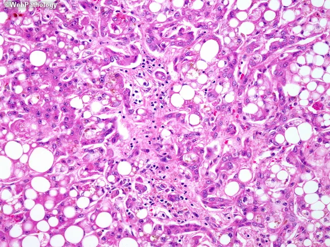
Alcoholic steatohepatitis manifested on liver biopsy as macrovesicular steatosis with hepatocellular injury, inflammatory infiltrates, and early fibrosis. This bright-field histology image, obtained from a paraffin-embedded liver biopsy and stained with Hematoxylin and Eosin, highlights lobular and portal changes. Large fat vacuoles displace hepatocyte nuclei; scattered ballooning degeneration accompanies hepatocellular damage. In the portal tracts, mixed inflammatory cells—neutrophils and lymphocytes including CD4+ and CD8+ T cells—are evident, often forming a neutrophilic infiltrate around ductular structures. A ductular reaction is visible as proliferating biliary-like cells at the limiting plate and portal-periportal interface. The surrounding parenchyma shows cytoplasmic rarefaction and mild-to-moderate inflammation, with early fibrotic bands encroaching on the portal triad region. Collectively, these features support an active alcoholic injury pattern rather than pure steatosis, differentiating from nonalcoholic fatty liver disease. Clinically, this image underpins diagnoses of alcoholic hepatitis/ASH in the appropriate history and contributes to assessments of disease activity and prognosis via fibrosis staging. It is relevant for medical education, histopathology libraries, and research on inflammatory liver diseases, fibrosis progression, and the hepatobiliary response to alcohol exposure. This image supports differential diagnosis with NAFLD, alcoholic hepatitis, DILI; useful for pathology education, case conferences, radiology-pathology correlation, and AI dataset curation, and clinical teaching.
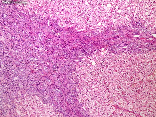
Comprehensive Description: This liver biopsy image illustrates chronic parenteral nutrition (TPN) associated liver disease with established cirrhosis. Modality is light microscopic histology with Hematoxylin and Eosin (H&E) staining. The hepatic parenchyma shows nodular arrangements demarcated by broad fibrous septa, consistent with cirrhotic remodeling. Within nodules, hepatocytes appear variably damaged, with cytoplasmic vacuolization and steatosis. Intersinusoidal and canalicular bile pigment is evident, with inspissated bile remnants filling dilated canaliculi and small ducts, indicating cholestasis. Prominent ductular reaction is present at the limiting plate and along fibrous bands, reflecting proliferative bile ductules responding to injury. The nodules are separated by fibrous bands that contain inflammatory cells and proliferating ductules, creating a characteristic irregular lobular architecture. The pattern is compatible with long-term TPN associated hepatopathy and may progress to macronodular or micronodular cirrhosis over time. Clinically, this constellation correlates with prolonged parenteral nutrition, cholestasis, and hepatocellular injury. The diagnostic significance lies in recognizing TPN associated liver fibrosis with steatosis and bile stasis, which bears risk for progressive liver dysfunction and cirrhosis. Differential diagnoses include alcoholic liver disease, nonalcoholic steatohepatitis, viral hepatitis, and biliary cholangiopathies; correlation with clinical history is essential for management. This image underscores the critical link between nutrition and hepatic structural integrity in patients.
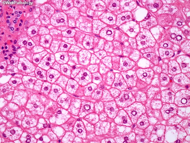
This is a liver biopsy histology image prepared for light microscopy, stained with hematoxylin and eosin (H&E). The primary subject is hepatocytes within the hepatic parenchyma showing features of alcoholic steatohepatitis. Macrovesicular steatosis is evident, with numerous large fat vesicles displacing the cytoplasm. More striking are ballooned hepatocytes, enlarged cells with pale, cytoplasmic clearing and disrupted cytoskeletal architecture. Within several ballooned cells, Mallory-Denk bodies (Mallory hyaline) appear as irregular eosinophilic, hyaline inclusions. The nuclear morphology is variably preserved, with occasionally pyknotic or shrunken nuclei in chronically damaged hepatocytes. The overall cellular morphology indicates hepatocellular injury with cytoskeletal disruption and intracellular inclusions consistent with steatohepatitis. Notably, CK8/18 immunostaining would reveal reduced or absent intermediate filament networks in ballooned hepatocytes, a feature that helps distinguish steatohepatitic ballooning from non-steatohepatitic ballooning seen in viral hepatitis where CK8/18 loss is less pronounced. In alcoholic liver disease, Mallory-Denk bodies and cytoplasmic clearing correlate with ongoing inflammation and progressive fibrosis risk, informing diagnostic significance and clinical management. This image is valuable for education on histologic differentiation between alcoholic steatohepatitis and other causes of hepatocellular ballooning, and for teaching pathology students about steatosis-related cytoskeletal disruption. Correlation with alcohol history and fibrosis staging enhances clinical decision making.
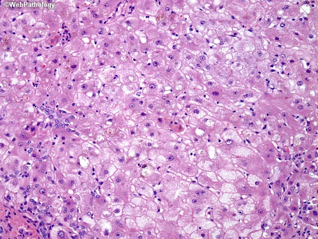
Imaging modality: light microscopy of a hematoxylin and eosin (H&E) stained liver biopsy section. The specimen demonstrates cirrhotic nodules with disrupted lobular architecture and bridging fibrosis within the hepatic parenchyma, compatible with advanced chronic liver disease. Hepatocytes display marked steatosis with macrovesicular fat and prominent ballooning degeneration, accompanied by densely eosinophilic cytoplasm. Focal cholestasis is evident, with canalicular bile and cholestatic pigment visible within affected cells. Large lipofuscin granules are scattered throughout hepatocytes, reflecting chronic oxidative injury linked to copper accumulation. Mallory bodies (Mallory‑Denk inclusions) are present in some hepatocytes, notably in the upper left field, indicating cytoskeletal damage. Collectively, these features illustrate progression from fatty change to chronic hepatitis and nodular cirrhosis in Wilson disease. The underlying etiology is copper overload due to ATP7B gene mutations causing impaired biliary copper excretion and hepatic copper deposition, with copper‑associated hepatocellular injury and oxidative stress. Diagnostic significance: when integrated with compatible serum studies (low ceruloplasmin, elevated hepatic copper) and genetic testing for ATP7B, histology supports Wilson disease diagnosis and guides management. Differential considerations include alcoholic liver disease, nonalcoholic fatty liver disease, cholestatic hepatitis, and other causes of cirrhosis. Clinical correlation with clinical presentation and copper metabolism testing is essential for accurate diagnosis and prognosis.
"alcoholic liver disease"[MeSH Terms] AND management
"alcohol-associated hepatitis" AND treatment
Alcoholic Liver Disease (ALD)
Disease Spectrum & Epidemiology
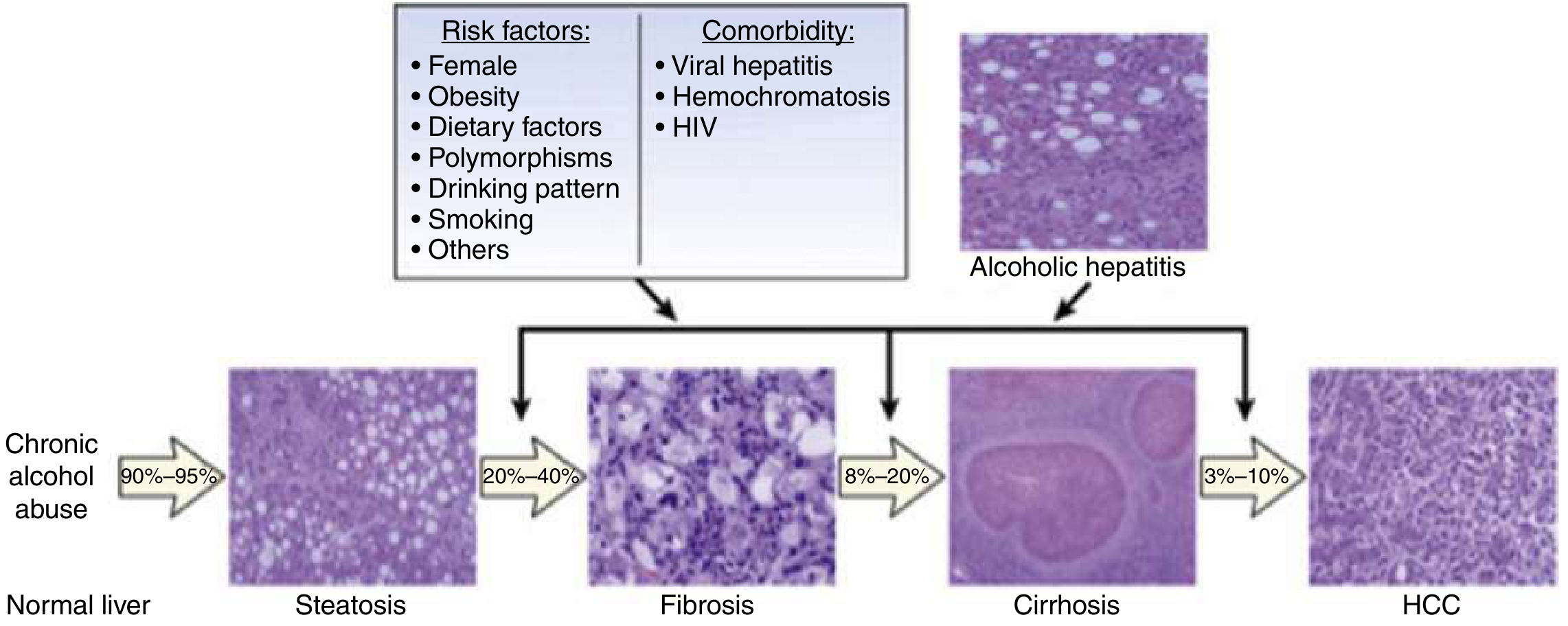
1. Pathophysiology
Ethanol Metabolism and Toxic Byproducts
- Alcohol dehydrogenase (ADH) pathway: Oxidizes ethanol to acetaldehyde, generating large amounts of NADH. The excess NADH shifts the redox balance in hepatocytes, suppressing fatty acid beta-oxidation and stimulating fatty acid synthesis and lipogenesis, leading to steatosis.
- Microsomal Ethanol Oxidizing System (MEOS) / CYP2E1: Responsible for ~10% of ethanol oxidation at low doses but is induced significantly at high intake. CYP2E1 generates reactive oxygen species (ROS) that damage cellular proteins, membranes, and mitochondria, promoting apoptosis and necrosis. CYP2E1 is concentrated in zone 3 (centrilobular/perivenular), which is also a zone of relative hypoxia and lowest glutathione levels - explaining why injury starts centrally.
- Acetaldehyde toxicity: Induces lipid peroxidation, disrupts cytoskeleton and membrane function, and forms acetaldehyde-protein adducts that may serve as neoantigens triggering immune responses. - Robbins & Cotran; Mulholland Surgery 7e
Steatosis Mechanisms
- NADH excess favors lipogenesis and blocks fatty acid oxidation
- Impaired lipoprotein (VLDL) assembly and secretion
- Enhanced peripheral fat catabolism, increasing hepatic lipid delivery
- Alcohol also impairs hepatic methionine metabolism, decreasing glutathione levels (sensitizing liver to oxidative injury) and contributing to homocysteine accumulation and endoplasmic reticulum stress. - Robbins & Cotran
Gut-Liver Axis and Inflammation
Fibrosis and Stellate Cell Activation
Risk Factors for Progression
| Factor | Detail |
|---|---|
| Sex | Women more susceptible dose-for-dose; estrogen increases gut permeability to LPS |
| Genetics | ALDH2 variant (50% of East Asians) - near-absent aldehyde dehydrogenase activity; ADH polymorphisms affect acetaldehyde accumulation |
| Comorbidities | HCV/HBV co-infection, MASLD, iron overload synergize with alcohol |
| Nutritional deficiency | Folate, thiamine, zinc, magnesium deficiencies accelerate injury |
| Drinking pattern | Binge vs. sustained drinking; drinking outside meal times |
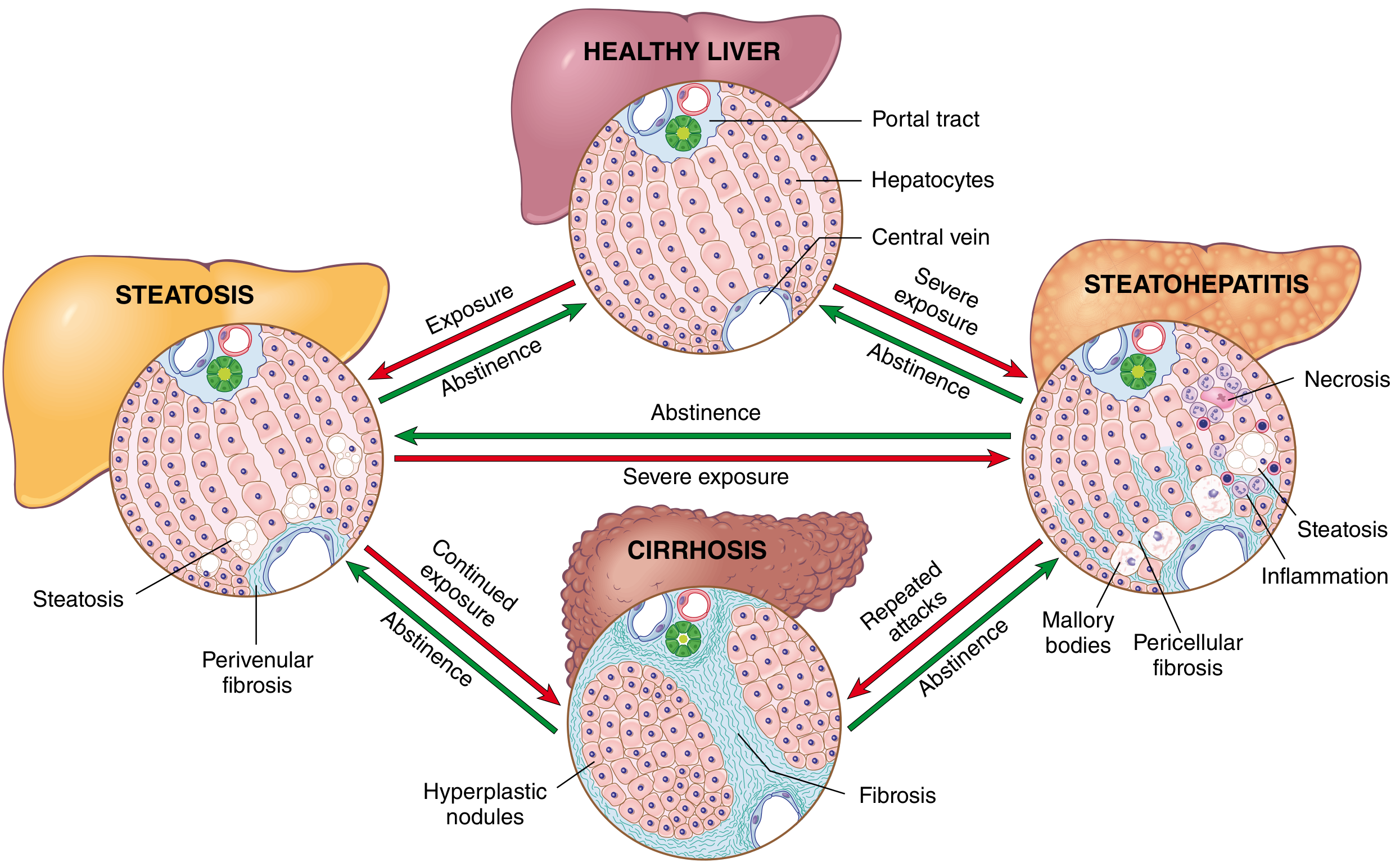
2. Morphology (Histopathology)
Stage 1 - Hepatic Steatosis (Fatty Liver)
- Macrovesicular steatosis predominates - large lipid droplets displace the nucleus to the periphery
- Liver is enlarged (up to 4-6 kg), soft, yellow, and greasy
- Completely reversible with abstinence (within ~2 weeks)
- Microvesicular (foamy) steatosis is an unusual variant associated with mitochondrial/ER damage
Stage 2 - Alcoholic Hepatitis (Steatohepatitis)
- Ballooned hepatocytes - swollen, pale cytoplasm with cytoskeletal damage
- Mallory-Denk bodies (Mallory hyaline) - eosinophilic skeins of ubiquitinated intermediate filaments (keratin 8/18) within ballooned hepatocytes; pathognomonic but not exclusive to ALD
- Neutrophilic infiltration surrounding ballooned hepatocytes (more prominent than in MASLD)
- Lobular lymphocytic infiltrates and portal inflammation
- Cholestasis and giant mitochondria ("cherry bodies") may be present
- Pericellular "chicken wire" fibrosis in zone 3
Stage 3 - Fibrosis and Cirrhosis
- Portal and perivenular fibrosis → fibrous septa → small regenerative nodules
- Diffuse interstitial fibrosis - collagen encircling individual hepatocytes without parenchymal regeneration
- Progressive perivenular fibrosis with dense zone 3 scar-like collagen
3. Clinical Features
Hepatic Steatosis
- Usually asymptomatic; found incidentally or with mild hepatomegaly
- Reversible with abstinence
- LFTs may be mildly elevated or normal
Alcoholic Hepatitis
- Acute illness in patients with chronic heavy alcohol use, often with recent binge
- Jaundice (often deep, with bilirubin >5 mg/dL)
- Fever, malaise, anorexia, nausea, vomiting
- Hepatomegaly (often tender)
- Right upper quadrant pain
- Hepatic bruit may be heard
- Complications: ascites, encephalopathy, coagulopathy, acute kidney injury
- AST elevated 2-6x normal; ALT may be less elevated
- AST:ALT ratio ≥ 2:1 (characteristic of ALD vs. viral hepatitis)
- Values rarely exceed 10x normal even in severe disease
- Leukocytosis, hyperbilirubinemia, prolonged PT/INR
- Hypoalbuminemia, thrombocytopenia
- Hypoglycemia (from suppressed gluconeogenesis and depleted glycogen stores)
- Electrolyte disturbances: hypomagnesemia, hypophosphatemia, hypokalemia
-
- Rosen's Emergency Medicine
Alcoholic Cirrhosis
- Ascites - often requiring repeated paracentesis
- Variceal bleeding (esophageal/gastric varices) - 10-15% acute mortality per episode
- Hepatic encephalopathy - ammonia-mediated cerebral dysfunction (precipitated by bleeding, infection, hypokalemia, dehydration)
- Hepatorenal syndrome (HRS) - heralded by rising creatinine in setting of liver failure; high mortality
- Spontaneous bacterial peritonitis (SBP)
- Coagulopathy (reduced clotting factor synthesis)
- Thrombocytopenia (portal hypertension-related hypersplenism)
- Spider nevi (cutaneous arterioles)
- Palmar erythema
- Gynecomastia and testicular atrophy (feminization from impaired estrogen metabolism)
- Dupuytren's contracture
- Parotid enlargement
- Leukonychia, clubbing
- Caput medusae
4. Severity Scoring
Maddrey Discriminant Function (MDF)
- MDF > 32 = severe disease, 1-month mortality ~35-40%, indicates potential benefit from corticosteroids
- Contraindications to corticosteroids (GI bleeding, active infection, renal failure) must be excluded before treatment - Rosen's EM; Goldman-Cecil Medicine
Other Scoring Systems
- MELD score > 21: Can substitute for MDF in guiding prednisolone use
- Lille score: Calculated at day 7 of corticosteroid therapy - Lille > 0.45 indicates non-response and treatment should be stopped
- Glasgow Alcoholic Hepatitis Score (GAHS): Score ≥ 9 predicts poor prognosis
- 3-month mortality ranges from 15% (mild) to 55% (severe alcoholic hepatitis)
5. Management
The Single Most Important Intervention: Abstinence
- Naltrexone 50 mg/day (up to 6 months+) - opioid antagonist reducing craving
- Acamprosate 333 mg tablets, 2 tablets TID for 1 year
- Nalmefene 20 mg/day (maintenance)
- Disulfiram generally avoided due to poor tolerability and hepatotoxicity
Nutritional Support
- All patients with alcoholic hepatitis and cirrhosis should be assessed for protein-calorie malnutrition and micronutrient deficiency
- Thiamine 100 mg IV should be given before any glucose-containing solution (to prevent Wernicke's encephalopathy)
- Empirical magnesium replacement (1 g IV MgSO4 or 200-1000 mg PO daily) - ethanol causes renal wasting
- High-calorie, vitamin-supplemented diet
- Enteral nutrition for severe decompensation
- Folate, zinc, and B-vitamin supplementation
Specific Treatment for Severe Alcoholic Hepatitis (MDF > 32)
- Prednisolone 40 mg/day PO for 4 weeks OR methylprednisolone 32 mg/day IV
- Indicated for MDF >32 or MELD >21, with encephalopathy but WITHOUT GI bleeding, active infection, or HRS
- Reduces 1-month mortality but likely not long-term mortality
- NAC (IV protocol: 150 mg/kg in 250 mL over 30 min, then 50 mg/kg in 500 mL over 4h, then 100 mg/kg in 1000 mL over 16h on Day 1; then 10 mg/kg/day on Days 2-5) added to prednisolone
- Significantly lower 1-month mortality vs. prednisolone alone; addresses oxidative stress - Goldman-Cecil Medicine
- May be used when corticosteroids are contraindicated
- Not superior to corticosteroids; does not improve 28-day survival vs. prednisolone
- No incremental benefit when added to corticosteroids
- If Lille >0.45 (non-response), steroids should be stopped
Management of Complications
| Complication | Management |
|---|---|
| Ascites | Sodium restriction, spironolactone ± furosemide; large-volume paracentesis with albumin 8 g/L removed; TIPS for refractory ascites |
| Variceal bleeding | IV octreotide/terlipressin; endoscopic band ligation; beta-blockers for prophylaxis; TIPS for refractory bleeding |
| Hepatic encephalopathy | Lactulose 30-60 g/day; rifaximin 400 mg TID; correct precipitants (electrolytes, bleeding, infection); branched-chain amino acids |
| HRS | Terlipressin + albumin; vasoconstrictors + albumin infusion; TIPS; dialysis bridge to transplant |
| SBP | Cefotaxime IV; prophylaxis with norfloxacin/ciprofloxacin |
| Coagulopathy with active bleeding | Cryoprecipitate 1 unit/10 kg (preferred over FFP); avoid ACE inhibitors/ARBs in decompensated disease |
Liver Transplantation
- For end-stage ALD not responding to medical therapy
- Traditionally required 6 months of abstinence ("6-month rule"), though evidence for this rule is limited
- Early liver transplantation for severe alcoholic hepatitis unresponsive to medical therapy has been performed in carefully selected patients (first-time liver disease, strong social support, absence of other organ failure, psychiatry clearance) - supported by the Mathurin et al. landmark trial (PMID: 22070476)
- The 2024 ACG Clinical Guideline (PMID: 38174913) provides updated recommendations
Emerging Therapies (Under Investigation)
- Gut microbiome modulation (fecal microbiota transplantation)
- IL-1 receptor antagonists (anakinra)
- Granulocyte colony-stimulating factor (G-CSF) for hepatic regeneration
- Anti-fibrotic agents targeting stellate cell activation
- Obeticholic acid and other FXR agonists
Summary Table
| Stage | Histology | Reversibility | Management |
|---|---|---|---|
| Steatosis | Macrovesicular fat, zone 3 | Fully reversible with abstinence | Abstinence only |
| Alcoholic hepatitis | Ballooned hepatocytes, Mallory bodies, neutrophils, pericellular fibrosis | Partially reversible | Abstinence + corticosteroids (if MDF>32) + NAC + nutrition |
| Cirrhosis | Micronodular, bridging fibrosis, vascular derangements | Rarely fully reversible | Abstinence + complication management + transplant listing |
- Robbins, Cotran & Kumar - Pathologic Basis of Disease (Chapter 18: Liver)
- Yamada's Textbook of Gastroenterology, 7e (Chapter 141)
- Mulholland & Greenfield's Surgery, 7e (Chapter on Alcohol)
- Goldman-Cecil Medicine (Chapter 138)
- Rosen's Emergency Medicine, 10e (Chapter 76)
- ACG Clinical Guideline: Alcohol-Associated Liver Disease, 2024 (PMID: 38174913)