
Station during delivery
fetal station ischial spines delivery diagram
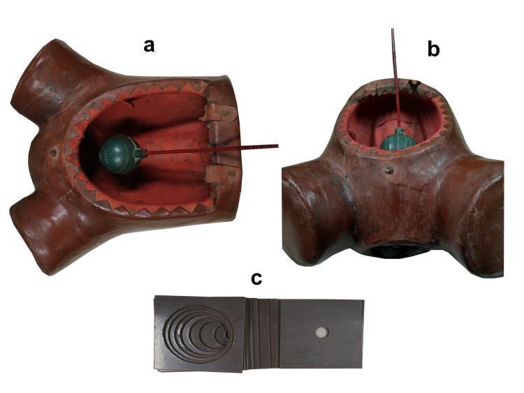
This composite image illustrates a three-part medical simulation training model designed for obstetric education, specifically for teaching cervical dilatation and fetal station assessment. Panels (a) and (b) show a female pelvic mannequin torso with an open abdominal aperture. Inside the pelvic cavity, a green spherical component represents the fetal head, attached to a graduated red rod. The rod features 1 cm markings to simulate and measure 'fetal station' relative to internal palpable landmarks (ischial spines). Panel (c) displays a set of dark, rectangular bakelite plates used to simulate the cervix. These plates feature central circular openings ranging from 1 to 10 cm in diameter, allowing trainees to practice digital vaginal examinations to determine cervical dilatation. The model serves as a low-cost, effective tool for labor and delivery simulation, enabling learners to develop tactile skills in assessing the progression of labor within a standardized clinical context.
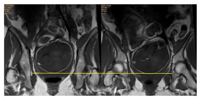
This diagnostic image consists of two side-by-side MRI scans in the coronal plane, focused on the maternal pelvis during late-term pregnancy. The images demonstrate the fetal head engaged within the pelvic canal. Key maternal anatomical landmarks include the iliac wings, the acetabula, and the ischial spines. A horizontal yellow reference line is drawn across the interspinal plane, connecting the maternal ischial spines. This line serves as a clinical marker for determining fetal station. The fetal skull is seen as a well-defined, dark, ovoid structure, with its leading part positioned at the level of the yellow interspinal line, visually confirming a fetal station of 0. This imaging is utilized in obstetrics to objectively assess the progress of fetal descent and engagement within the birth canal, comparing clinical examination findings with high-resolution radiological evidence.
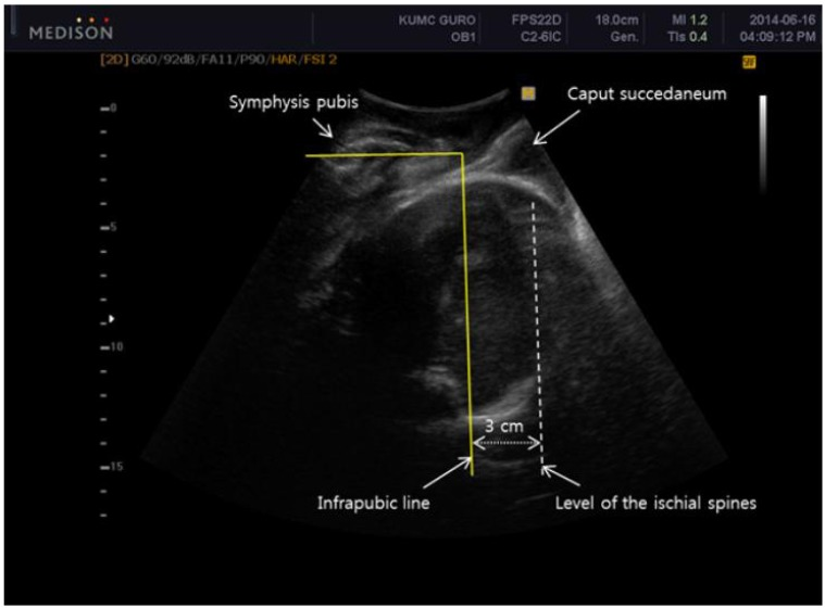
**Imaging Modality:** Two-dimensional (2D) transperineal ultrasound. **Anatomical Region:** Maternal pelvis and fetal head during labor. **Observed Anatomy & Landmarks:** - **Maternal Landmarks:** The symphysis pubis is visible superiorly as a hyperechoic structure. The "infrapubic line" is represented by a solid yellow line drawn perpendicular to the long axis of the symphysis pubis. - **Fetal Landmarks:** The fetal head is visible within the pelvic canal. A "caput succedaneum" (localized scalp edema) is identified as a hypoechoic soft tissue swelling superficial to the fetal skull. **Key Measurements & Findings:** The image demonstrates the spatial relationship used to determine fetal station via ultrasound. A dotted vertical line marks the "level of the ischial spines," situated 3 cm caudal (posterior in this orientation) to the solid "infrapubic line." This 3 cm distance serves as a reference for quantifying the descent of the fetal head relative to the pelvic mid-plane. **Clinical Significance:** This intrapartum ultrasound assessment provides an objective measurement of fetal head engagement and station, differentiating the true fetal skull position from soft tissue findings like caput succedaneum.
Station During Delivery
The Station Scale
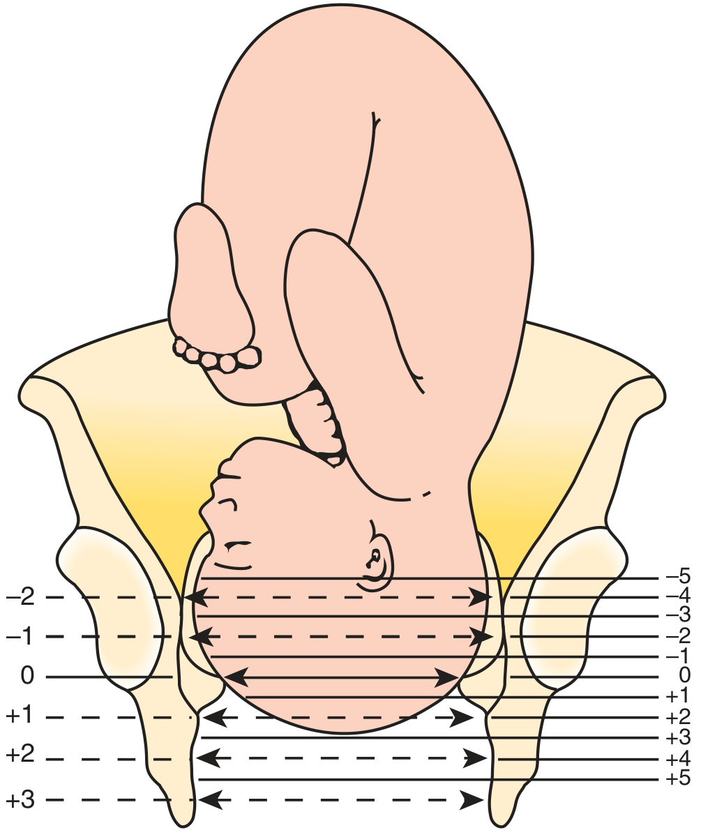
| Station | Meaning |
|---|---|
| 0 | Presenting part is exactly at the level of the ischial spines |
| -1 to -5 | Presenting part is above the ischial spines (not yet engaged at -5) |
| +1 to +5 | Presenting part is below the ischial spines (deeper in the pelvis); +5 = delivery at the perineum |
Old vs. New Classification
- Old (3-point) system: Used only -3, -2, -1, 0, +1, +2, +3 (subjective and less precise).
- New (5-point) system: Uses -5 through 0 through +5, with each unit = 1 cm. This is the current standard and is more objective.
Engagement
Clinical Significance
- Effacement - thinning of the cervix (0-100%)
- Dilatation - cervical opening in cm (0-10 cm)
- Position - relationship of the presenting part to the birth canal (e.g., occiput anterior)
- Presentation - which fetal part leads (vertex in 95% of labors)
- Progress of labor - descent from negative to positive stations during the active phase and second stage
- Readiness for operative delivery - vacuum or forceps are generally applied at station 0 or below (engaged head)
- Risk of obstructed labor - failure of descent despite adequate contractions suggests cephalopelvic disproportion