
Give in detail about acromioclavicular joint injury according to orthopaedics text books
acromioclavicular joint injury classification Rockwood

A composite of four bilateral anteroposterior (AP) stress radiographs of the shoulder girdle, illustrating different grades of acromioclavicular (AC) joint injuries according to the Rockwood classification system. These views are obtained under a 10-kg axial load to assess ligamentous stability. Image (a) represents a Rockwood Type I injury, showing normal anatomical alignment of the clavicle, acromion, and coracoid process. Image (b) corresponds to a Rockwood Type II injury, demonstrating mild superior displacement of the distal clavicle and slight widening of the AC joint space. Image (c) shows a Rockwood Type III injury, characterized by moderate superior displacement of the clavicle with increased coracoclavicular (CC) distance, indicating disruption of both AC and CC ligaments. Image (d) depicts a Rockwood Type V injury, featuring severe superior elevation of the distal clavicle relative to the acromion, resulting in a significantly increased CC distance and total loss of joint congruity. This series serves as an educational diagnostic tool for orthopedic evaluation and surgical decision-making in AC joint instability.
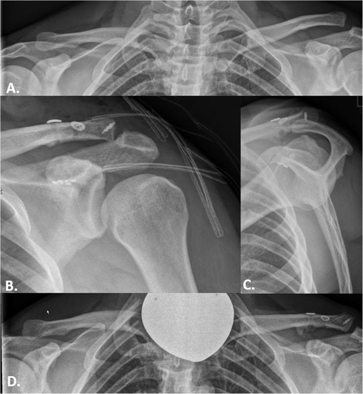
A series of conventional radiographs detailing the management of a Rockwood type IIIb acromioclavicular (AC) joint injury in a 42-year-old patient. Panel A is a preoperative bilateral panoramic view showing a significant superior displacement of the distal clavicle relative to the acromion on the left side, with an increased coracoclavicular (CC) distance. Panels B and C represent immediate postoperative anterior-posterior and Alexander views, respectively, demonstrating surgical reduction and fixation using coracoclavicular suspensory implants (buttons and high-strength sutures). The alignment of the acromion and distal clavicle is restored, and surgical hardware is visible superior to the clavicle and inferior to the coracoid process. Panel D is a 67-month follow-up panoramic radiograph showing stable maintenance of the AC joint reduction. Key features include subtle heterotopic ossification in the coracoclavicular ligament region and intact implants without evidence of migration or secondary dislocation. The comparison illustrates successful surgical stabilization and long-term osseous remodeling in orthopaedic trauma management.
This set of diagnostic X-ray radiographs demonstrates the preoperative and postoperative evaluation of an acromioclavicular (AC) joint injury in a 26-year-old male. Radiograph A (panoramic view) shows a subtle superior displacement of the distal clavicle, initially suggesting a Rockwood type II injury. Radiograph B (Alexander view/scapular lateral) reveals significant horizontal instability with the distal clavicle overriding the acromion posterior-superiorly, confirming a Rockwood type IV injury. Radiograph C (postoperative panoramic view) displays the surgical correction using two dog-bone fixation devices bridging the coracoid process and the clavicle to stabilize the joint. Radiograph D (postoperative Alexander view at 6 weeks) confirms the resolution of horizontal instability with anatomic restoration of the acromioclavicular alignment. This series highlights the clinical significance of specialized radiographic views, such as the Alexander view, in identifying horizontal instability that may be masked in standard AP or panoramic projections, ultimately guiding orthopedic surgical intervention.
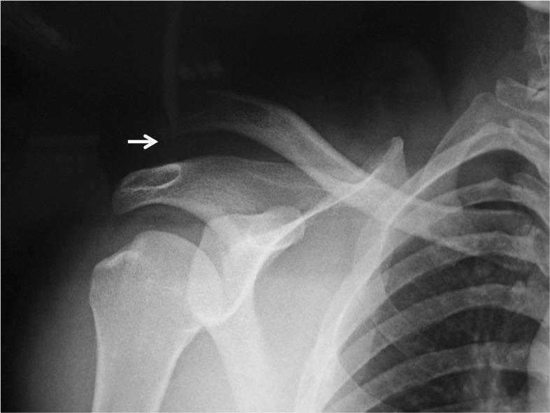
Anteroposterior (AP) radiograph of the right shoulder demonstrating a complete acromioclavicular (AC) joint separation, consistent with a Rockwood Type III injury. The image shows significant superior displacement of the distal clavicle relative to the acromion, with a white arrow highlighting the widened vertical distance between these two structures. This indicates a complete rupture of both the acromioclavicular and coracoclavicular (CC) ligaments. The glenohumeral joint is seen in a reduced state, with the humeral head properly aligned within the glenoid fossa, indicating successful reduction following a prior dislocation. Visible skeletal structures include the humerus, scapula (coracoid process, acromion, and glenoid), clavicle, and the superior ribs. No acute fractures are identified in the visualized field. This diagnostic image is used to educate on the radiographic findings of shoulder trauma, joint alignment, and the classification of AC joint injuries.
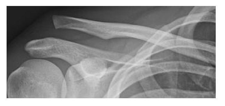
This diagnostic X-ray captures an anteroposterior (AP) view of the shoulder girdle, demonstrating a high-grade acromioclavicular (AC) joint dislocation, consistent with a Rockwood Type V injury. The primary pathological finding is a significant superior displacement of the distal clavicle relative to the acromion of the scapula. The normal alignment of the AC joint is completely disrupted, with the clavicle overriding the acromion and showing a markedly increased coracoclavicular distance, indicating complete rupture of both the acromioclavicular and coracoclavicular ligaments. Anatomical landmarks clearly visible include the clavicular shaft, the acromion process, the coracoid process, the glenoid fossa, and the proximal humerus (head and greater tuberosity). This image is essential for teaching orthopedic assessment of shoulder trauma, joint stability, and the radiographic classification of ligamentous injuries. It serves as a classic clinical example for medical students and residents studying musculoskeletal radiology and sports medicine.
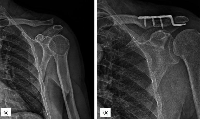
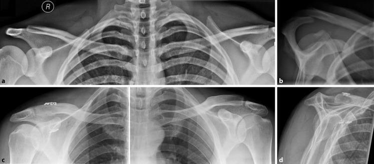
This composite educational resource consists of two anteroposterior (AP) shoulder radiographs demonstrating the management of a high-grade acromioclavicular (AC) joint injury. Image (a) shows a preoperative view of a Rockwood Type III AC joint dislocation. Key findings include superior displacement of the distal clavicle relative to the acromion, indicating a complete rupture of the acromioclavicular and coracoclavicular ligaments. The coracoclavicular distance is significantly widened, while the glenohumeral joint remains aligned. Image (b) shows the same shoulder six months postoperatively following open reduction and internal fixation (ORIF). An orthopedic hook plate is visible, with the plate body secured to the superior distal clavicle by three cortical screws and the hook portion positioned beneath the acromion process. This hardware successfully maintains vertical stability and joint reduction, evidenced by the restored alignment of the inferior borders of the distal clavicle and acromion. These images illustrate the radiological diagnosis and surgical stabilization of traumatic AC joint instability using a hook plate construct.
Acromioclavicular (AC) Joint Injury
1. Anatomy
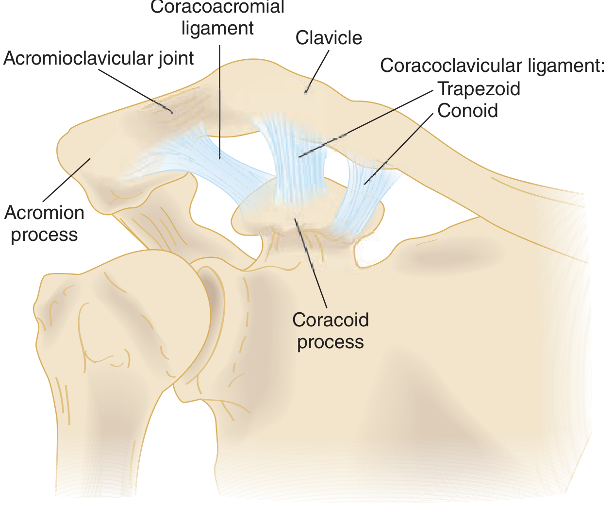
- Fibrocartilaginous intra-articular disc: Present in two forms - complete and partial (meniscoid). It undergoes degeneration by the fourth decade of life. - Rockwood & Green's, p.1113
- Joint inclination: The joint may be nearly vertical or inclined downward and medially, with the clavicle overriding the acromion by up to 50 degrees. Approximately 50% of joints have incongruent articular surfaces. - Rockwood & Green's, p.1113
- Nerve supply: Branches of the axillary, suprascapular, and anterolateral pectoral nerves supply the AC joint.
Stabilizing Ligaments
| Ligament | Role |
|---|---|
| AC ligaments (capsular) | Primary restraint to horizontal (anteroposterior) translation |
| Coracoclavicular (CC) ligaments | Primary restraint to vertical (superoinferior) translation |
- Trapezoid ligament (lateral) - resists axial compression and anterior-posterior translation
- Conoid ligament (medial) - resists superior displacement most strongly
2. Epidemiology and Mechanism of Injury
Mechanism
- Direct force (most common): A fall on the point of the shoulder with the arm adducted drives the acromion downward while the clavicle remains held up by the sternocleidomastoid, creating a shearing force across the joint.
- Indirect force (less common): A fall on the outstretched hand transmits force up the arm to the AC joint.
- The scapula and shoulder complex droop inferomedially while the clavicle appears to displace superiorly. - Rockwood & Green's, p.1113
3. Rockwood Classification (Tossy-Rockwood)
| Type | Ligament Injury | Radiographic Finding | Clinical Exam |
|---|---|---|---|
| I | AC ligaments sprained (intact). CC ligaments intact. Deltotrapezial fascia intact | Normal radiograph | Tenderness over AC joint only. No step-off |
| II | AC ligaments ruptured. CC ligaments sprained (stretched). Deltotrapezial fascia intact | Slight widening of AC joint; clavicle elevated 25-50% above acromion; may be slight widening of CC interspace | Mild step-off deformity; horizontal instability present; vertical stability preserved |
| III | AC ligaments ruptured. CC ligaments ruptured. Deltoid and trapezius partially detached | Clavicle elevated 100% above acromion; CC interspace widened 25-100% | Distal end of clavicle prominent; shoulder droops; the "piano key" sign may be present |
| IV | All supporting structures ruptured. Clavicle posteriorly displaced into or through the trapezius | May appear similar to II or III on AP view; axillary view required to visualize posterior displacement | Posterior displacement of clavicle; skin tenting may occur posteriorly |
| V | All supporting structures ruptured (more severe form of III). Deltotrapezial fascia completely stripped | CC interspace widened 200-300% compared to normal shoulder; clavicle almost subcutaneous | Gross deformity; severe pain; clavicle nearly subcutaneous |
| VI | AC and CC ligaments disrupted. Deltoid and trapezius disrupted | AC joint dislocated; clavicle displaced inferiorly (subcoracoid or subclavicular position) | Rare, high-energy injury. Severe swelling; multiple associated injuries; inferior clavicle displacement |
4. Clinical Features
History
- Fall on the point of the shoulder or FOOSH (fall on outstretched hand)
- Pain at the top of the shoulder, localized to the AC joint
- Difficulty lifting the arm overhead or across the body
Physical Examination
- Point tenderness directly over the AC joint
- Step-off deformity (prominence of the distal clavicle) in Types III-V
- Cross-arm adduction test: Arm elevated to 90 degrees, then adducted across the chest with elbow at 90 degrees. Positive test = pain specifically at AC joint. O'Brien et al. reported 88% sensitivity for AC joint abnormalities. - Rockwood & Green's, p.1095
- Piano key sign: In acute complete dislocations, the clavicle can be pushed down and springs back up when released
- Range of motion: May be limited depending on severity; a high painful arc (last 20-30° of elevation) is common. - Bailey & Love's, p.2252
- Cross-body adduction reproduces pain in AC joint arthritis
5. Imaging
Plain Radiographs
- AC-specific views require one-third to one-half the beam penetration of standard shoulder films. Always request "AC joint" radiographs, not "shoulder" radiographs, to avoid overpenetration. - Rockwood & Green's, p.1095
- Zanca view: 10-15 degree cephalic tilt AP projection - removes the spine of the scapula from the image; the preferred view for AC joint evaluation.
- Axillary lateral view: Mandatory when posterior dislocation (Type IV) is suspected.
- Bilateral comparison views: Allow direct comparison of CC distance.
Stress Radiographs
- Weighted stress views (10 kg) were historically used to differentiate Type III from Type V injury, but are no longer routinely recommended because:
- They have not been validated to correlate with intraoperative findings
- They do not reliably change injury grade or treatment decision
- They cause significant pain in the acute setting - Rockwood & Green's, p.1095
Normal Radiographic Measurements
- AC joint width: 1-3 mm (joint space)
- CC distance: 11-13 mm (conoid to trapezoid tuberosity)
-
25% side-to-side difference in CC distance = significant ligament injury
Advanced Imaging
- CT scan: For complex injuries, suspected fractures, or when plain films are inconclusive
- MRI: Useful for evaluating associated labral, rotator cuff, or soft tissue injuries
- Ultrasound: Can demonstrate joint contour and dynamic instability; less useful than CT for SC joint injuries
6. Associated Injuries
- Rotator cuff tears
- Distal clavicle fractures
- Coracoid process fractures
- Neurovascular injuries
- Pneumothorax or rib fractures in severe trauma
- Glenohumeral dislocation
7. Treatment
Non-Operative Management
- Acute (0-2 weeks): Rest, ice, analgesics, sling immobilization. A simple sling is the most convenient and effective initial treatment.
- Subacute (2-6 weeks): Gradual return to range-of-motion exercises starting at 7-14 days for Types I and II.
- Return to sport: As early as 10-15 days for low-grade (Type I/II) injuries in competitive athletes, though pain-free return may take longer.
- Local anesthetic injections at the AC joint are an accepted adjunct for competitive athletes (Australian Rules football and rugby data show mean of 5.7 games using injections). - Rockwood & Green's, p.1118
Operative Management
- Types IV, V, and VI (operative treatment is the standard of care)
- Type III failing 3-6 months of supervised rehabilitation
- Specific Type III subgroups: young overhead athletes, manual laborers, polytrauma patients
- Chronic symptomatic instability
Surgical Techniques
1. Hook Plate Fixation
- Currently the preferred acute surgical technique according to Rockwood & Green's
- Plate body secured to superior distal clavicle with cortical screws; hook portion passes beneath the acromion
- Supported by retrospective studies and biomechanical data
- Reliable for preventing loss of reduction in the acute setting
- Requires secondary surgery to remove the hook plate to avoid subacromial impingement - Rockwood & Green's, p.1124
2. Coracoclavicular Suspensory Fixation (Button/Loop Techniques)
- Uses high-strength suture or synthetic ligament (e.g., TightRope, Surgilig) through bone tunnels in the clavicle and around/through the coracoid process
- Advantages: minimally invasive; avoids need for hardware removal
- Disadvantages: risk of coracoid/clavicle fracture at tunnels; loss of reduction in 10-30% of cases
3. CC Ligament Reconstruction (Weaver-Dunn and Modifications)
- Involves transfer of the coracoacromial ligament to reconstruct the CC ligament
- Often combined with graft augmentation (e.g., gracilis or semitendinosus autograft) for chronic reconstructions
4. Historical Techniques (largely abandoned or used selectively)
- K-wire fixation across the AC joint: Significant risk of pin migration, breakage, and neurovascular injury - no longer recommended
- Bosworth screw: CC fixation with a large screw; risk of hardware failure and need for removal
- Phemister technique: Temporary Kirschner wire fixation
5. Arthroscopic-Assisted Reconstruction
- Gaining popularity for chronic AC joint instability
- Allows concurrent intra-articular procedures (labral repair, etc.)
- Techniques include arthroscopic-assisted coracoclavicular loop fixation
8. Complications
Post-Operative Complications - Rockwood & Green's, p.1141
| Complication | Notes |
|---|---|
| Loss of reduction | Most common; 10-30% incidence across literature. Complex loading through a small fixation area in high-grade injuries |
| Clavicle or coracoid fracture | Occurs at bone tunnels, particularly with button/loop techniques |
| Hardware failure | Pins, K-wires especially prone to migration; screw breakage |
| Hardware prominence/pain | Particularly with hook plates; requires removal surgery |
| Infection/osteomyelitis | Uncommon but serious |
| Graft resorption or rupture | With ligament reconstruction techniques |
| Posttraumatic AC arthritis | Even with successful reduction; may require distal clavicle excision |
| Cosmetic deformity | Late degenerative changes or residual bump |
Non-Operative Complications
- Posttraumatic osteolysis of the distal clavicle: Seen after repetitive stress or single acute injury
- Persistent horizontal instability: Particularly after Type II injuries with unrecognized posterior capsular disruption
- AC joint arthritis: Common long-term sequela requiring corticosteroid injection or distal clavicle excision
9. AC Joint Arthritis (Chronic/Late Presentation)
- Common in males aged 20-50 years
- Inferior osteophytes can impinge on the underlying rotator cuff
- Symptoms: pain worse with overhead activity and cross-body adduction; high painful arc (last 20-30°)
- Treatment: Intra-articular corticosteroid injection (diagnostic and therapeutic); surgical excision of the lateral 0.5-1 cm of the distal clavicle (Mumford procedure) - arthroscopic or open - gives reliable pain relief
10. Summary - Treatment Algorithm
AC Joint Injury
     │
     ├── Type I & II ──► Conservative (sling, analgesia, early ROM)
     │
     ├── Type III ──► Conservative first (3-6 months)
     │                    │
     │                    └── Failure / young overhead athlete ──► Surgery
     │
     ├── Type IV ──► Surgery (open reduction)
     │
     ├── Type V ──► Surgery (preferred: hook plate or CC reconstruction)
     │
     └── Type VI ──► Surgery (assess and treat associated injuries)
Key Teaching Points
- The AC joint is stabilized by two separate ligament systems: the AC ligaments (horizontal stability) and the CC ligaments (vertical stability). Each must be assessed independently.
- The Rockwood classification (Types I-VI) drives treatment decisions. The critical distinction is Type III vs Type V, determined by whether the deltotrapezial fascia is stripped.
- AC-specific radiographs are mandatory - standard shoulder technique overpenetrates and misses subtle fractures. The Zanca view (10-15° cephalic tilt) is preferred.
- Weighted stress views are no longer routinely recommended.
- Type III injuries remain controversial: current evidence supports a trial of nonoperative management for most patients.
- Types IV, V, and VI are surgical injuries. Hook plate fixation is currently the preferred acute technique per Rockwood & Green's.
- Loss of reduction (10-30%) is the most common surgical complication.
Radiographic Images - Rockwood Classification
Shoulder dislocation in details according to orthopaedics text books
shoulder dislocation anterior posterior glenohumeral Bankart Hill-Sachs
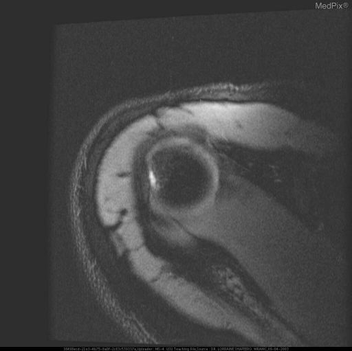
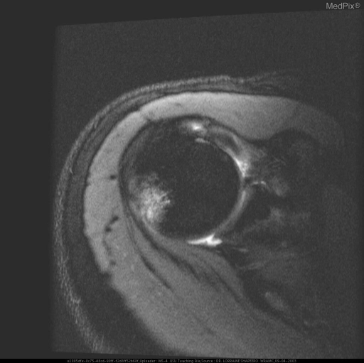
**Imaging Modality:** Magnetic Resonance Imaging (MRI), axial T1-weighted or proton density (PD) sequence. **Anatomical Region:** Right shoulder girdle at the level of the glenohumeral joint. **Observed Pathology:** Post-traumatic changes consistent with anterior shoulder instability. - **Hill-Sachs Deformity:** There is a visible cortical depression or "flattening" on the posterolateral aspect of the superior humeral head. - **Bankart Lesion:** Disruption or attenuation of the anterior-inferior glenoid labrum is present, consistent with an avulsion injury following an anterior glenohumeral dislocation. **Characteristic Visual Features:** - A focal loss of the normal circular contour of the posterior humeral head, appearing as an impaction fracture (Hill-Sachs lesion). - Signal abnormality at the anterior glenoid rim indicating labral detachment. - The glenohumeral relationship is currently reduced, but with evidence of chronic or subacute osseous and soft tissue sequelae. **Key Diagnostic Features:** The combination of a posterolateral humeral head impaction fracture and anterior-inferior labral injury serves as a classic diagnostic marker for recurrent anterior shoulder instability and prior dislocation events.
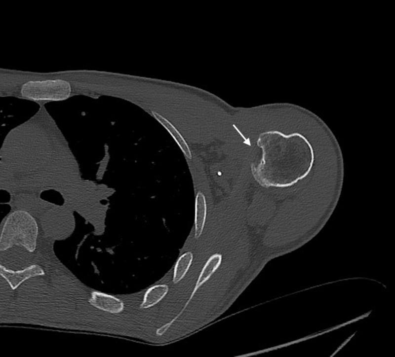
This diagnostic axial CT image of the shoulder demonstrates a posterior glenohumeral dislocation or subluxation in a 39-year-old patient. The humeral head is displaced posteroinferiorly relative to the glenoid fossa. A prominent impaction fracture is visible on the anteromedial aspect of the humeral head (indicated by the white arrow), characteristic of a reverse Hill-Sachs lesion. This type of bony defect typically occurs when the anterior humeral head impacts against the posterior glenoid rim during a posterior dislocation event. Additionally, small radiopaque hyperdensities are visible in the anterior periarticular soft tissues, representing metallic suture anchors from a prior McLaughlin-type remplissage surgical procedure. The posterior glenoid rim appears relatively well-preserved, without evidence of a significant bony Bankart lesion. This image serves as a clinical example of chronic posterior shoulder instability and the associated characteristic secondary bony changes often seen in patients with repetitive trauma or seizure-related dislocations.
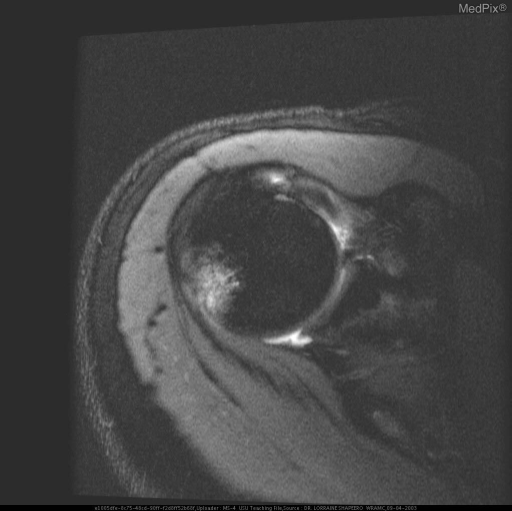
**Imaging Modality:** Magnetic Resonance Imaging (MRI), axial plane. **Anatomical Region:** Shoulder joint (glenohumeral joint). **Observed Pathology:** * **Hill-Sachs Lesion:** A prominent impaction fracture is visible on the posterolateral aspect of the humeral head. This is characterized by a cortical depression and subchondral marrow edema, appearing as an area of increased signal intensity on this fluid-sensitive sequence. * **Bankart Lesion:** Evidence of injury to the anteroinferior glenoid labrum. There is disruption of the labral-ligamentous complex, consistent with an avulsion of the labrum from the glenoid rim. * **Joint Effusion:** Increased intra-articular signal intensity indicates presence of joint fluid/effusion. **Characteristic Visual Features:** The "hatchet" deformity of the posterior humerus and the blunting/detachment of the anterior labrum are classic markers of shoulder instability. **Clinical Context:** These findings are pathognomonic for sequelae of recurrent anterior glenohumeral dislocation. **Diagnostic Differentiating Features:** The co-occurrence of a posterolateral humeral head defect (Hill-Sachs) and an anteroinferior labral tear (Bankart) confirms a mechanism of anterior instability, distinguishing it from posterior dislocation patterns.
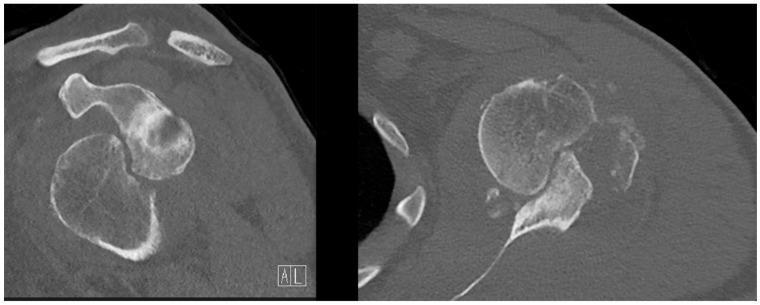
This diagnostic imaging set consists of two preoperative Computed Tomography (CT) scans of a human shoulder, presented in coronal (left) and transversal (right) views. The images demonstrate significant orthopedic pathology following an anterior glenohumeral dislocation. The transversal view clearly shows a large Hill-Sachs deformity, characterized by a substantial, irregular cortical depression or compression fracture on the posterolateral aspect of the humeral head. In the coronal view, a bony Bankart lesion is evident as a cortical discontinuity and bone loss at the anterior-inferior glenoid rim. These combined findings represent bipolar bone loss, which is clinically significant for determining shoulder stability and surgical planning. The CT highlights the morphological changes in the glenohumeral joint, providing essential detail for classifying the lesions as 'off-track' and assessing the need for procedures like a Latarjet or Remplissage.
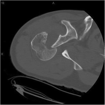
This diagnostic image is an axial computed tomography (CT) scan of the right shoulder, showing the glenohumeral joint. The image demonstrates a posterior shoulder dislocation, characterized by the humeral head being displaced posteriorly relative to the glenoid fossa. A significant impaction fracture is visible on the anteromedial aspect of the humeral head, representing a reverse Hill-Sachs lesion, which commonly occurs when the anterior humeral head strikes the posterior glenoid rim during a posterior dislocation event. The glenoid fossa itself appears to have an intact cortical outline without evidence of an associated bony Bankart lesion or significant fracture in this view. This scan is educationally significant for identifying the typical radiological findings of chronic or locked posterior shoulder instability and the secondary osseous defects resulting from mechanical impaction.
**Imaging Modality:** Magnetic Resonance Imaging (MRI), axial view. **Anatomical Region:** Glenohumeral joint (shoulder). **Observed Pathology:** The image demonstrates findings consistent with sequelae of anterior shoulder dislocation, specifically a bony Bankart lesion and an associated Hill-Sachs deformity. **Characteristic Visual Features:** * **Bankart Lesion:** Evidence of a fracture and fragmentation at the anterior-inferior aspect of the glenoid rim. There is associated disruption of the anterior labrum. * **Hill-Sachs Deformity:** A cortical depression and wedge-shaped defect are visible on the posterolateral aspect of the humeral head. * **Signal Intensity:** High T2-weighted/STIR signal intensity is noted within the humeral head and adjacent glenoid, indicating subchondral bone marrow edema or bruising (bone marrow contusion) at the impact sites. **Clinical Context:** These findings are pathognomonic for recurrent or acute anterior glenohumeral instability. The "kissing lesions" represent the forceful impact between the humeral head and the glenoid during the dislocation event. **Key Diagnostic Features:** Posterolateral humeral head compression fracture (Hill-Sachs) and anterior-inferior glenoid rim osseous avulsion (Bony Bankart).
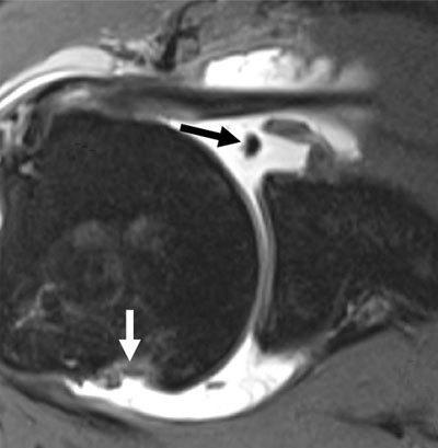
This diagnostic image is an axial MR arthrogram of the shoulder joint, highlighting the glenohumeral anatomy in a patient with chronic antero-inferior instability. The image demonstrates two hallmark findings of anterior shoulder dislocation. First, a soft-tissue Bankart lesion is visible at the antero-inferior aspect of the glenoid (indicated by a black arrow), characterized by the detachment of the fibrocartilaginous labrum from the glenoid rim with a concurrent tear of the glenoid periosteum. Second, a Hill-Sachs lesion is present (indicated by a white arrow) as an impaction fracture at the postero-superior aspect of the humeral head. Intra-articular contrast material fills the joint space, accentuating the discontinuity between the labrum and the glenoid bone. This image is a key educational resource for orthopedic radiology, illustrating the common sequelae of shoulder dislocation and the importance of identifying both soft tissue and bony defects that contribute to joint instability.
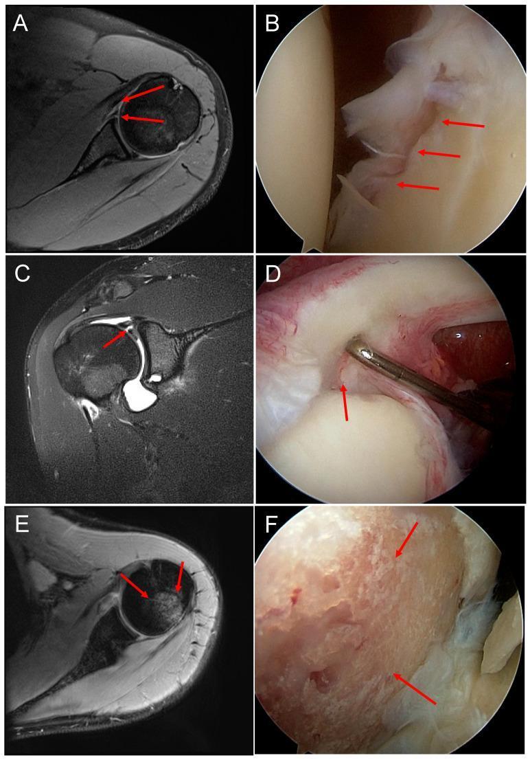
This composite educational image provides a side-by-side comparison of magnetic resonance (MR) and arthroscopic imaging for common shoulder pathologies resulting from anterior traumatic shoulder dislocation. (A, B) A Bankart lesion: the axial MR image shows an anterior-inferior labral tear, while the corresponding arthroscopic view shows direct visualization of the detached and frayed ventral labrum. (C, D) Superior Labrum Anterior to Posterior (SLAP) lesion: the MR arthrogram displays high signal intensity in the superior labrum, while the intraoperative image reveals a physical tear at the biceps tendon anchor point, being evaluated with a probe. (E, F) Hill-Sachs lesion: the axial MR image demonstrates a compression fracture of the posterolateral humeral head (bony defect), which is visualized arthroscopically as an area of eroded cartilage and subchondral bone with a coarse, uneven texture. These panels highlight the correlation between radiological findings and direct surgical visualization in orthopedic surgery, specifically focusing on glenohumeral instability and intra-articular damage.
Shoulder (Glenohumeral) Dislocation
1. Anatomy and Biomechanics of Glenohumeral Stability
Static Stabilizers
| Structure | Role |
|---|---|
| Glenoid labrum | Fibrocartilaginous bumper attached circumferentially around the glenoid. Deepens the socket and anchors the glenohumeral ligaments. Labral tears directly reduce the "concavity-compression" effect |
| Glenohumeral ligaments (GHL) | Superior GHL (SGHL) - resists inferior translation at 0° abduction; Middle GHL (MGHL) - resists anterior translation at 45°-90° abduction; Inferior GHL complex (IGHL) - hammock-like structure with anterior band, posterior band, and axillary pouch - the primary restraint at 90° abduction |
| Joint capsule & intra-articular vacuum | Negative pressure resists inferior translation. Loss of this vacuum (e.g., capsular venting) results in significant inferior translation |
| Articular version | Normal glenoid retroversion ~5°; humeral retroversion ~20-30° |
Dynamic Stabilizers
- Rotator cuff muscles (supraspinatus, infraspinatus, teres minor, subscapularis): Provide stability via three mechanisms: (1) concavity-compression - compressing the humeral head into the glenoid; (2) coordinated contraction to guide the head onto the glenoid center; (3) dynamization of the GHL through their attachments. A 50% reduction in rotator cuff forces increases anterior displacement by 46% and posterior displacement by 31%. - Rockwood & Green's, p.1272
- Long head of biceps: Contributes to anterior stability, particularly in the ABER position
- Scapulothoracic musculature: Positions the scapula to optimize glenohumeral stability; scapulothoracic dyskinesia is especially important in MDI
2. Epidemiology
- GH dislocation is the most common dislocation in the body - accounts for approximately 50% of all dislocations
- Incidence: ~17 per 100,000 person-years
- Anterior dislocations account for 95-97% of all GH dislocations
- Peak incidence in males aged 20-30 years (sports-related) and in the elderly (falls)
- Recurrence rate after first-time anterior dislocation approaches 80% in males under 20 years old - Rockwood & Green's, p.1262
3. Classification Systems
A. Matsen & Thomas Classification (TUBS / AMBRII) - Rockwood & Green's, p.1267
- Traumatic
- Unidirectional
- Bankart lesion present
- Surgery usually required for recurrent instability
- Atraumatic
- Multidirectional
- Bilateral laxity
- Rehabilitation is first-line treatment
- If surgery necessary, tighten the Inferior capsule and rotator Interval
B. Bailey & Love Classification - Bailey & Love's, p.2323
- Traumatic: Unidirectional, involuntary - surgery usually successful
- Atraumatic: Multidirectional, painful, involuntary - responds to surgery
- Habitual: Voluntary, ligament laxity, painless - surgery usually contraindicated
C. FEDS Classification (Kuhn) - Rockwood & Green's, p.1265
- Frequency (solitary / occasional / frequent)
- Etiology (traumatic / atraumatic)
- Direction (anterior / inferior / posterior)
- Severity (subluxation / dislocation)
D. OTA Classification
- 10-A1: Anterior dislocation
- 10-A2: Posterior dislocation
- 10-A5: Inferior dislocation (luxatio erecta)
E. Stanmore Triangle Classification - Rockwood & Green's, p.1267
- Polar Type 1: Traumatic, structural (acute / persistent / recurrent)
- Polar Type 2: Atraumatic, structural (recurrent)
- Polar Type 3: Muscle patterning, non-structural (no labral damage, abnormal EMG)
F. ABC Classification (Posterior Instability - Moroder & Scheibel)
- A: First-time (A1 = subluxation, A2 = locked dislocation)
- B: Dynamic recurrent (B1 = functional, B2 = structural with Bankart/bone loss)
- C: Chronic static (C1 = constitutional, C2 = acquired)
4. Anterior Shoulder Dislocation
Mechanism
- Arm raised in abduction + external rotation (throwing, tackling)
- Fall on outstretched hand (FOOSH) with arm abducted
Subtypes of Anterior Dislocation (by position of humeral head)
- Subcoracoid - most common; head lies inferior to coracoid process
- Subglenoid - head lies inferior to glenoid
- Subclavicular - rare; head lies medial to coracoid, under clavicle
- Intrathoracic - very rare; head displaced into chest between ribs
Clinical Features
- Severe shoulder pain and inability to use the arm
- Arm held in slight abduction and external rotation
- Shoulder has a "squared off" appearance - loss of normal rounded deltoid contour
- Humeral head palpable anteriorly below the coracoid
- Hollow beneath the acromion (empty glenoid)
- Patient resists adduction and internal rotation
- Cannot touch contralateral shoulder with the affected hand (Dugas test positive)
- Neurovascular examination is mandatory - axillary nerve is most commonly injured (test sensation over deltoid patch)
Associated Pathological Lesions
| Lesion | Description | Significance |
|---|---|---|
| Bankart lesion | Avulsion of the anterior-inferior glenoid labrum from the glenoid rim (with or without bone) | Present in ~80-90% of traumatic anterior dislocations. The primary lesion preventing healing and causing recurrence |
| Hill-Sachs lesion | Compression fracture of the posterolateral humeral head caused by impaction against the anterior glenoid rim during dislocation | Present in 47-80% of anterior dislocations; up to 93% in recurrent dislocations |
| Bony Bankart | Osseous avulsion of the anterior-inferior glenoid rim | Present in ~50% of recurrent cases; "inverted pear" glenoid when >25-27% width lost |
| ALPSA lesion | Anterior labroligamentous periosteal sleeve avulsion - labrum stripped medially off the glenoid with intact periosteum | Can heal in malposition, predisposing to recurrence |
| HAGL lesion | Humeral avulsion of the glenohumeral ligament | Occurs in ~9% of cases; must be identified as Bankart repair alone will fail |
| Rotator cuff tear | Most common in patients >40 years | Axillary nerve or suprascapular nerve injury may accompany |
| Greater tuberosity fracture | Occurs in ~15-35% of anterior dislocations in older patients | Often reduces with joint reduction |

5. Posterior Shoulder Dislocation
Key Points
- Accounts for only 2-4% of all GH dislocations but is the most commonly missed dislocation in clinical practice - Bailey & Love's, p.2414
- Often initially missed on an AP radiograph, which may appear deceptively near-normal
Mechanism
- Electric shock or epileptic seizure (massive simultaneous contraction of internal rotators)
- "Half-Nelson" position (forced internal rotation with arm behind the back)
- Direct anterior blow to the shoulder
- Heavy fall with arm across the body
Clinical Features
- Arm held in adduction and internal rotation (opposite of anterior dislocation)
- Shoulder appears flattened anteriorly, prominent posteriorly
- Loss of external rotation (a key diagnostic sign - patient cannot externally rotate past neutral)
- AP radiograph may appear near-normal to the unwary; requires high clinical suspicion
Radiographic Signs of Posterior Dislocation
- "Light bulb sign" (or "drum-stick sign"): On AP view, the humeral head appears symmetrically rounded due to fixed internal rotation, lacking the normal convex/biconcave shadow of the lateral head - Bailey & Love's, p.2460
- "Trough line sign": A vertical line of cortical compression (reverse Hill-Sachs / McLaughlin lesion) on the anteromedial humeral head, visible on AP view
- Widening of the joint space >6 mm
- Axillary or scapular Y view confirms the dislocation and is mandatory - Tintinalli's
Associated Lesions
- Reverse Hill-Sachs (McLaughlin) lesion: Impaction fracture of the anteromedial humeral head against the posterior glenoid rim
- Reverse Bankart lesion: Posterior labral tear
- If >20-40% of the articular surface is involved, the head locks on the posterior glenoid rim ("locked posterior dislocation")
6. Inferior Dislocation (Luxatio Erecta)
- Extremely rare; arm is locked in full abduction overhead (patient cannot bring arm down)
- Mechanism: hyperabduction forcing the humeral head inferiorly, levering the neck against the acromion
- Associated with high rates of neurovascular injury (brachial plexus, axillary artery)
- Classified as OTA 10-A5
- Treatment: Traction in the direction of the arm (upward), then adduction
7. Imaging Protocol
Plain Radiographs (Minimum Three Views)
- AP (True AP / Grashey view): Standard; 35-40° oblique to the plane of the scapula
- Axillary lateral view: Gold standard for confirming direction of dislocation; mandatory when posterior dislocation is suspected
- Scapular Y view ("Y view"): Useful when axillary view cannot be obtained
Advanced Imaging
- CT scan: Best for quantifying glenoid bone loss and Hill-Sachs lesion size; used for surgical planning. 3D CT reconstruction with en-face glenoid view allows measurement of bone defect using the "circle method"
- MRI / MR arthrogram: Best for soft tissue lesions - labral tears (Bankart, ALPSA, HAGL), IGHL damage, rotator cuff tears, cartilage lesions. MRI circle method accuracy is within 1.3% of 3D CT for glenoid bone loss calculation. - Rockwood & Green's, p.1263
Critical Glenoid Bone Loss - "The 25% Rule"
- Loss of >25-27% of the anterior-inferior glenoid width = inverted pear glenoid
- This amount of bone loss exceeds the critical threshold and predicts failure of soft-tissue repair (Bankart) alone
- Requires a bony augmentation procedure (Latarjet or bone block) - Rockwood & Green's, p.1263
8. Reduction Techniques - Anterior Dislocation
1. Stimson (Hanging-Weight) Method
- Patient prone, affected arm hanging off the table, 5-10 kg weight attached
- Gravity and muscle fatigue allow spontaneous reduction over 15-30 minutes
- Gentle and atraumatic; useful when patient cooperation is good
2. Cunningham (Muscle Massage) Technique
- Patient seated, examiner massages the deltoid, biceps, and trapezius while guiding the arm into adduction and external rotation
- No analgesia required in many cases; high success rate
3. External Rotation Method (Leidelmeyer / Eachempati)
- Patient supine, elbow flexed to 90°, arm slowly externally rotated in the plane of the body without traction
- Gentle; avoids forceful manipulation; success rate ~80-90%
4. Milch Technique
- Arm abducted overhead with simultaneous external rotation; thumb pressure on humeral head guides it back into the glenoid
- Can be done without sedation
5. Kocher Method (historical; now used cautiously)
- Elbow flexed 90°; traction then external rotation, adduction across chest, internal rotation
- Risk of humeral fracture with forceful application; no longer the first choice in most centres
- Contraindicated if fracture suspected
6. Hippocratic Method
- Operator places foot in axilla as counter-traction while applying longitudinal traction on the arm
- Risk of axillary nerve/vessel injury with the foot in the axilla; largely superseded
7. FARES (Fast, Reliable, Safe) Method
- Patient supine; longitudinal traction applied with oscillatory vertical movements as arm is progressively abducted to >90°; external rotation applied at approximately 90°
- High success rates without sedation in multiple studies
Posterior Dislocation Reduction
- Gentle traction on the adducted, internally rotated arm with anteriorly directed pressure on the posterior humeral head
- If locked (reverse Hill-Sachs engages the posterior glenoid), closed reduction under general anesthesia is required; locked lesions >20-40% articular involvement need open surgery - Bailey & Love's, p.2444
- Post-reduction: immobilize in external rotation brace to allow stretched posterior structures to heal
Post-Reduction Protocol
- Neurovascular check immediately after reduction
- Postreduction radiographs to confirm reduction and identify any new fractures
- Sling immobilization for comfort; early ROM exercises
- Physical therapy focused on rotator cuff strengthening
9. Natural History and Recurrence Risk
| Age Group | Recurrence Rate |
|---|---|
| <20 years | 66-88% |
| 20-30 years | ~55-67% at 5 years |
| >40 years | ~10-15% |
10. Surgical Treatment
Indications for Surgery
- Acute: First-time dislocation in young athletes with high recurrence risk (relative indication); locked or irreducible dislocations; associated displaced fracture; neurovascular injury
- Recurrent instability: After failed conservative management; most common indication
- Significant bone loss: Inverted pear glenoid (>25% loss) mandates bony procedure
A. Arthroscopic Bankart Repair (ABR) - Standard for Soft Tissue Instability
- Patient in lateral decubitus or beach chair position with arm in traction
- Posterior portal entry (2 cm below, 1 cm lateral to posterolateral acromion edge)
- Two anterior portals: anterolateral (near supraspinatus) and anteroinferior/5:30 portal (above subscapularis) for drilling and suture passing
- Diagnostic arthroscopy evaluates all intra-articular structures
- Bankart lesion elevated and mobilised; anterior glenoid rim prepared with burr
- Minimum 3 suture anchors (2.9-mm or 3-mm) placed along anterior glenoid rim
- Labral tape shuttled and loaded onto knotless PushLock anchors creating a "bumper" effect
- Associated posterior tears repaired via accessory posterolateral portal
B. Open Bankart Repair
- Beach chair position; deltopectoral approach
- Superior half-to-two-thirds subscapularis tenotomy (1 cm from bicipital groove)
- T-capsulotomy
- Bankart lesion elevated; anterior glenoid rim abraded
- Anchors placed on glenoid rim; capsule shifted and repaired with arm at 30° flexion and 30° external rotation
- Subscapularis tenotomy repaired
C. Latarjet Procedure - For Significant Glenoid Bone Loss
- Bony block: Coracoid transfer restores the anterior glenoid arc
- Sling effect: The conjoined tendon (coracobrachialis + short head biceps) acts as a dynamic sling across the anterior capsule in the ABER position
- Capsular reinforcement: The coracoacromial ligament is repaired to the anterior capsule
- Deltopectoral approach (incision over coracoid to axilla)
- Cephalic vein identified; retracted laterally with deltoid
- Pectoralis minor released from medial coracoid; coracoacromial ligament resected from acromion and preserved
- Coracoid osteotomy at base; inferior surface flattened
- Horizontal subscapularis split; anterior capsule exposed
- Coracoid fixed to anterior glenoid neck with two screws (5-6 mm medial to glenoid surface)
- Coracoacromial ligament repaired to capsule; subscapularis split closed
D. Remplissage Procedure - For "Engaging" Hill-Sachs Lesion
- Indication: Large Hill-Sachs lesion that "engages" the anterior glenoid rim in the ABER position (off-track lesion), performed in conjunction with ABR
- Capsulotenodesis of the infraspinatus tendon into the Hill-Sachs defect using suture anchors
- Fills the defect, preventing engagement
- Results in slight loss of external rotation (~10°) - Rockwood & Green's, p.1281
E. Posterior Labral Repair (for Recurrent Posterior Instability)
- Repair of posterior Bankart lesion with anchor fixation
- Posterior capsular tightening/plication
- McLaughlin procedure or modified McLaughlin (subscapularis transfer into reverse Hill-Sachs lesion) for locked posterior dislocation with <40% articular involvement
- Humeroplasty or humeral head arthroplasty when >40% articular surface involved
F. Inferior Capsular Shift (Neer & Foster) - for MDI
- Open procedure addressing the patulous inferior capsule
- Can also be performed arthroscopically (capsular plication + rotator interval closure)
11. Complications
Early
- Axillary nerve injury: Most common nerve injury (~9-18%); transient neuropraxia is common; test sensation over deltoid patch
- Axillary artery injury: Rare (<1%); more common in elderly; presents with expanding hematoma or absent radial pulse
- Brachial plexus injury: Traction injury; most recover spontaneously
- Rotator cuff tear: Especially in patients >40 years; incidence increases with age (up to 30-35% in patients >40 years)
- Greater tuberosity fracture: ~15% of anterior dislocations
- Recurrence: Most common complication overall, especially in young males
Late
- Recurrent instability: Natural history; surgical stabilization required
- Glenohumeral arthritis: Hovelius found ~2/3 of patients had arthritis at mean 25-year follow-up - Rockwood & Green's, p.1262
- Distal clavicle/glenoid osteolysis: Posttraumatic
- Bankart repair failure: Loss of reduction; often related to bone loss not addressed at initial surgery
- Stiffness: Particularly after overly tight capsular repair; loss of external rotation
Surgical Complications
- Loss of reduction (10-30%)
- Anchor pullout or anchor-related complications
- Coracoid nonunion (Latarjet)
- Coracoid screw complications: Neurovascular proximity (musculocutaneous nerve at risk during Latarjet)
- Subscapularis failure after open procedures
12. Apprehension Test (Key Clinical Examination Finding)
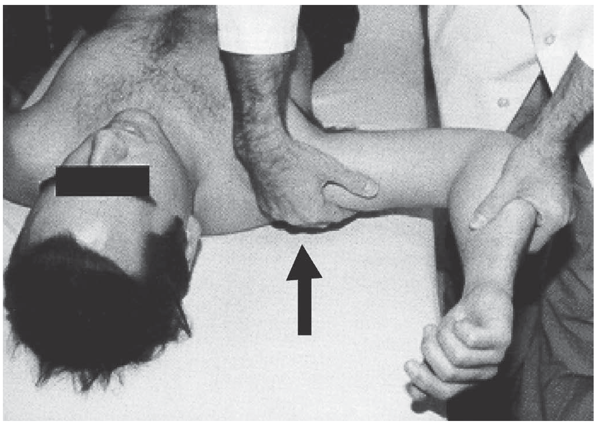
- Sulcus sign: Inferior traction on the humerus creates a sulcus under the acromion (inferior laxity/MDI)
- Load-and-shift test: Translates the humeral head anteriorly/posteriorly while in the glenoid; grades translation
- O'Brien's active compression test: 88% sensitivity for AC joint and labral pathology
13. Atraumatic Instability (MDI)
- No history of an initiating injury
- Multidirectional - instability in >1 direction
- Associated with generalised ligament laxity (Beighton score)
- Sulcus sign positive; apprehension tests positive in multiple directions - Bailey & Love's, p.2484
- Initial treatment: Supervised rehabilitation - rotator cuff and scapular stabiliser strengthening, proprioception training. Most patients respond without surgery.
- Surgery (inferior capsular shift / arthroscopic plication) reserved for failures after a dedicated 6-month rehabilitation program
14. Summary Treatment Algorithm
SHOULDER DISLOCATION
│
├── ACUTE DISLOCATION
│    ├── Pre-reduction X-ray (rule out fracture)
│    ├── Closed reduction (method depends on clinical factors)
│    ├── Post-reduction X-ray
│    ├── Neurovascular check
│    └── Immobilize → supervised rehabilitation
│
├── RECURRENT ANTERIOR INSTABILITY
│    ├── No significant bone loss (<25%) ──► Arthroscopic Bankart Repair
│    │                                       ± Remplissage (if engaging Hill-Sachs)
│    └── Significant bone loss (>25%) ──────► Latarjet Procedure
│         or failed prior repair
│
├── RECURRENT POSTERIOR INSTABILITY
│    ├── Conservative (rehab) first
│    └── Structural lesion / failure ──────► Posterior Bankart Repair
│         Locked posterior (McLaughlin) ──► McLaughlin / Open Reduction
│
└── MDI (MULTIDIRECTIONAL)
     ├── Rehabilitation (6 months) first
     └── Failure ──────────────────────────► Inferior Capsular Shift / Plication
Key Teaching Points
- The GH joint sacrifices bony constraint for mobility. Only 25-30% of the humeral head contacts the glenoid at any time; stability depends primarily on the IGHL complex and rotator cuff.
- Anterior dislocation accounts for 95-97% of cases; the arm is held in abduction and external rotation.
- Posterior dislocation is easily missed - key clue is the history (seizure, electric shock, half-Nelson). The "light bulb sign" and loss of external rotation are diagnostic hints.
- The Bankart lesion (anteroinferior labral avulsion) is the primary pathological lesion in traumatic anterior instability. A Hill-Sachs lesion (posterolateral humeral head impaction) accompanies it in up to 80%.
- Recurrence risk is highest in young males in contact sports (~80%). Age is the single biggest predictor.
- Critical glenoid bone loss (>25%) requires a bony procedure (Latarjet). Soft-tissue repair alone will fail.
- The Latarjet procedure provides a triple blocking mechanism: bone, sling effect (conjoined tendon), and capsular reinforcement.
- The remplissage is added for "engaging" (off-track) Hill-Sachs lesions to prevent engagement with the glenoid rim.