
I have my university exam of 1 st year of subject physiology These are the important questions give me answer of each question "Plasma proteins. " Follow University exam pattern Use easy language
Reading File
Now let me search the medical library for plasma proteins content:
Finding Sources
Finding Sources
Good. Let me now read the most relevant physiology textbook sections in parallel:
Reading File
Reading File
Reading File
Now let me get more detailed content on albumin, globulins, fibrinogen, functions and electrophoresis:
Finding Sources
Finding Sources
Reading File
Reading File
I now have comprehensive information from the textbooks. Let me also fetch the electrophoresis diagram image:
Loading Image
I now have all the information needed to write a comprehensive university exam answer. Here is the full answer:
PLASMA PROTEINS
(University Exam Answer - 1st Year Physiology)
Definition
Plasma proteins are the proteins present in the fluid part of the blood (plasma). They are large molecules that normally stay inside the blood vessels and cannot freely pass through capillary walls.
- Total plasma protein concentration: 7 to 7.5 g/dL
- Plasma proteins remain in the blood and help maintain its normal composition.
Types of Plasma Proteins (Classification)
Plasma proteins are divided into 3 main fractions - seen clearly on electrophoresis:
| Fraction | Normal Level | Examples |
|---|---|---|
| Albumin | 3.5 - 5.0 g/dL | Albumin |
| Globulins | 2.3 - 3.5 g/dL | α1, α2, β, γ globulins |
| Fibrinogen | 0.2 - 0.4 g/dL | Fibrinogen |
A/G Ratio (Albumin : Globulin ratio) = normally 1.2 : 1 to 1.5 : 1
Site of Synthesis (Origin)
This is a very common exam question!
- Liver synthesizes: Albumin, Fibrinogen, α and β globulins, clotting factors, complement proteins (70-80% of all plasma proteins are made in the liver)
- Lymphocytes / Lymphoid tissue synthesizes: γ-globulins (immunoglobulins / antibodies)
- Vascular endothelium: von Willebrand factor
Simple rule to remember: "Liver makes all except γ-globulins"
Functions of Plasma Proteins
1. Maintenance of Oncotic Pressure (Most Important Function)
- Plasma proteins, mainly albumin, create an osmotic pressure of about 25 mm Hg across the capillary wall
- This is called colloid osmotic pressure or oncotic pressure
- It pulls water back from tissues into the blood
- This prevents fluid from leaking out of blood vessels (prevents edema)
2. Transport / Carrier Function
- Many substances are transported in the blood attached to plasma proteins:
- Albumin carries: fatty acids, bilirubin, drugs, calcium, hormones
- Transferrin (β-globulin) carries: iron
- Ceruloplasmin carries: copper
- Haptoglobin carries: hemoglobin
- Thyroxine-binding globulin (TBG) carries: thyroid hormones
- Transcortin (CBG) carries: cortisol
3. Buffering Function
- Plasma proteins contribute about 15% of the total blood buffering capacity
- They act as buffers because of their free -COOH (acidic) and -NH₂ (basic) groups
- At normal blood pH 7.4, proteins are mostly in the anionic (negatively charged) form
4. Defense / Immunity
- Gamma globulins (Immunoglobulins) - IgG, IgM, IgA, IgE, IgD - are antibodies that protect the body against infection
- Complement proteins help in immune reactions
5. Blood Clotting (Coagulation)
- Fibrinogen is converted to fibrin during clotting, which forms the clot structure
- All clotting factors (except factor VIII) are synthesized in the liver
- Clotting factors are proteins (globulin fractions)
6. Reserve Amino Acid Source
- When body tissues become depleted of proteins, plasma proteins can be broken down and used as a source of amino acids
- Tissue macrophages can take in plasma proteins by pinocytosis, break them down, and release amino acids for tissue use
7. Viscosity of Blood
- Plasma proteins help maintain normal blood viscosity
- They also influence erythrocyte sedimentation rate (ESR)
Plasma Protein Electrophoresis
Electrophoresis is a technique used to separate plasma proteins based on their size and electrical charge using an electric field.
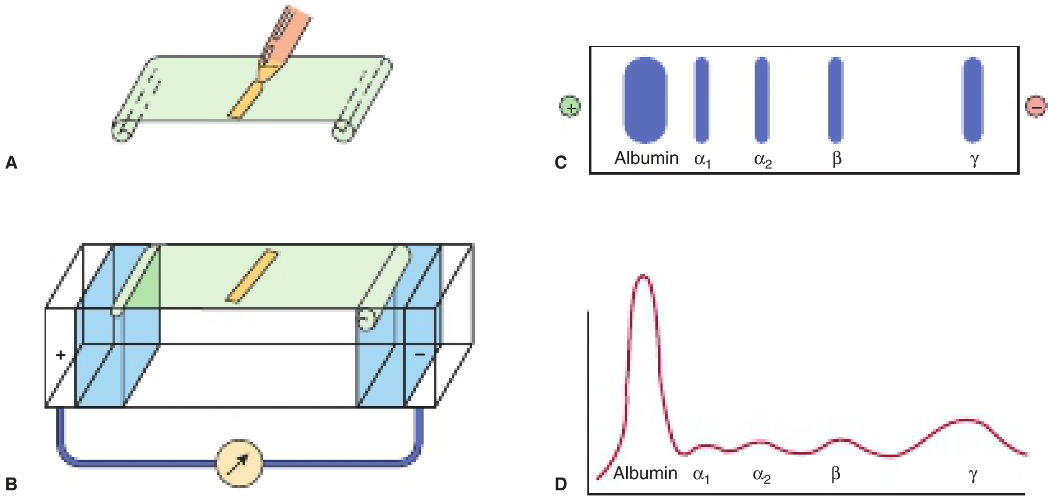
Steps:
- Serum is applied to cellulose acetate strip
- Electric current is passed through buffer
- Proteins migrate toward the positive pole (anode) at different speeds
- Staining shows separate bands
- Densitometer scanning shows relative amounts
Order from anode (+) to cathode (-):
Albumin → α1 → α2 → β → γ (slowest)
Albumin migrates the fastest (largest peak) because it is the most abundant and moves fastest toward the positive pole.
Hypoproteinemia (Decreased Plasma Proteins)
Definition: Decreased plasma protein levels
Causes:
- Decreased synthesis:
- Liver disease (cirrhosis, hepatitis) - liver cannot make enough proteins
- Malnutrition / starvation - not enough amino acids
- Increased loss:
- Nephrotic syndrome - albumin lost in urine
- Burns - protein lost from burned skin
- Malabsorption syndromes - intestinal disease
Effect: Decreased oncotic pressure → water leaks into tissues → EDEMA
Acute Phase Proteins
- In inflammation or injury, the liver produces more of certain proteins called acute phase proteins (positive acute phase reactants)
- Examples: C-reactive protein (CRP), fibrinogen, alpha-1 antitrypsin, haptoglobin, ceruloplasmin
- Albumin is a negative acute phase reactant - its level decreases during inflammation
Important Individual Proteins - Quick Summary
| Protein | Main Function | Clinical Note |
|---|---|---|
| Albumin | Oncotic pressure, transport | Decreased in liver disease, nephrotic syndrome |
| Fibrinogen | Blood clotting | Converted to fibrin; absent in afibrinogenemia |
| Immunoglobulins (γ-globulins) | Antibodies, immunity | Increased in infections/myeloma |
| Transferrin | Iron transport | Decreased in iron deficiency anemia |
| Ceruloplasmin | Copper transport | Decreased in Wilson's disease |
| Haptoglobin | Binds free Hb | Decreased in hemolytic anemia |
| Alpha-1 antitrypsin | Protease inhibitor | Deficiency causes lung and liver disease |
| C-reactive protein (CRP) | Acute phase protein | Marker of inflammation |
Key Points to Remember for Exam
- Total plasma proteins = 7 - 7.5 g/dL
- Albumin is the most abundant plasma protein (3.5-5 g/dL)
- Liver is the main site of synthesis (makes 70-80%)
- γ-globulins are made by lymphocytes (not liver)
- Oncotic pressure = 25 mm Hg (mainly due to albumin)
- Decreased protein → decreased oncotic pressure → edema
- Electrophoresis order: Albumin, α1, α2, β, γ
- A/G ratio = normally 1.2-1.5 : 1 (reversal seen in liver disease, myeloma)
Sources: Ganong's Review of Medical Physiology (26th Ed.), Guyton & Hall Textbook of Medical Physiology, Harper's Illustrated Biochemistry (32nd Ed.), Medical Physiology (Boron & Boulpaep)
This is a shared conversation. Sign in to Orris to start your own chat.