
What to expect after septoplasty
septoplasty recovery what to expect postoperative care
septoplasty nasal septum surgery procedure
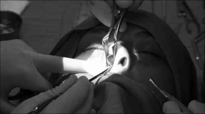
This monochrome clinical photograph captures an intraoperative view of a nasal septoplasty or rhinoplasty procedure. The surgical field focuses on the endonasal cavity under bright focal illumination. A Killian or Cottle nasal speculum is being used to retract the nasal alae and provide visualization of the internal nasal structures. A secondary surgical instrument, likely a periosteal elevator or suction-lifter, is positioned to manipulate the nasal septum. The image illustrates the resection of an anterior deviated portion of the septal cartilage while maintaining the structural integrity of the supratip and nasal dorsum regions to prevent postoperative saddle nose deformity. The surrounding area shows sterile draping and gloved hands, characteristic of an Otolaryngology (ENT) operative setting. This visual serves as an educational tool for understanding conservative septal resection techniques and the preservation of dorsal support during reconstructive nasal surgery.
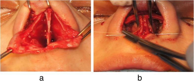
Two-panel clinical photograph illustrating intraoperative steps of a rhinoplasty or septoplasty procedure focusing on the nasal tip and septum. Panel A shows an inferior view of the nasal base with detachment of the membranous septum. This surgical exposure reveals the cartilaginous septum positioned between the medial crura of the lower lateral cartilages. Retractors are used to maintain the surgical field, showing raw, vascularized tissue. Panel B demonstrates the reconstruction phase, where suturing is performed to approximate the medial crura to the caudal septum. A surgical needle holder is visible, placing white suture material to stabilize the nasal tip. The procedure illustrates the anatomical relationship between the septal cartilage and the medial crural footplates, highlighting techniques used to modify nasal tip projection and rotation. This image serves as an educational resource for plastic surgery and otolaryngology residents to understand the 'tongue-in-groove' technique or similar septocolumellar stabilization methods.
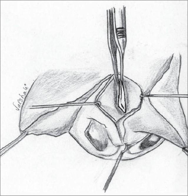
Anatomical diagram and surgical illustration detailing a key step in primary nasal correction for cleft lip or septoplasty. The drawing depicts the surgical field of the nasal septum from an anterior view. It illustrates the septal cartilage being meticulously dissected and freed from the surrounding mucoperichondrium on both sides. Additional separation is shown at the junctions with the vomer and the perpendicular plate of the ethmoid. A surgical scalpel is positioned centrally to perform precise dissection or scoring of the cartilage. Retraction sutures are visible on the lateral edges of the surgical site to maintain exposure of the deep structures. This procedure is clinically relevant for addressing septal deviation and repositioning the nasal midline during reconstructive surgery, such as the Millard procedure for unilateral cleft lip. The illustration serves as an educational tool for plastic surgery and otolaryngology trainees to understand the planes of dissection required for nasal septal mobilization.
After Septoplasty: What to Expect
Day of Surgery
- Septoplasty is an outpatient procedure - you go home the same day once anesthesia has worn off.
- The procedure is done entirely inside the nose, so there is no change to the external appearance.
- Internal splints or nasal packing may be placed to support the septum during early healing.
Immediate Post-Op (First 24-48 Hours)
- Nasal congestion - significant stuffiness is normal and expected; the nose will feel blocked.
- Mild bleeding/oozing - a nasal drip pad is applied. Some bloody discharge is normal. Bleeding typically tapers in the first 24 hours. As noted in Cummings Otolaryngology, "mild postoperative oozing is common and not necessarily a complication during the early phase but rather something to be expected."
- Nausea - swallowing blood during surgery makes nausea very common in nasal procedures.
- Head cold symptoms - increased mucus drainage, mild headache, throat irritation.
- Soreness inside the nostrils, at the tip of the nose, or in the upper teeth.
- Fatigue is normal.
- Sleep with your head elevated on 2+ pillows (or a recliner) for at least 3 days to reduce swelling.
First Week
- Splint/packing removal: Internal splints are usually removed around day 5-7. Expect immediate relief from congestion after removal.
- Pain: Usually mild. Prescription pain medication is given; avoid aspirin and ibuprofen for at least 2 weeks as they thin the blood.
- Nasal saline rinses: Start as early as the day after surgery. Saline irrigation (e.g., NeilMed, Simply Saline) is strongly recommended to clear crusts and surgical debris and keep mucosa moist.
- No nose blowing: Avoid for at least 1 week. If you need to sneeze, keep your mouth open.
- No hot or steamy showers for several days.
- Antibiotics: Often prescribed while packing is in place.
- Most patients return to desk work or school within 2 days.
Weeks 2-4
- Activity restrictions: No heavy lifting, bending, straining, or stooping for at least 2 weeks - these raise blood pressure and can trigger bleeding.
- Light exercise can resume around 10 days post-op.
- Strenuous exercise/contact sports: wait 4+ weeks.
- Swelling gradually reduces. The initial significant swelling typically subsides within 2-3 weeks.
- Continued rhinorrhea (runny nose) may persist for a few months - this is normal.
Months 1-3 (Full Recovery)
- Full benefits of improved breathing are typically felt within 3 months, as internal healing completes.
- The nose "settles" fully around the 3-month mark.
- Some patients take up to 6 months for complete resolution of all symptoms.
Possible Complications to Watch For
| Complication | Notes |
|---|---|
| Persistent obstruction | Most common; often from inadequately addressed posterior septal spurs |
| Septal hematoma | Blood pooling between cartilage and mucosa; needs urgent drainage |
| Septal perforation | Hole in the septum from unrepaired mucosal tears |
| Shape changes | Tip drooping or dorsal saddle deformity from over-resection |
| Synechiae (adhesions) | Scar tissue bridging septum and turbinate |
| Infection | Rare |
When to Call Your Surgeon Immediately
- Heavy, uncontrolled bleeding
- Fever above 38.5°C (101.3°F)
- Severe pain not controlled by medication
- Swelling of the nose becoming tense (may indicate hematoma)
- Visible deformity or collapse of nasal bridge
Key "Dos and Don'ts" Summary
| Do | Don't |
|---|---|
| Use saline rinses daily | Blow your nose for the first week |
| Elevate head when sleeping | Take aspirin or ibuprofen |
| Take prescribed antibiotics and pain meds | Do heavy lifting or strenuous exercise for 2+ weeks |
| Sneeze with mouth open | Pick at or disturb the nose |
| Attend all follow-up appointments | Smoke (delays healing significantly) |
- Cummings Otolaryngology Head and Neck Surgery, Complications of Septoplasty section
- Scott-Brown's Otorhinolaryngology Head & Neck Surgery Vol 1, Septoplasty chapter
- American Society of Plastic Surgeons - Septoplasty Recovery
I had septoplasty 5 days ago And now I have congestion sometime in left and sometimes in right nostril and there is discharge in my nose that I can't blow out and thel smell is also bad
septoplasty day 5 bad smell discharge congestion infection hematoma warning signs
⚠️ Contact Your Surgeon Today
What's Likely Happening
- Alternating left/right congestion is actually very normal - it's called the nasal cycle, and the post-op swelling makes it more noticeable.
- Thick discharge and crusting inside the nose at day 5 is expected as the healing tissue weeps and dries.
- You cannot blow it out because you're not supposed to blow your nose yet - that's correct.
- Early infection - bacteria colonizing the healing wound or nasal packing area
- Infected crusting - dried blood and mucus getting infected
- Septal hematoma (less likely at day 5 but possible) - pooled blood getting infected
Check Yourself Right Now
| Symptom | If YES, call urgently |
|---|---|
| Fever above 38°C / 100.4°F | Yes - call now |
| Pain getting worse, not better | Yes - call now |
| Nose feels more swollen/tense than day 1-2 | Yes - call now |
| Yellow-green thick discharge | Yes - call today |
| Just the bad smell, otherwise improving | Call today - non-urgent but same day |
What To Do Right Now
-
Call your surgeon's office today - even if it's just to describe the symptoms over the phone. They may want to see you, or they may prescribe antibiotics if you're not already on them.
-
Do NOT try to blow the discharge out - this risks bleeding and disrupting healing tissue.
-
Do saline rinses if you were given them - gently irrigate with saline to loosen crusting. This is the safe way to clear discharge at this stage. Lean forward, let it drain passively.
-
Do not use cotton swabs inside the nose - only gentle external dabbing with tissue.
-
Do not panic - infection after septoplasty is uncommon (~3%) and when caught early, it responds well to antibiotics. But "early" is the key word.
Bottom Line
The smell is not foul smelling but it's decreasing my smell and more noticable while expire
- A smell you notice when you breathe out (not a foul infection odor)
- Reduced sense of smell (hyposmia)
septoplasty reduced smell sense hyposmia day 5 crusting discharge smell when exhaling normal recovery
What You're Experiencing - and Why
1. The smell you notice when breathing OUT
- Dried blood and mucus crusting inside the nose
- Normal healing tissue and secretions
- Possibly the material from splints or sutures
2. Reduced sense of smell (hyposmia)
3. Alternating congestion left/right
What Helps Right Now
- Use a NeilMed squeeze bottle or similar nasal rinse twice daily
- This loosens and flushes out the crusting and dried discharge you can't blow out
- It will reduce the exhale smell noticeably within a day or two of regular use
- Lean forward and let it drain passively - do not sniff it back or blow forcefully
Reassurance Summary
| Your symptom | Meaning |
|---|---|
| Smell noticeable on exhale | Normal - crusting/discharge in the healing nose |
| Reduced sense of smell | Normal - physically blocked by swelling, resolves in 1-2 months |
| Alternating congestion | Normal nasal cycle, exaggerated by post-op swelling |
| Discharge you can't blow out | Normal - don't blow, use saline rinse instead |
But what's the benefit of septoplasty if still I have nose blockage
You Haven't Seen the Benefit Yet - And That's Normal
How blocked you feel:
Day 0 (surgery) ──► Days 2-5 ──────► Week 2 ──► Month 1 ──► Month 3
  Blocked          MOST BLOCKED      Better    Much better  Full benefit
                   (you are HERE)
What Septoplasty Actually Fixed
- Noticeably easier breathing through both nostrils
- Better sleep quality and reduced snoring
- Fewer sinus infections (drainage pathways open properly)
- Reduced headaches from sinus pressure
- Better sense of smell (air can reach receptors properly)
When Will You Actually Feel It?
| Timeframe | What changes |
|---|---|
| Week 1-2 | Splints out, swelling starts dropping, first glimpse of improvement |
| Week 3-4 | Noticeably easier to breathe, congestion much reduced |
| Month 1-2 | Majority of benefit felt, smell returns |
| Month 3-6 | Full healing, maximum benefit |
One Important Caveat
- The blockage also involved swollen turbinates (which may need separate treatment)
- There was additional valve narrowing not addressed at surgery
- The septum shifts slightly during healing