
Post laryngectomy stomal stenosis causes and management in detail in simplified manner and foreign bodi in bronchus in detailed simplified manner according to standard ENT textbook
foreign body bronchus chest X-ray hyperinflation
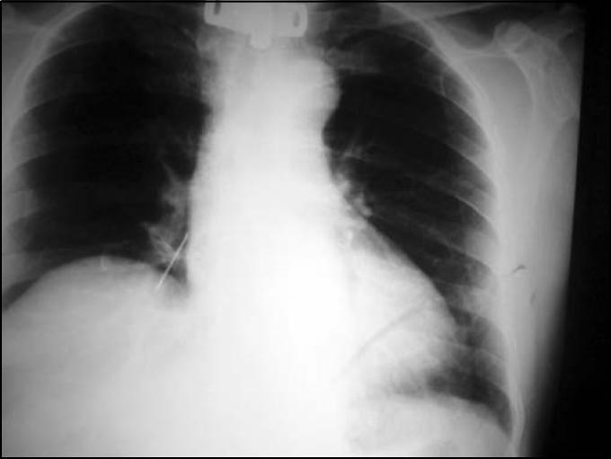
This diagnostic image is a posteroanterior (PA) chest X-ray demonstrating a radiopaque foreign body aspiration. The primary finding is a linear, high-density metallic object located in the right lower lung field, consistent with the anatomical position of the right inferior lobar or segmental bronchus. The surrounding lung parenchyma appears largely clear, with no immediate evidence of distal atelectasis, obstructive hyperinflation, or consolidation in this projection. The cardiac silhouette and mediastinum are within normal limits of size and position. A tracheostomy cannula or related medical hardware is partially visible at the top of the image in the midline tracheal position. The bony structures, including the ribs and clavicles, appear intact. This radiograph serves as a clinical example of foreign body localization in the tracheobronchial tree, highlighting the use of plain film radiography in identifying aspirated metallic materials and assessing for secondary pulmonary complications like pneumonia or collapse.
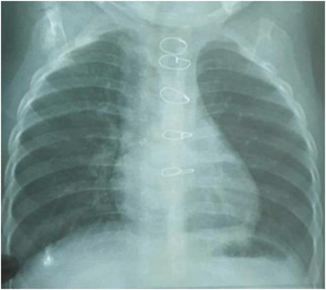
Anterior-posterior (AP) pediatric chest radiograph demonstrating post-surgical changes in an infant. The image reveals multiple radiopaque cerclage wires (sternal wires) along the midline, indicative of a previous sternotomy for cardiac repair, such as for Tetralogy of Fallot. The lung fields appear relatively clear without overt focal consolidation, pleural effusion, or pneumothorax visible at this resolution. The cardiac silhouette is visible, though its assessment is limited by the AP projection and pediatric anatomy. The trachea is midline. Notably, while the image was taken in the context of suspected foreign body aspiration (sunflower seed in the right main bronchus), the object itself is not radiopaque and is not clearly visualized on this X-ray, highlighting the clinical educational point that organic foreign bodies are often radiolucent and may not be detected through standard radiography without secondary signs like localized hyperinflation or atelectasis.
laryngectomy stoma tracheostomy
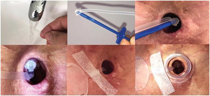
This clinical photographic series illustrates a six-step procedural sequence for the insertion and securing of a tracheostomy plug, commonly used in post-laryngectomy or tracheostomy care. The first frame shows the preparation phase, involving the rinsing or lubrication of a flexible silicone plug under a water stream. The second frame depicts the plug attached to a blue, ergonomic insertion tool (mandrel). The third and fourth frames show the clinical application: the insertion of the plug into a well-healed tracheostomy stoma. The fifth frame demonstrates the securement phase, where a transparent safety strap is taped to the peristomal skin using medical-grade adhesive tape. The final frame shows the completed assembly, with the plug fully seated and an adhesive baseplate or housing applied over the stoma site to facilitate the attachment of additional heat and moisture exchangers (HME) or speaking valves. This series serves as an educational guide for patient self-care or clinician training in stoma management and the prevention of stoma stenosis.
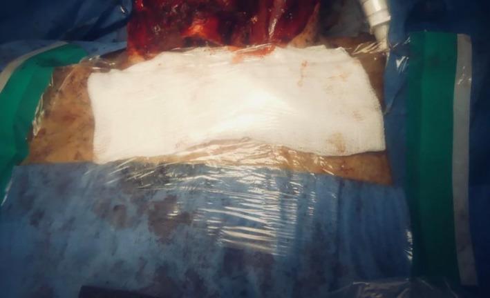
A clinical photograph depicting a post-operative surgical field following a tracheostomy procedure. The central focus is a closed airway system where a white gauze pad is placed over the stoma site, secured and sealed with a large Tegaderm transparent film dressing to minimize aerosolization. A J-shaped Rusch endotracheal tube is visible, extending into the stoma to maintain ventilation. The surgical site is surrounded by sterile blue and green surgical drapes. Superior to the dressing, the operative field shows exposed tissue with visible blood and skin markings, consistent with a laryngectomy or major neck surgery. The image illustrates infection control protocols, specifically the use of physical barriers and transparent dressings to maintain a closed circuit and prevent the spread of droplets or fomites during high-risk airway maneuvers in an operating room environment.
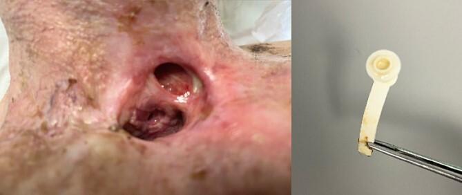
This composite clinical image illustrates complications following a total laryngectomy, specifically a pharyngocutaneous fistula and a degraded tracheoesophageal prosthesis (TEP). The left image is a clinical photograph of the anterior neck, showing a permanent tracheostomy stoma. Superior to the primary stoma, there is a second, smaller opening consistent with a pharyngocutaneous fistula. The surrounding peristomal skin is erythematous, friable, and exhibits signs of chronic irritation and potential infection. Internal mucosal surfaces are visible, showing varied coloration from healthy pink to dark, congested tissue. The right image displays a removed TEP held by forceps against a neutral background. The prosthesis, constructed of off-white polymer, shows significant extrinsic staining and structural degradation. The distal end appears irregular and frayed, suggesting erosion or material failure. This visual pair demonstrates the clinical presentation of TEP-related complications, including fistula formation and hardware failure, which are critical in otolaryngology and head and neck surgery education.
PART 1: Post-Laryngectomy Stomal Stenosis
What Is a Laryngeal Stoma?
Causes of Stomal Stenosis
1. Poor Surgical Technique at Creation
- The stoma must be created with adequate tracheal diameter from the start.
- If the trachea is not beveled, the base not elongated, and exposed cartilage not covered with mucosa/skin, the stoma heals with scarring and contracts.
- Cummings notes: "If the stoma is created as described, stomal stenosis is uncommon" - meaning most stenosis is preventable with good surgical technique.
2. Small-Diameter Trachea
- Patients (especially women) with naturally narrower tracheas are at higher risk.
- Women have a higher likelihood of stomal stenosis after laryngectomy.
3. Scar Contracture
- Wound healing after laryngectomy involves fibrosis. If scar tissue forms circumferentially around the stoma, it gradually contracts and narrows the opening.
4. Post-Radiation Changes
- Prior or postoperative radiotherapy causes tissue fibrosis, poor healing, and ischemia. This increases the tendency for scar formation and contraction at the stoma.
5. Wound Infection / Pharyngocutaneous Fistula
- Infection in the postoperative period causes excess granulation tissue and fibrosis.
- A pharyngocutaneous fistula draining near the stoma introduces chronic inflammation, leading to scarring.
6. Cartilage Remnants
- Remnants of thyroid/cricoid cartilage left in the wound can cause inflammatory reactions and scar tissue that narrows the stoma.
7. Stoma Trauma
- Pressure from ill-fitting laryngectomy tubes or prostheses; repeated instrumentation.
8. Tumor Recurrence
- Always must be excluded before treating presumed benign stenosis.
9. Level of Stenosis (Montgomery's Classification)
- Suprastomal - above the stoma (at the tracheal wall just inside)
- Stomal - at the skin-trachea junction itself
- Infrastomal - below (rare in laryngectomy, more common in tracheostomy complications)
Clinical Features
| Feature | Description |
|---|---|
| Slowly progressive dyspnea | Worsening breathing difficulty over months |
| Stridor | Inspiratory or biphasic depending on degree |
| Difficulty fitting stoma buttons | Stoma becomes too narrow for prosthesis |
| Difficulty voicing (if using TEP) | Tracheoesophageal voice requires at least 1 cm stoma diameter |
| Visible narrowing | On direct inspection |
Management of Stomal Stenosis
Prevention (Best Approach)
- Bevel the trachea (cut at an angle to maximize opening)
- Elongate the base of the stoma (create a longer skin flap inferiorly)
- Cover all exposed cartilage with skin or mucosa (bare cartilage causes inflammatory stenosis)
- Use a triangular skin flap ("fish-mouth" technique) especially in irradiated patients
Conservative/Simple Measures
- Laryngectomy tube (stoma button): A silicone or rubber tube placed in the stoma to dilate and maintain the lumen. Required long-term in some cases.
- Regular dilation: Progressively larger stoma buttons used to stretch the narrowed opening.
Surgical Management
a) V-Y Advancement Plasty
- A V-shaped flap of skin around the stoma is advanced to create a larger circular opening.
- The skin tension is redistributed so the stoma remains open.
b) Z-Plasty
- Classic technique to break up linear scar contractures.
- Interdigitating triangular flaps are transposed, reorienting the scar and enlarging the stoma.
c) "Fish-Mouth" Stomoplasty
- The superior or inferior edge of the stoma is incised and a triangular flap of skin is inset into the tracheal wall (like inserting a triangular flap into a cut in the tracheal wall).
- This technique is especially preferred when prior radiotherapy has been given (to break the line of scar contracture).
d) Excision of Cartilage Remnants
- Any residual thyroid or cricoid cartilage fragments contributing to stenosis are removed.
- Preferable to radical excision (which removes too much tissue).
e) Revision Stomaplasty Using Original Technique
- When the stoma was not created correctly initially, the best revision is to redo it using the proper technique (bevel + elongate base + cover cartilage).
f) Prolonged Stoma Tube Use
- In refractory cases, a laryngectomy tube may be worn indefinitely to keep the stoma patent.
PART 2: Foreign Body in the Bronchus
Introduction
- 25% of cases: the foreign body has been present for over 2 weeks before diagnosis.
- The aspiration may be observed or unobserved - any suspected aspiration must be investigated.
Types of Foreign Bodies
| Category | Examples |
|---|---|
| Organic/food (most common) | Peanuts, seeds, beans, popcorn, grapes (especially in children) |
| Inorganic | Coins, small toys, pins, safety pins, beads, pen caps |
| Dental | Tooth fragments, crowns, filling material |
- Organic FBs (especially peanuts and beans) are particularly dangerous because:
- Peanuts release oil, causing intense chemical inflammation and granulation tissue.
- Dried beans absorb water and swell, increasing obstruction.
- Both cause more tissue reaction and are harder to remove.
Sites of Lodgement (in Decreasing Frequency)
| Location | Frequency |
|---|---|
| Right main bronchus | 60% |
| Left main bronchus | 30% |
| Hypopharynx | 2-5% |
| Trachea | 3-12% |
| Larynx | 1-7% |
- More vertical (less angled from the trachea)
- Wider in diameter
- In direct line with the trachea
Pathophysiology: The Ball-Valve Effect
INSPIRATION:
  Intrathoracic pressure falls → Bronchus widens → Air enters past the FB
                                          ↓
                                   Lung inflates normally

EXPIRATION:
  Intrathoracic pressure rises → Bronchus compresses around the FB
                                          ↓
                               Air CANNOT get out → Lung stays hyperinflated
Symptoms
By Phase
| Phase | Symptoms |
|---|---|
| Initial (aspiration phase) | Sudden choking, coughing, gagging, sputtering - usually brief as FB moves past cords |
| Asymptomatic phase | Symptoms settle; can last hours to weeks - danger of missed diagnosis |
| Reactive/late phase | Cough, wheeze, fever, recurrent infections, bronchiectasis |
By Location
| Location | Symptoms |
|---|---|
| Larynx | Change in voice, cough, odynophagia, airway obstruction (most dramatic) |
| Trachea | Palpable thud (felt on neck), audible thud, expiratory wheeze |
| Bronchus | Unilateral wheeze, cough, reduced air entry on one side |
Diagnosis
1. History
- Sudden onset of choking in a child (especially < 4 years)
- History alone may be sufficient to prompt bronchoscopy, even if X-ray is normal.
2. Physical Examination
- Reduced air entry on one side
- Unilateral wheeze
- "Palpable thud" if in trachea (can be felt by placing hand on trachea)
3. Chest X-Ray (AP + Lateral)
| Finding | Notes |
|---|---|
| Radiopaque FB visible | Less than 25% of cases (most FBs are radiolucent - organic material) |
| Hyperinflation of one lung | Classic ball-valve effect - most important sign |
| Mediastinal shift | Shifts AWAY from the side with FB (because that lung is over-distended) |
| Elevated contralateral hemidiaphragm | |
| Postobstructive collapse/atelectasis | In late/chronic cases |
| Pneumothorax | Complication |
| Pneumomediastinum | Complication |
4. Inspiratory-Expiratory / Lateral Decubitus Films
- A dependent lung normally collapses in the dependent position.
- If a FB is blocking a bronchus, the dependent lung cannot collapse - it stays hyperinflated.
- This finding is only present in ~50% of patients (specific but not sensitive).
5. CT / Virtual Bronchoscopy
- Highly sensitive for identifying FB location.
- Caveat: If CT requires general anesthesia, most surgeons prefer to proceed directly to rigid bronchoscopy rather than CT first (patient may deteriorate under anesthesia for CT, requiring bronchoscopy anyway).
- CT may be appropriate in asymptomatic patients where diagnosis is uncertain, or after a negative bronchoscopy to look for fragments in subsegmental bronchi.
6. Bronchoscopy (Gold Standard)
- Rigid bronchoscopy is the definitive diagnostic AND therapeutic procedure.
Management
First Aid / Emergency (if complete obstruction)
- Heimlich maneuver (abdominal thrusts) in children >1 year and adults
- Back blows + chest thrusts in infants <1 year
- If unresponsive: direct laryngoscopy with Magill forceps to remove visible FB
Definitive Management: Rigid Bronchoscopy
Why Rigid, Not Flexible?
- Flexible bronchoscope cannot ventilate the patient and causes significant airway obstruction.
- Rigid bronchoscope allows ventilation (via side holes), better visualization, and more secure FB retrieval.
- Flexible bronchoscopy may be used in older children and adults without GA, but rigid is the mainstay.
Anesthetic Technique
- Spontaneous ventilation with inhalation anesthesia is preferred in children.
- Reason: Positive pressure ventilation may force FB fragments deeper; paralysed patients have apnoeic periods, making retrieval difficult.
- No paralysis until airway is assessed and secured.
- No endotracheal tube before bronchoscopy (ETT may push a laryngeal/subglottic FB to cause complete obstruction).
- Topical anesthetic (lignocaine) on larynx to prevent laryngospasm.
- Topical adrenaline on mucosa around FB to decongest and reduce bleeding, especially if granulation tissue is present.
Retrieval Technique
- Sharp-edged FBs (pins, needles) are carefully withdrawn INTO the bronchoscope tip before removing to protect the airway mucosa.
- Optical forceps via a Hopkins rod are used to grasp the FB.
- After removal: immediate second look bronchoscopy to ensure no retained fragments.
- Steroids (IV or IM) are given post-procedure to reduce mucosal oedema from airway instrumentation.
Timing of Bronchoscopy
- Urgent if: unstable symptoms, peanut or organic FB, battery, pin, or broken seeds.
- Can be delayed to morning if: child is well with a history of possible FB and stable overnight (remains controversial).
Adjuncts
- A urological stone basket via flexible bronchoscope can be used if rigid bronchoscopy fails.
- Flexible bronchoscopy with urological basket has been reported as a fallback option when rigid fails.
When Endoscopy Fails
- Tracheal fissure (incision into trachea) for tracheal FBs that cannot be retrieved endoscopically.
- Thoracotomy for bronchial FBs that are impacted and cannot be removed endoscopically.
Complications of Missed or Delayed FB
| Complication | Mechanism |
|---|---|
| Recurrent pneumonia | Retained secretions distal to obstruction |
| Lung abscess | Secondary infection |
| Bronchiectasis | Chronic infection + bronchial wall damage (most serious long-term complication) |
| Atelectasis | Complete distal lung collapse |
| Granulation tissue formation | Especially with organic/oily FBs like peanuts |
| Pneumothorax/Pneumomediastinum | Air leak complication |
Risk of the Procedure Itself
- Reported mortality during endoscopy for FB removal: 0.42-1.8%
- Risk is higher with impacted organic FBs and very young children
Key Comparison: Stomal Stenosis vs Foreign Body Bronchus
| Feature | Stomal Stenosis | FB in Bronchus |
|---|---|---|
| Setting | Post-laryngectomy adults | Children < 4 years primarily |
| Onset | Gradual (weeks to months) | Sudden (acute) |
| Main danger | Progressive airway compromise | Acute obstruction, asphyxia |
| Key investigation | Clinical examination | Chest X-ray + Bronchoscopy |
| Definitive treatment | Stomoplasty (Z-plasty, fish-mouth) | Rigid bronchoscopy + retrieval |
| Prevention | Good surgical technique at TL | Supervision of children, keep small objects away |