
effector mechanisms of cell mediated immunity
Reading File
Now let me search the medical library for comprehensive content on cell-mediated immunity effector mechanisms.
Finding Sources
Finding Sources
Finding Sources
Finding Sources
Finding Sources
The DB is temporarily overloaded. Let me retry and also search the web in parallel.
Finding Sources
Searching the Web
effector mechanisms of cell mediated immunity T cells cytotoxic helper
Now let me read the most relevant textbook sections - Cellular and Molecular Immunology and Janeway's Immunobiology:
Reading File
Reading File
Excellent content retrieved. Now let me get the detailed effector mechanism sections from Janeway's and also from the Jawetz microbiology book:
Reading File
Reading File
Let me get the detailed effector mechanisms section from Janeway's - specifically the cytotoxicity and macrophage activation parts:
Finding Sources
Finding Sources
Finding Sources
Finding Sources
Excellent! I have rich material. Now let me read the key sections from Roitt's (CTL mechanisms) and Robbins (Th1/Th17 cytokine effectors):
Reading File
Reading File
Let me get the Roitt's CD8+ effector section and the Fas/FasL pathway details, plus the image of the CTL killing diagram:
Reading File
Reading File
Now let me get the Fas/FasL killing pathway completion and the Robbins Type IV hypersensitivity section:
Reading File
Reading File
I now have comprehensive information. Let me fetch the key diagram images to include:
Loading Image
Loading Image
I now have all the information needed for a thorough, well-illustrated answer. Let me compile the comprehensive response.
Effector Mechanisms of Cell-Mediated Immunity (CMI)
Cell-mediated immunity (CMI) is carried out by T lymphocytes that recognize peptide antigens displayed on MHC molecules of other cells. Unlike humoral immunity, CMI does not involve antibodies - it depends on direct cell-cell interactions and secreted cytokines. There are two broad arms: CD4+ helper T cell-mediated responses and CD8+ cytotoxic T lymphocyte (CTL)-mediated killing.
Overview: Why CMI is Needed
Certain pathogens evade humoral immunity by living inside host cells - viruses replicating in the cytosol, and bacteria such as Mycobacterium tuberculosis or Listeria that survive in phagocytic vesicles. Antibodies cannot reach these intracellular organisms. CMI evolved specifically to handle these threats by either activating phagocytes to destroy intravesicular organisms or directly killing infected cells.
- Cellular and Molecular Immunology, p. 49-50
- Robbins & Kumar Basic Pathology, p. ~147
Step 1: Generation of Effector T Cells
Naive T cells circulate through secondary lymphoid organs (lymph nodes, spleen, MALT). When a naive T cell encounters its cognate antigen - presented as a peptide:MHC complex on a dendritic cell - it receives two signals:
- Signal 1: TCR engagement with peptide-MHC complex
- Signal 2 (costimulation): CD28 on the T cell binds B7 (CD80/CD86) on the APC
This drives clonal expansion and differentiation into effector T cells, which no longer need costimulation for activation and redistribute to sites of infection in peripheral tissues. - Janeway's Immunobiology 10e, p. 368
Effector Mechanism 1: CD8+ Cytotoxic T Lymphocyte (CTL) Killing
CTLs recognize peptides presented on MHC class I molecules (expressed on virtually all nucleated cells). This allows them to survey for any cell that has been "hijacked" by an intracellular pathogen. Once a CTL recognizes its target, it kills via two distinct pathways, both culminating in apoptosis of the target cell.
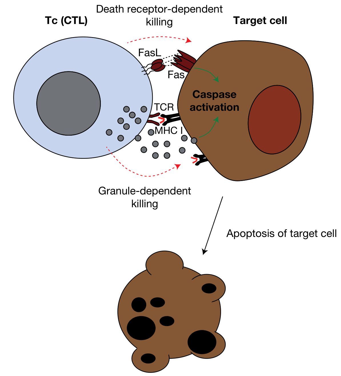
A. Perforin/Granzyme Pathway (Granule-Dependent Killing)
This is the dominant CTL killing mechanism, especially against virally infected cells.
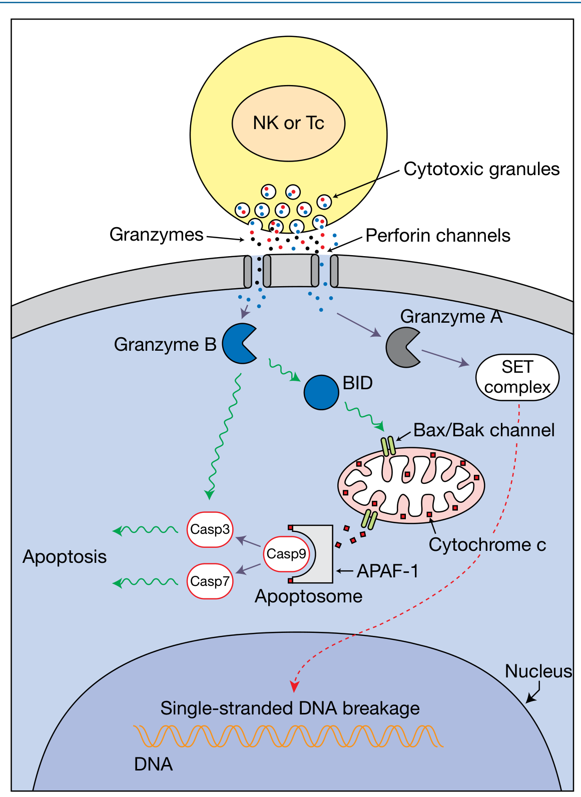
Sequence of events:
- TCR recognition: CTL binds target cell displaying peptide-MHC I complex
- Immunological synapse formation: The cytoskeleton of the CTL polarizes its cytotoxic granules toward the point of contact at high speed (up to 1.2 µm/s)
- Directional degranulation: Cytotoxic granules (modified lysosomes) are exocytosed at the synapse - this directionality prevents killing of innocent bystander cells
- Perforin (a pore-forming protein similar to complement C9) oligomerizes in the target cell membrane, forming pores that permit entry of granzymes into the cytosol (or facilitates endocytosis/escape of granule contents from endosomes)
- Granzyme B (serine protease) cleaves and activates:
- BID → triggers Bax/Bak pore in mitochondria → release of cytochrome c → formation of the apoptosome (with APAF-1/caspase-9) → activation of caspase-3 and caspase-7
- Caspases 3 & 7 directly (shortcut route)
- Granzyme A cleaves the SET complex (ER-associated), releasing NM23-H1 nuclease into the nucleus → single-stranded DNA breaks
- CTL disengages ("kiss of death") and seeks another target - it is a serial killer with rapid synthesis of new granules
Perforin deficiency in humans causes type 2 familial hemophagocytic lymphohistiocytosis (FHL), a fatal disorder of immune hyperactivation. - Roitt's Essential Immunology, p. 273
B. Fas/Fas Ligand (FasL) Pathway - Death Receptor-Dependent Killing
- CTLs express Fas ligand (FasL/CD95L) on their surface
- FasL engages the trimeric Fas receptor (CD95) on the target cell
- Fas clustering recruits the adaptor protein FADD to its cytoplasmic tail
- FADD recruits and activates caspase-8 (initiator caspase)
- Caspase-8 then:
- Directly activates downstream caspases (caspase-3, -7) - Type I pathway
- Cleaves BID → cytochrome c release → apoptosome → caspase activation - Type II pathway
- Either route leads to rapid apoptosis of the target cell
Both killing pathways account for most CTL killing activity; TNF secreted by CTLs contributes a minor third component. - Roitt's Essential Immunology, p. 274
Key point: CTLs also secrete IFN-γ and TNF, which have direct antiviral effects and activate macrophages, respectively.
Effector Mechanism 2: CD4+ T Helper Cell-Mediated Responses
CD4+ T cells recognize antigens on MHC class II molecules (expressed on professional APCs - macrophages, dendritic cells, B cells). After priming, CD4+ effector cells differentiate into functionally distinct subsets depending on the cytokine milieu at the time of activation.
A. Th1 Cells - Macrophage Activation and Classical Inflammation
Driving cytokine: IL-12 (from macrophages/DCs)
Signature cytokines secreted: IFN-γ, TNF, IL-2
Effector functions:
- IFN-γ is a potent macrophage activator - it upregulates MHC II expression on APCs (amplifying the T cell response) and, combined with CD40L-CD40 signaling, drives classical macrophage activation
- Classical activation induces macrophages to produce:
- Reactive oxygen intermediates (superoxide, H₂O₂)
- Reactive nitrogen intermediates (nitric oxide via iNOS)
- Lysosomal enzymes and hydrolases → This leads to destruction of ingested intracellular microbes (e.g., M. tuberculosis, Leishmania)
- Th1 cytokines (especially IFN-γ) stimulate production of IgG2a antibodies (opsonizing antibodies)
- Sustained Th1 responses with macrophage activation → granuloma formation (e.g., in tuberculosis, sarcoidosis)
- Th1 cells can also directly kill some target cells by expressing FasL (inducing Fas-mediated apoptosis)
Robbins & Kumar Basic Pathology (p. 147): "Th1 cells secrete the cytokine IFN-γ, which is a potent macrophage activator. The combination of CD40- and IFN-γ-mediated activation results in 'classical' macrophage activation, leading to the induction of microbicidal substances in macrophages."
B. Th17 Cells - Neutrophilic/Monocytic Inflammation
Driving cytokines: IL-6, TGF-β, IL-23
Signature cytokines: IL-17A, IL-17F, IL-22
Effector functions:
- IL-17 acts on stromal cells and epithelial cells to recruit neutrophils and monocytes
- Particularly important against extracellular bacteria and fungi (e.g., Candida, Staphylococcus)
- Involved in mucosal barrier defense
- Implicated in inflammatory diseases (psoriasis, ankylosing spondylitis, IBD)
Robbins & Kumar Basic Pathology (p. 147): "Th17 cells recruit neutrophils and monocytes, which destroy some extracellular bacteria and fungi and are involved in certain inflammatory diseases."
C. Th2 Cells - Eosinophil Activation and Anti-Helminth Defense
Driving cytokine: IL-4, IL-33, TSLP
Signature cytokines: IL-4, IL-5, IL-9, IL-13
Effector functions:
- IL-4: Stimulates B cells to produce IgE; promotes Th2 differentiation (positive feedback)
- IL-5: Activates and recruits eosinophils → eosinophil degranulation kills helminths (e.g., Schistosoma) too large to be phagocytosed
- IL-13: Activates mucosal epithelial cells to secrete mucus; drives alternative macrophage activation (associated with tissue repair and fibrosis)
- Th2 responses are anti-inflammatory relative to Th1 and underlie allergic diseases when dysregulated
D. T Follicular Helper (Tfh) Cells - B Cell Help
Tfh cells (CXCR5+, PD-1+) migrate to B cell follicles in lymphoid organs and provide help for:
-
Germinal center formation
-
Affinity maturation via somatic hypermutation (selecting highest-affinity clones)
-
Isotype class switching (via CD40L-CD40 and IL-21, IL-4)
-
Long-lived plasma cell and memory B cell generation
-
Roitt's Essential Immunology, p. 290
Effector Mechanism 3: Regulatory T Cells (Tregs) - Suppression of CMI
Tregs (CD4+, CD25+, FoxP3+) are an important negative regulatory arm:
- Suppress excessive T cell responses
- Use the granzyme-perforin system to kill activated NK and CTL cells
- Modulate APC activation
- Secrete IL-10 and TGF-β to dampen inflammation
Summary Table
| Effector Cell | MHC Restriction | Key Mechanism | Target/Purpose |
|---|---|---|---|
| CD8+ CTL | MHC Class I | Perforin/granzyme + Fas/FasL → apoptosis | Virus-infected cells, tumor cells, intracytosolic bacteria |
| Th1 (CD4+) | MHC Class II | IFN-γ + CD40L → classical macrophage activation | Intravesicular bacteria (TB, Leishmania) |
| Th17 (CD4+) | MHC Class II | IL-17 → neutrophil/monocyte recruitment | Extracellular bacteria, fungi |
| Th2 (CD4+) | MHC Class II | IL-5 → eosinophil activation; IL-4 → IgE | Helminths, allergy |
| Tfh (CD4+) | MHC Class II | CD40L, IL-21 → B cell help | Antibody production, affinity maturation |
| Tregs (CD4+) | MHC Class II | IL-10, TGF-β, Granzyme-Perforin | Suppress excessive responses |
Clinical Correlates
- Perforin deficiency → Familial hemophagocytic lymphohistiocytosis (FHL) - fatal immune hyperactivation
- Defective CMI (HIV, post-transplant immunosuppression) → susceptibility to intracellular pathogens (Mycobacteria, Listeria, CMV, Pneumocystis)
- Granuloma formation (TB, sarcoidosis, leprosy) = Th1-driven macrophage activation with failure to clear antigen
- Delayed-type hypersensitivity (DTH) = Type IV hypersensitivity - CD4+ Th1/Th17-mediated inflammatory tissue damage (e.g., tuberculin skin test, contact dermatitis)
- Checkpoint blockade (anti-PD-1, anti-CTLA-4) works by releasing CTLA-4/PD-1-mediated inhibition of CTLs to enhance anti-tumor CMI
Sources:
- Cellular and Molecular Immunology (Abbas), p. 49-50
- Roitt's Essential Immunology, p. 273-274
- Robbins & Kumar Basic Pathology, p. 147
- Janeway's Immunobiology 10e, p. 367-368
- Jawetz, Melnick & Adelberg's Medical Microbiology, p. 146
This is a shared conversation. Sign in to Orris to start your own chat.