
Humerus shaft fracture in details according to rockwood
humeral shaft fracture classification radial nerve injury
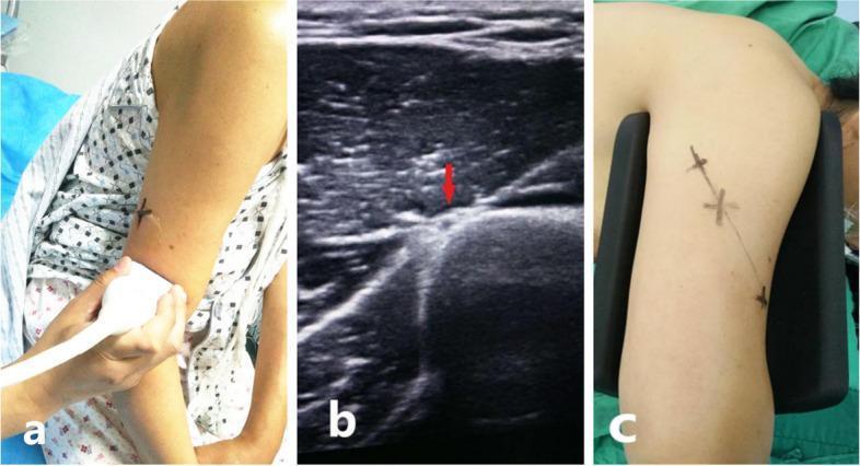
This composite of three panels (a, b, and c) illustrates the preoperative ultrasound-guided localization of the radial nerve in the posterior upper arm, typically performed before surgical management of a humeral shaft fracture. Panel (a) shows a clinical photograph of a high-resolution linear ultrasound transducer being applied to the posterior aspect of the mid-humerus in a seated patient. Panel (b) presents a grayscale ultrasound (US) image in the transverse plane, where a red arrow identifies the radial nerve as a distinct hypoechoic, oval structure situated in the spiral groove between the triceps muscle bellies and the hyperechoic humeral cortex. Panel (c) displays the final clinical result, with the anatomical course of the radial nerve demarcated on the patient's skin using surgical marker 'X' annotations connected by a line. This technique aims to facilitate safer surgical exposure and reduce the risk of iatrogenic radial nerve injury during orthopedic procedures like open reduction and internal fixation (ORIF).
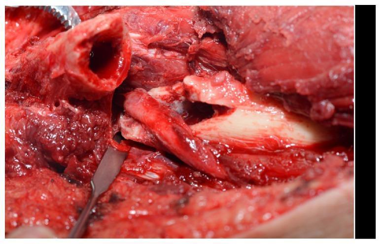
An intraoperative clinical photograph showing a surgical exploration of the mid-humerus via an anterolateral approach. The image demonstrates a humeral shaft fracture with a jagged, irregular fracture line. Centrally, the radial nerve is clearly visualized and is significantly interposed—or trapped—within the fracture site between the bone fragments. The nerve appears erythematous and swollen, consistent with a localized contusion or compression injury. Surrounding the fracture is exposed muscular tissue and a surgical retractor is visible at the left margin, highlighting the operative field. This visual provides high-level educational evidence of nerve entrapment as a potential complication of humeral fractures, demonstrating the anatomical relationship between the radial nerve and the humeral diaphysis.
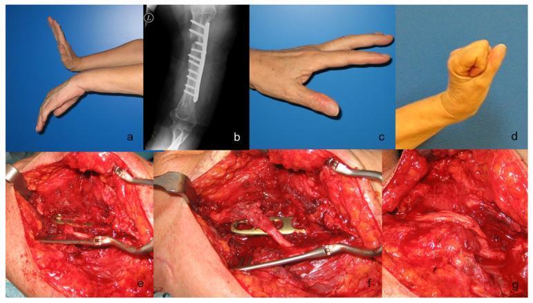
This composite figure illustrates a clinical case of radial nerve injury and subsequent surgical reconstruction following humeral fracture fixation. (a) Clinical photograph shows a classic preoperative 'wrist drop' deformity with inability to extend the wrist and fingers, characteristic of radial nerve palsy. (b) Anteroposterior radiograph demonstrates an osteosynthesis plate with multiple screws on the humeral shaft. (c, d) Postoperative photographs show successful functional recovery with active finger extension and wrist dorsiflexion. (e) Intraoperative view reveals the radial nerve severely compressed and pinched beneath the metallic humeral plate. (f) After removing a screw and elevating the plate, the nerve is found to be completely disrupted (neurotmesis). (g) Surgical reconstruction is shown using four cables of sural nerve grafts to bridge the 5 cm neural defect, secured with microsurgical sutures. This sequence demonstrates the identification, release, and grafting of a post-traumatic and iatrogenic nerve injury, highlighting the importance of clinical-radiographic correlation and microsurgical repair techniques in peripheral nerve surgery.
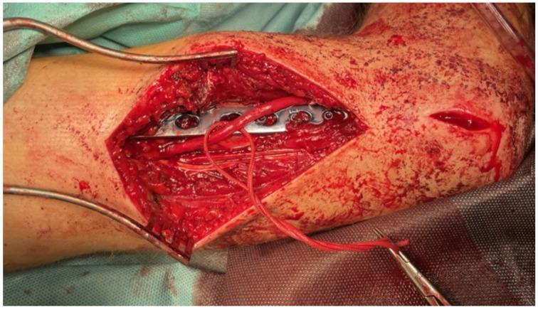
This intraoperative clinical photograph captures a surgical procedure for open reduction and internal fixation (ORIF) of a humeral shaft fracture. The surgical field reveals a long, metallic internal fixation plate with approximately 8-10 visible screw holes positioned directly on the bone. The radial nerve is prominently exposed and mobilized to prevent entrapment or injury during the fixation; it appears as a continuous, reddish-pink tubular structure. A red vessel loop or suture is used to gently retract and identify the nerve, which is held by a surgical instrument. Self-retaining retractors are positioned to maintain the surgical window through the muscle and soft tissue layers. The image demonstrates the critical relationship between orthopedic hardware and adjacent neurovascular bundles in the distal third of the humerus. This visual is significant for illustrating nerve exploration techniques used to manage radial nerve palsy associated with humeral fractures. Key educational concepts include surgical anatomy, fracture management, and peripheral nerve preservation during orthopedic trauma surgery.
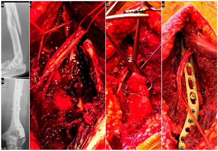
This composite educational image illustrates the surgical management of a humerus fracture with associated radial nerve involvement. (a) Preoperative lateral X-ray displays a displaced, multi-fragmentary humeral shaft fracture. (b) Postoperative X-ray shows internal fixation using a long locking compression plate and screws. (c) Intraoperative clinical photograph via a posterior approach demonstrates the radial nerve (*) being carefully mobilized away from the fracture fragments (**). (d) The fracture is shown undergoing temporary reduction, secured by a reduction clamp and cerclage wires, while the nerve (*) is protected and kept tension-free. (e) Final stabilization phase reveals the radial nerve (*) crossing superficially over the metallic locking plate (***). This sequence highlights key orthopedic principles: anatomical reduction, stable internal fixation, and the critical preservation of neurovascular structures during trauma surgery. The educational focus is on the spatial relationship between orthopedic hardware and the radial nerve to prevent iatrogenic injury during humerus osteosynthesis.
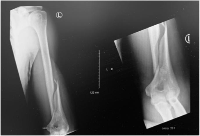
This diagnostic image consists of two radiographic views of the left upper extremity, specifically targeting the humerus. The x-rays reveal a displaced fracture of the distal third of the humeral shaft, identified as a Holstein-Lewis fracture (AO/OTA classification 12A1.3). The fracture pattern is primarily spiral or long oblique with significant lateral displacement and angulation of the distal fragment. Visible soft tissue swelling is noted adjacent to the injury site. The proximal humerus and the elbow joint, including the articulation of the distal humerus with the radius and ulna, appear otherwise intact with preserved joint spaces. A clinical marker 'L' denotes the left side. This case is highly relevant for orthopaedic education, illustrating a classic mechanism-of-injury fracture (torsional force from arm wrestling) that carries a high clinical association with radial nerve neuropraxia due to the proximity of the nerve to the spiral groove in the distal third of the humerus.
1. EPIDEMIOLOGY
- Constitute 1-2% of all fractures in the body and 13-14% of all humeral fractures
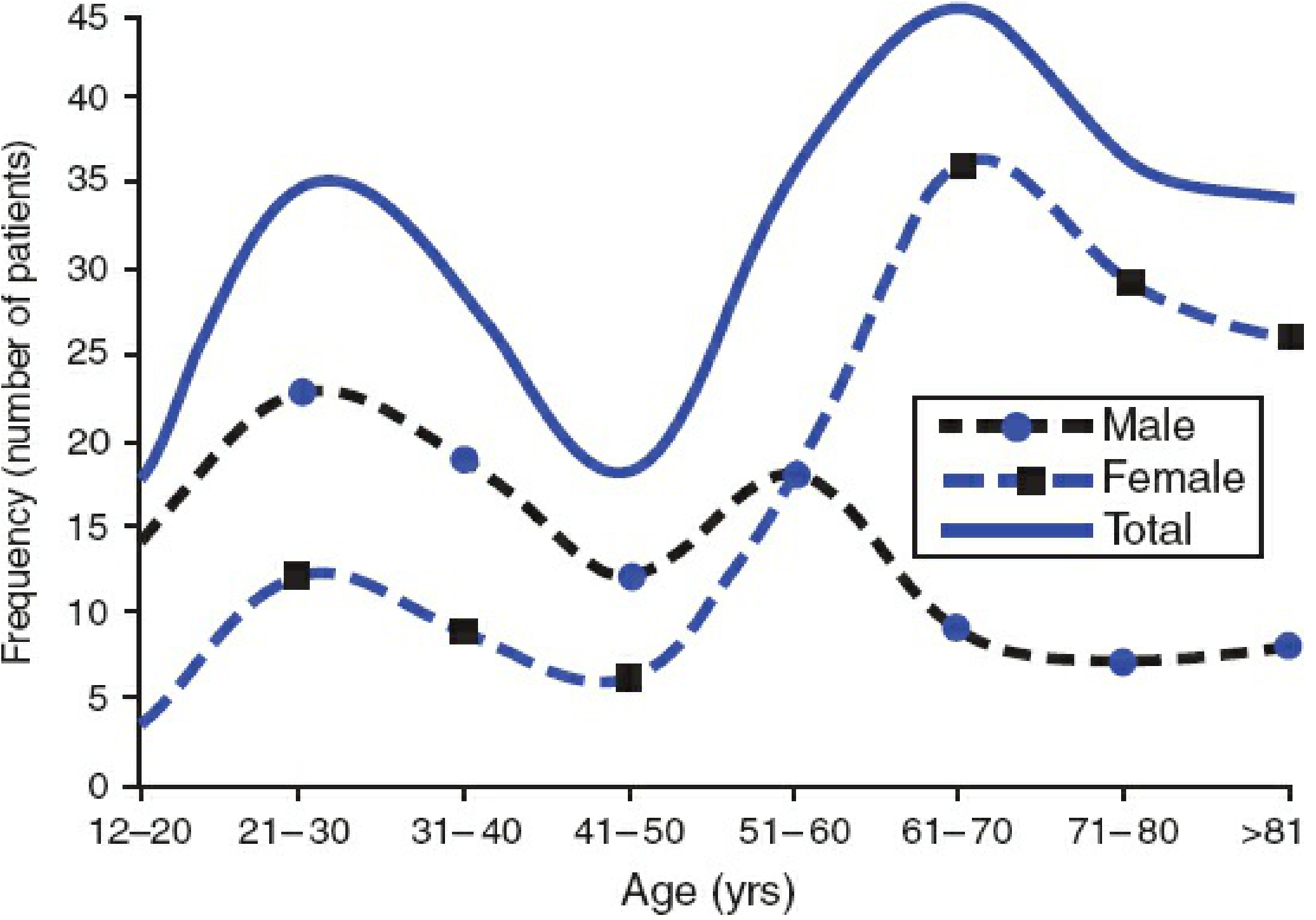
- Bimodal age-gender distribution: young men (peak 21-30 yrs from high-energy trauma) and older women (peak 61-70 yrs from low-energy falls)
- Ground-level fall is the most common mechanism (simple fall:RTA ratio = 9:1 in the USA; 3.5:1 in the UK)
- Incidence is projected to nearly double by 2030 compared with 2008
2. MECHANISMS OF INJURY
| Mechanism | Notes |
|---|---|
| Ground-level fall | Most common overall; typically older women |
| Road traffic accident | High-energy; typically younger men |
| Sports/falls from height | 5-8% depending on geography |
| Direct blow | Transverse or comminuted fracture pattern |
| Twisting/torsional force | Spiral fracture pattern (classic: arm wrestling) |
3. ANATOMY AND APPLIED PATHOLOGY
- Proximal: superior border of pectoralis major insertion
- Distal: superior border of the supracondylar ridges (or olecranon fossa)
- Radial nerve runs in the spiral groove of the posterior humerus and is the most vulnerable structure - at risk in middle and distal third fractures
- Brachial artery and median nerve lie anteromedially
- Musculocutaneous nerve penetrates the coracobrachialis proximally
- Holstein-Lewis fracture (distal third spiral fracture) classically entraps the radial nerve at the lateral intermuscular septum
| Fracture Level | Proximal Fragment | Distal Fragment |
|---|---|---|
| Above pectoralis major | Abducted + externally rotated (deltoid/rotator cuff) | Displaced medially (pec major) |
| Between pec major and deltoid insertion | Adducted medially (pec major) | Proximally + abducted (deltoid) |
| Below deltoid insertion | Abducted (deltoid) | Proximal pull (biceps/triceps) |
4. CLASSIFICATION
AO/OTA Classification (most widely used)
- Type A - Simple (A1: spiral, A2: oblique, A3: transverse)
- Type B - Wedge (B1: spiral wedge, B2: bending wedge, B3: fragmented wedge)
- Type C - Complex/comminuted (C1: spiral, C2: segmental, C3: irregular)
AO classification by bone loss / soft tissue damage:
- Open fractures graded using the OTA/OFS Open Fracture Classification (Skin / Muscle / Arterial / Contamination / Bone Loss - each graded 1-3)
- Closed fractures: Tscherne-Gotzen classification (Grade 0-III)
5. ASSOCIATED INJURIES
Radial Nerve Injury
- Occurs in 10-12% of closed humeral shaft fractures - the most common associated injury
- Clinical features: wrist drop (inability to dorsiflex wrist and digits), numbness over dorsoradial hand and dorsal aspect of radial 3.5 digits
- Holstein-Lewis fracture (distal spiral) has highest risk of radial nerve entrapment
- Primary (immediate) palsy at time of injury: Initial expectant (wait-and-watch) treatment is preferred. ~70% recover spontaneously within 3-6 months
- Secondary palsy (after closed manipulation or open fractures): Explore immediately
- If no recovery by 3-4 months - electrophysiologic studies (EMG/NCS) and exploration if needed
- Shao et al. (systematic review 1964-2004): Confirmed policy of initial conservative observation is preferred
Vascular Injury
- Less common but limb threatening; brachial artery injury is the most critical
- Clinical: absent distal pulses, expanding hematoma, limb ischemia
- Requires emergency vascular exploration and repair
Other Associated Injuries
- Ipsilateral forearm fractures ("floating elbow") - absolute indication for surgery
- Shoulder and elbow intra-articular extension
- Brachial plexus injury with high-energy or shoulder-dislocating mechanisms
6. IMAGING
- Standard: AP and lateral radiographs of the whole humerus (including shoulder and elbow joints)
- CT scan: if intra-articular extension is suspected or fracture lines approach joints
- Fracture union (Sarmiento's definition): osseous bridging between main fragments on at least one radiograph with no pain at fracture site
- CT is not routine - reserved for suspected nonunion or delayed union not progressing on plain films
7. NON-OPERATIVE TREATMENT
Acceptable Alignment Criteria
| Parameter | Acceptable Limit |
|---|---|
| Varus/valgus angulation | Up to 20-30° |
| Anterior angulation | Up to 20° |
| Shortening | Up to 3 cm |
| Rotation | Up to 15° |
Methods (Progression over time)
- Coaptation/U-Slab splint - Initial immobilization for swelling
- Hanging arm cast - Traction by gravity; patient must remain upright; risk of distraction if used too long
- Functional Brace (Sarmiento brace) - The gold standard for non-operative treatment
- Applied at 1-2 weeks post-injury when pain/swelling allows
- Hydrostatic compression of soft tissues around bone
- Allows shoulder and elbow mobility
- Union rates: 85-95% for mid-shaft fractures
- Higher nonunion risk: AO type A fractures, proximal third fractures, large gap, absence of bridging callus at 6 weeks
- Low-intensity pulsed ultrasound (LIPUS)
- Vitamin D supplementation
8. OPERATIVE TREATMENT
Absolute Indications for Surgery
- Inability to maintain satisfactory reduction by closed means
- Multiple injuries (polytrauma)
- Bilateral fractures
- Floating elbow (ipsilateral forearm fracture)
- Intra-articular fracture extension
- Progressive nerve palsy or nerve palsy after closed manipulation
- Significant vascular injury
- Neurologic deficit after penetrating injury
- Nonunion / infected nonunion
- Open fractures
- Pathologic fractures
Relative Indications for Surgery
- Segmental fractures
- Noncompliant patients
- Obesity or large-breasted patients (preclude brace use)
- Periprosthetic fractures
9. SURGICAL OPTIONS
A. Open Reduction Internal Fixation (ORIF) with Plate
Surgical Approaches:
- Anterior (anterolateral) - Henry approach; exposes entire humerus; radial nerve identified in distal third; suitable for middle and distal third fractures
- Posterior - Triceps-splitting or triceps-sparing (paratricipital); best for distal shaft and fractures involving radial nerve exploration; provides widest access to radial nerve in spiral groove
- Lateral - Less commonly used; intermediate option
Plate Selection:
- Traditional 4.5 mm narrow DCP or LC-DCP - minimum 3-4 screws proximal and distal; 8-10 hole for simple fractures; longer bridging plates for comminuted fractures
- Locking compression plates (LCP) - superior for osteoporotic bone; better pull-out resistance
- Dual plating - orthogonal 3.5 mm plates (9-hole anterior + 7-hole lateral) - superior rotation stiffness; useful for narrow humerus or combined diaphyseal-articular fractures
MIPO (Minimally Invasive Plate Osteosynthesis)
- Anterior or posterior approach with 2 small "windows"
- Avoids fracture hematoma disturbance; radial nerve must be identified and protected
- Suitable for middle and distal diaphyseal fractures
B. Intramedullary Nailing (IMN)
Antegrade IMN
- Entry point: tip of greater tuberosity at medullary canal axis (1 cm medial to greater tuberosity tip)
- Patient positioned: supine or beach chair, arm hanging off table
- Nail length/diameter: measured on preoperative AP + lateral radiographs
- Minimum 3 interlocking screws proximally and distally
- Specific complications:
- Nail protrusion and shoulder impingement
- Shoulder dysfunction (rotator cuff damage from entry)
- "Backing out" of proximal locking screws
- Iatrogenic proximal humeral fracture
Retrograde IMN
- Entry: 1 × 2 cm window in posterior supracondylar cortex via olecranon fossa
- Patient positioned: prone or lateral decubitus
- Skin incision: 4-5 cm from olecranon tip proximally; triceps split
- Specific complications:
- Iatrogenic supracondylar fracture at entry site
- Elbow stiffness
Plating vs. Nailing Comparison
| Feature | ORIF Plate | IMN |
|---|---|---|
| Shoulder dysfunction | Lower | Higher (antegrade) |
| Radial nerve iatrogenic injury | Higher (direct exposure) | Lower |
| Elbow complications | Lower | Higher (retrograde) |
| Rotation control | Good (dual plate superior) | Moderate |
| Preference | Most centers worldwide | Certain cases: segmental, osteoporotic |
C. External Fixation
- Rarely used as definitive treatment
- Indicated in: contaminated open fractures, massive soft tissue injury, temporary stabilization in polytrauma
- Complications: pin track infection, refracture after removal
10. AUTHOR'S PREFERRED TREATMENT (Rockwood Chapter Authors)
- Most closed humeral shaft fractures: Functional brace (Sarmiento) as primary treatment
- Operative treatment reserved for the absolute and relative indications listed above
- If surgery is needed: ORIF with plate (posterior or anterior approach) is preferred over IMN in most cases due to lower shoulder dysfunction rates
- Radial nerve palsy in closed fractures: expectant observation initially; explore only if secondary palsy or no recovery at 3-4 months
11. COMPLICATIONS AND ADVERSE OUTCOMES
General Complications (all methods)
Nonunion
- Incidence: 1-10% after non-operative and 10-15% after surgical management
- Higher after surgery likely reflects selection bias (more complex fractures operated)
- Risk factors: open fractures, segmental, transverse, comminuted, significant displacement, bone loss, smoking, diabetes, NSAIDs, malnutrition, noncompliance
- Nonunion after surgery often due to technical error (inadequate fixation, fracture gap distraction)
- Most are atrophic nonunions
- Standard: ORIF + autologous bone graft (iliac crest)
- If bone defect: autograft, allograft, BMPs, or shortening considered
- Technique: wide exposure, remove previous implants, debride all fibrous tissue and dead bone until healthy bleeding bone on both sides, compress fragments, stable fixation
Infection
- Higher risk with open fractures and revision surgery
- Requires debridement + antibiotics; may require staged procedures
Secondary Neurologic Injury
- Iatrogenic radial nerve injury during ORIF (posterior approach)
- Risk reduced by careful nerve identification and protection
Implant-Specific Complications
| Implant | Specific Complications |
|---|---|
| Plate | Loss of fixation (especially osteoporotic bone) |
| Antegrade nail | Nail protrusion + impingement, shoulder dysfunction, proximal screw back-out |
| Retrograde nail | Supracondylar fracture at entry, elbow stiffness |
| External fixation | Pin track infection, refracture after removal |
12. SPECIAL CONSIDERATIONS
Open Fractures
- Classified by OTA/OFS system (Skin / Muscle / Artery / Contamination / Bone Loss)
- Require urgent surgical debridement and irrigation
- Antibiotic coverage; delayed wound closure or flap coverage if needed
- Definitive stabilization: plate or external fixator depending on contamination
Osteoporotic Fractures
- Higher risk of nonunion and fixation failure
- LCP preferred over conventional plates; consider bone grafting at index surgery
- Nutritional optimization (vitamin D, calcium) important
Pathologic Fractures
- Most commonly from metastatic disease (breast, lung, kidney, prostate)
- Prophylactic fixation if at risk; intramedullary nailing preferred for metastatic lesions (load-sharing implant)
- Postoperative radiotherapy often required
Periprosthetic Fractures
- Around shoulder or elbow prostheses
- Classification and treatment depend on prosthesis stability and bone stock
- Stable prosthesis + adequate bone: ORIF with plate and cerclage wires
- Loose prosthesis: revision arthroplasty + fixation
SUMMARY TABLE
| Aspect | Key Point |
|---|---|
| Incidence | 1-2% of all fractures |
| Bimodal distribution | Young men (trauma) + older women (falls) |
| Most common mechanism | Ground-level fall |
| Most common nerve injury | Radial nerve (10-12%) |
| High-risk fracture for radial nerve | Holstein-Lewis (distal spiral) |
| Gold standard non-operative Rx | Sarmiento functional brace |
| Acceptable angulation | Up to 20-30° varus/valgus, 20° anterior |
| Nonunion rate non-op | 1-10% |
| Nonunion rate operative | 10-15% |
| Preferred operative Rx | ORIF with plate (anterior or posterior approach) |
| Radial nerve palsy - primary | Observe; 70% spontaneous recovery |
| Radial nerve palsy - secondary | Immediate exploration |
Supracondylar humerus fracture in details according to orthopaedics text books and recent literature
"supracondylar humerus fracture" AND children
supracondylar humerus fracture Gartland classification pediatric elbow
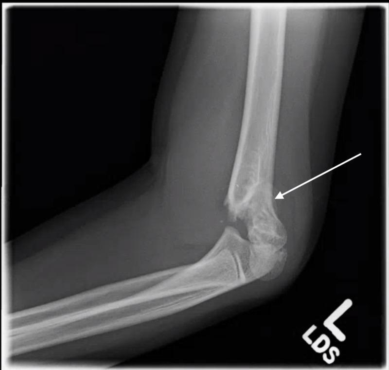
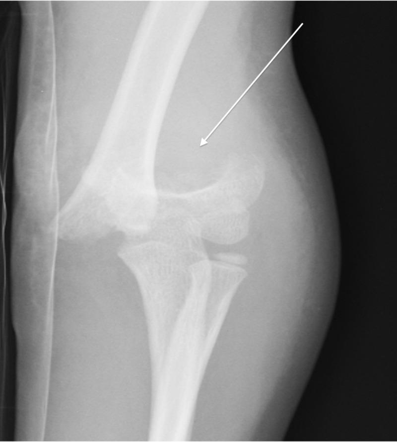
This diagnostic image is a lateral plain radiograph of the elbow in a pediatric patient, demonstrating a supracondylar fracture of the distal humerus, classified as a Gartland Type 2 fracture. A white arrow points to the fracture site at the supracondylar ridges. The radiograph shows a complete fracture with clear cortical disruption. There is a visible posterior displacement and posterior angulation of the distal humeral fragment relative to the humeral shaft. While the posterior cortex remains partially intact (hinged), providing the Type 2 classification, the normal anterior humeral line is disrupted and does not bisect the capitellum. The image also depicts associated soft tissue haziness consistent with swelling. This clinical image is highly relevant for pediatric orthopedics and emergency medicine, illustrating common mechanisms of injury such as a fall on an outstretched hand (FOOSH).
This diagnostic image is an anteroposterior (AP) view radiograph of a pediatric elbow, demonstrating a severe supracondylar humerus fracture, classified as a Gartland Type 3. The imaging shows a complete cortical discontinuity in the distal humeral metaphysis, immediately proximal to the epicondyles. A white arrow highlights the fracture site where there is significant posterolateral displacement and misalignment of the distal fragment relative to the humeral shaft. The normal anatomical relationship between the distal humerus, proximal radius, and ulna is disrupted, suggesting associated joint instability or dislocation. Pediatric growth plates are visible at the proximal radius and ulna, consistent with the patient's age. This radiograph illustrates a common orthopedic emergency in children, typically resulting from a fall on an outstretched hand (FOOSH), and is used to teach fracture classification, assessment of neurovascular risk, and the necessity of surgical reduction.
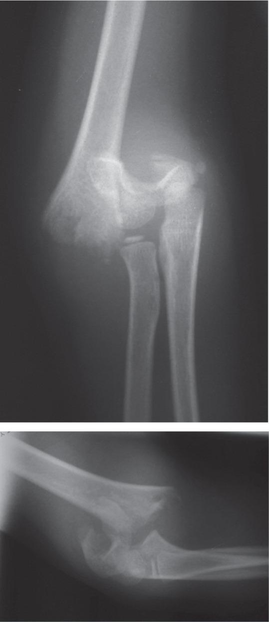
This diagnostic image consists of two radiographic views (anteroposterior and lateral) of a pediatric elbow joint. The X-rays demonstrate a complete, severely displaced supracondylar fracture of the distal humerus, consistent with a Gartland type III classification. The anteroposterior view shows a transverse fracture line through the supracondylar region with significant medial and posterior displacement of the distal humeral fragment. This displacement results in a disruption of the normal anatomical relationship between the distal humerus and the proximal radius and ulna. In the lateral view, there is evidence of posterior angulation and complete lack of cortical contact between the proximal and distal segments. The epiphyses of the distal humerus, radial head, and olecranon are visible, confirming the pediatric nature of the patient. There is associated soft tissue swelling surrounding the elbow joint. Clinically, this imaging represents an orthopedic emergency due to the high risk of neurovascular compromise involving the brachial artery, radial nerve, or median nerve.
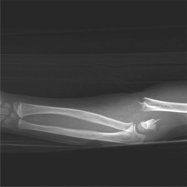
This lateral view X-ray of a pediatric upper extremity demonstrates a severe supracondylar fracture of the distal humerus, classified as a Gartland Type III injury. The radiographic findings show a complete, multi-fragmentary disruption of the humeral shaft just proximal to the epicondyles. There is significant posterior and superior displacement of the distal humeral epiphysis and the articulating forearm bones (radius and ulna) relative to the proximal humeral shaft. The anterior humeral line does not intersect the capitellum, indicating significant extension-type angulation. While the distal forearm appears grossly intact with preserved alignment between the radius, ulna, and proximal carpals, the elbow joint shows total loss of anatomical congruity. This clinical image is representative of high-energy pediatric trauma and is of high educational value for orthopedic classification and assessing associated neurovascular risks, such as brachial artery injury or compartment syndrome.
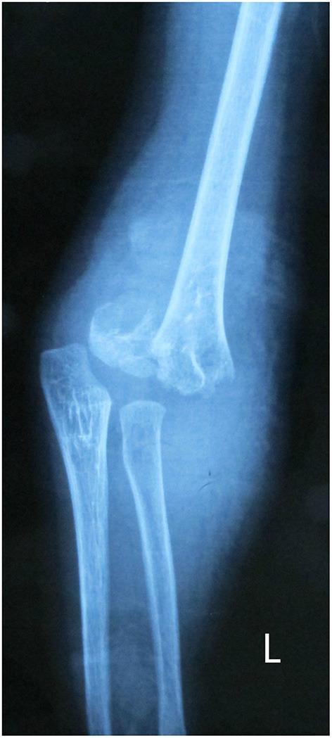
A lateral x-ray radiography of a pediatric elbow demonstrating a severe supracondylar humerus fracture, classified as a Gartland type III. The imaging reveals a complete transverse fracture line across the distal humerus, superior to the condyles. There is significant posterior displacement and angulation of the distal humeral fragment relative to the proximal humeral shaft. The anatomical alignment of the elbow joint is heavily disrupted; the normal radiocapitellar line is lost as the capitellum is displaced with the distal humeral fragment. The proximal radius and ulna remain articulated with the distal humerus but are posteriorly translated alongside it. The image serves as a clinical example of a common pediatric orthopedic emergency requiring urgent reduction to prevent neurovascular compromise or long-term deformity such as cubitus varus. This visual is typical of injuries seen in the context of a fall on an outstretched hand in children.
"supracondylar humerus fracture" AND "pin fixation" AND outcomes
PMID: 37395427
PMID: 41076056