
Generate 50 highly yield MCQ question and answer covering all important information from pyq for orthopaedic subject for neet pg preparation
NEET PG orthopaedics most repeated PYQ topics high yield
High-Yield Orthopaedics MCQs for NEET PG (PYQ-Based)
| Topic | Classification |
|---|---|
| Femoral neck fracture | Garden (I-IV) |
| Growth plate injury | Salter-Harris (I-V) |
| Open fractures | Gustilo-Anderson (I, II, IIIA/B/C) |
| Peripheral nerve injury | Seddon (neurapraxia/axonotmesis/neurotmesis) & Sunderland (1-5) |
| Hip fracture displacement/femoral head AVN risk | Garden III/IV highest risk |
Generate pdf from this question if possible add 20 image based questions also and add ans and explanation in last
Colles fracture distal radius X-ray dinner fork deformity
Monteggia and Galeazzi fracture forearm X-ray
Supracondylar fracture of humerus in children X-ray
Salter Harris physeal growth plate fracture classification X-ray
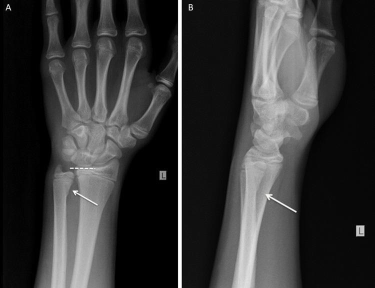
Diagnostic imaging of a pediatric left wrist featuring two X-ray views: Anteroposterior (A) and Lateral (B). The images demonstrate a Salter-Harris type II fracture of the distal ulnar epiphysis in a skeletally immature patient. View A shows a clear horizontal lucency through the ulnar growth plate with a small metaphyseal fragment (Thurston Holland sign), indicated by a white arrow. View B confirms the posterior displacement of the distal ulnar epiphysis relative to the shaft, also marked by an arrow. The adjacent distal radius appears intact without evidence of cortical disruption or physeal widening. The carpal bones, metacarpals, and visualized phalanges show normal alignment and ossification for the developmental age. This clinical image is illustrative of ulnar-sided physeal injuries typically resulting from a fall on an outstretched hand (FOOSH). Educational value lies in identifying Salter-Harris classification patterns and distinguishing physeal fractures from normal growth plate appearances in pediatric orthopedic trauma.
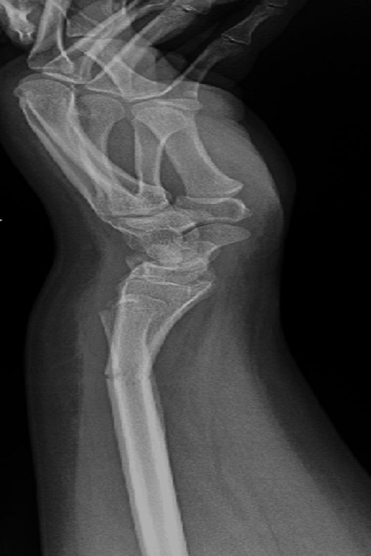
A lateral view X-ray of the wrist and distal forearm demonstrating a Galeazzi-equivalent fracture pattern in a pediatric patient. The radiograph reveals two distinct fractures: a transverse fracture in the distal third of the radial shaft with approximately 21 degrees of dorsal apex angulation, and a concomitant fracture of the distal ulna. The ulnar injury is characterized as a Salter-Harris type II physeal fracture involving the distal ulnar epiphysis, showing approximately 2.5 mm of minimal volar displacement. The anatomical alignment between the radius and ulna is disrupted at the level of the fractures, representing a pediatric variant of the Galeazzi injury where a distal ulnar physeal fracture occurs instead of a distal radioulnar joint (DRUJ) dislocation. Pronounced soft tissue swelling is visible around the wrist and distal forearm. This diagnostic image is intended for educational purposes in orthopedics and trauma radiology, focusing on pediatric fracture classification and the management of forearm shaft injuries involving the growth plate.
congenital talipes equinovarus clubfoot clinical photograph
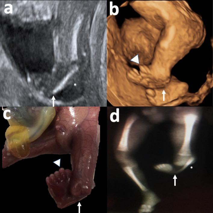
A multi-panel medical image illustrating congenital lower limb malformations across different imaging modalities and specimens. Panel (a) is a 2D grayscale ultrasound of a fetal leg showing a hypoplastic fibula (*) and clubfoot (arrow). Panel (b) depicts a 3D ultrasound reconstruction of the same limb, highlighting the inverted position of the clubfoot and pre-axial polydactyly (arrowhead). Panel (c) provides a clinical photograph of the post-mortem fetal specimen, clearly showing the supernumerary digits of the pre-axial polydactyly and the severe equinovarus deformity (clubfoot). Panel (d) shows a postnatal X-ray of the lower limbs; the left leg demonstrates a hypoplastic, shortened fibula and abnormal bony alignment of the foot consistent with clubfoot, while the right leg displays a normal tibia and fibula. The collective findings demonstrate tibial hemimelia and associated distal limb defects including polydactyly and talipes equinovarus, serving as a comparative educational resource for prenatal diagnosis and pathological correlation.
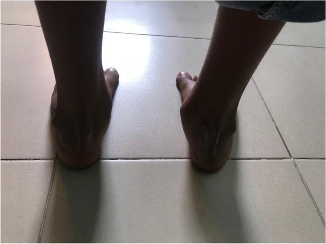
This clinical photograph shows a posterior view of both feet and ankles in a weight-bearing position on a tiled surface. The image demonstrates the post-operative results of a pediatric bilateral posteromedial release (PMR) with cuboid osteotomy. The right foot features a prominent vertical surgical scar extending over the posterior aspect of the calcaneus (heel), consistent with a Carroll surgical approach for clubfoot correction. The left foot appears relatively smooth with less visible scarring. Both heels demonstrate improved alignment from a presumed pre-operative equinovarus deformity, though the right heel shows a slightly different contour compared to the left. The feet are positioned in a neutral weight-bearing axis relative to the floor. This image is used to evaluate long-term outcomes of surgical intervention for congenital talipes equinovarus (clubfoot) in a pediatric patient, specifically focusing on skin integrity, scar formation, and hindfoot alignment.
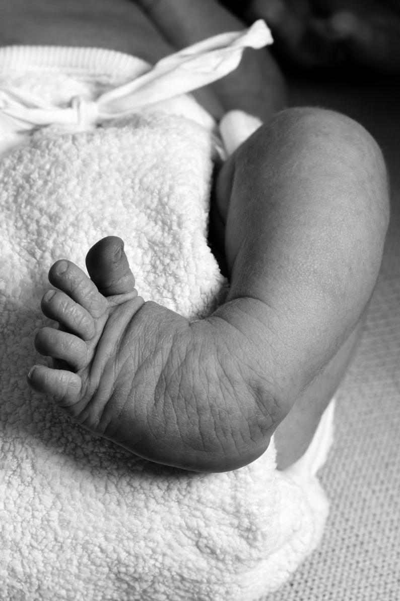
A grayscale clinical photograph of an infant's foot and lower leg demonstrating Congenital Talipes Equinovarus (CTEV), commonly known as clubfoot. The image captures the foot in a characteristic position of adduction, inversion, and plantarflexion. Key anatomical features include a distinctly rounded lateral border of the foot and a poorly defined, soft-appearing heel. The dorsum of the foot and the ankle region exhibit prominent deep skin creases and transverse folds, which are classic clinical markers of this deformity. The toes appear relatively short and fan out slightly from the metatarsophalangeal joints. This photograph is used for educational purposes to illustrate the physical signs of clubfoot and the associated challenges in anatomical landmark identification for clinical measurement and diagnosis in pediatric orthopedics.
developmental dysplasia of hip and Perthes disease X-ray
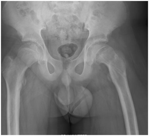
osteosarcoma Codman triangle sunburst Ewing sarcoma onion skin X-ray
giant cell tumor bone soap bubble osteoid osteoma nidus X-ray
multiple myeloma punched out lesions skull Paget disease bone X-ray
Medical education schematic diagram of Garden classification of femoral neck fractures, showing four stages labeled I, II, III, IV with increasing displacement of the femoral head fracture
Medical education schematic diagram of Gustilo-Anderson classification of open fractures, showing types I, II, IIIA, IIIB, IIIC with increasing wound size and soft tissue damage
Medical education diagram of brachial plexus anatomy showing the site of injury in Erb's palsy at C5-C6 upper trunk and Klumpke's palsy at C8-T1 lower trunk
Medical education diagram of lower limb nerve dermatomes and motor distribution comparing common peroneal nerve palsy causing foot drop versus tibial nerve injury
Medical education illustration of osteosarcoma X-ray appearance schematic showing Codman triangle periosteal reaction and sunburst pattern in distal femur
Medical education illustration of Ewing sarcoma X-ray appearance schematic showing onion-skin lamellated periosteal reaction in a long bone diaphysis
Medical education anatomical diagram of the knee joint showing anterior cruciate ligament, posterior cruciate ligament, medial collateral ligament, and lateral collateral ligament
Medical education diagram comparing hand joint involvement pattern in rheumatoid arthritis affecting MCP and PIP joints versus osteoarthritis affecting DIP joints with Heberden nodes
Medical education illustration of spine X-ray schematic showing bamboo spine appearance of ankylosing spondylitis with syndesmophytes bridging vertebrae
Medical education illustration of thoracolumbar spine schematic showing tuberculous spondylitis Pott disease with vertebral body destruction, disc space narrowing, and kyphotic gibbus deformity
mkdir -p /tmp/workspace/ortho_mcq/images && ls -la /tmp/workspace/
~/ortho_mcq/download_images.py
import urllib.request
import os

images = {
    "img01_colles.png": "https://cdn.orris.care/cdss_images/BAILY_1760539929138_c81fd1b2-f67c-4435-8525-9898449ccc70.png",
    "img02_plate_fixation.png": "https://cdn.orris.care/cdss_images/BAILY_1760539864924_5845fda4-0366-4436-939e-f7f49ebb5f65.png",
    "img03_sh2_ulna.jpg": "https://cdn.orris.care/cdss_images/pmc_clinical_VQA_298c399fac2c48d568ff563b10c8308b1a6f148b2b096fd15b2a07164091bac2.jpg",
    "img04_galeazzi.jpg": "https://cdn.orris.care/cdss_images/pmc_clinical_VQA_54059be2e5d6b2b8cad96f41aff5dfec746c61f4de72329139e1e212433f8480.jpg",
    "img05_monteggia_fixed.jpg": "https://cdn.orris.care/cdss_images/pmc_clinical_VQA_0007fe145d9a793169d4b72eb8a187fe8a4712ab582e578399e0d3f953b1fc43.jpg",
    "img06_olecranon_ct.jpg": "https://cdn.orris.care/cdss_images/pmc_clinical_VQA_018506d7fd3c1d097caf2415423b66e6954422f637833d73d74dbe6df17b1339.jpg",
    "img07_ctev_infant.jpg": "https://cdn.orris.care/cdss_images/pmc_clinical_VQA_58a1f16ca9fe6f72effa7c60df7cdbb75ff1c64b9dc7000deffbda2f8adc3ec6.jpg",
    "img08_ctev_postop.jpg": "https://cdn.orris.care/cdss_images/pmc_clinical_VQA_6c0880b23e2173e0f4ad1aee86e19b56a1b9a75c9796fed2ae7ffff3893e1462.jpg",
    "img09_scfe.jpg": "https://cdn.orris.care/cdss_images/pmc_clinical_VQA_390a8900af74af5e8d4582e49da8744bd2bfd6987a7a783b76607a41378de627.jpg",
    "img10_avn_mri.png": "https://cdn.orris.care/cdss_images/medpix_vqa_358d864b-5faf-4900-b4f7-edb4a13e5556_1766169691220.png",
    "img11_garden.png": "https://cdn.orris.care/image-gen/b7942a50d8d448f49b9ad685a456f15c.png",
    "img12_gustilo.png": "https://cdn.orris.care/image-gen/347f5ab0caaf4ae38071bfc0592deb6b.png",
    "img13_brachial_plexus.png": "https://cdn.orris.care/image-gen/330aefee2ee948529c638ca684eaab6f.png",
    "img14_peroneal_tibial.png": "https://cdn.orris.care/image-gen/96181eae931547628e3141681a3de33e.png",
    "img15_osteosarcoma.png": "https://cdn.orris.care/image-gen/8c8d283a69e64149879d8e31e88366c2.png",
    "img16_ewing.png": "https://cdn.orris.care/image-gen/eddf8ef9cfce49b8a35983ae3f4ad4eb.png",
    "img17_knee_ligaments.png": "https://cdn.orris.care/image-gen/27d036dd94f94077afcdfa8f88fdc57b.png",
    "img18_ra_vs_oa.png": "https://cdn.orris.care/image-gen/3bbee62157484f189a8d9406374407a5.png",
    "img19_bamboo_spine.png": "https://cdn.orris.care/image-gen/6f09e0f5078f4d43a4b0ae79afa3450b.png",
    "img20_pott_disease.png": "https://cdn.orris.care/image-gen/894861a70e9d4dc1bd3c63ca2fc0a228.png",
}

outdir = "/tmp/workspace/ortho_mcq/images"
os.makedirs(outdir, exist_ok=True)
headers = {"User-Agent": "Mozilla/5.0"}

ok, fail = 0, 0
for fname, url in images.items():
    path = os.path.join(outdir, fname)
    try:
        req = urllib.request.Request(url, headers=headers)
        with urllib.request.urlopen(req, timeout=30) as resp, open(path, "wb") as f:
            f.write(resp.read())
        size = os.path.getsize(path)
        print(f"OK  {fname}  ({size} bytes)")
        ok += 1
    except Exception as e:
        print(f"FAIL {fname}: {e}")
        fail += 1

print(f"\nDone. ok={ok} fail={fail}")
cd /tmp/workspace/ortho_mcq && python3 download_images.py
~/ortho_mcq/build_pdf.py
# -*- coding: utf-8 -*-
import os
from reportlab.lib.pagesizes import A4
from reportlab.lib.units import cm
from reportlab.lib import colors
from reportlab.lib.styles import getSampleStyleSheet, ParagraphStyle
from reportlab.platypus import (SimpleDocTemplate, Paragraph, Spacer, Image,
PageBreak, Table, TableStyle, KeepTogether)
from reportlab.lib.enums import TA_CENTER, TA_LEFT
IMGDIR = "/tmp/workspace/ortho_mcq/images"
OUT = "/tmp/workspace/ortho_mcq/Orthopaedics_NEET_PG_MCQs.pdf"
styles = getSampleStyleSheet()
title_style = ParagraphStyle("TitleBig", parent=styles["Title"], fontSize=20, spaceAfter=6)
sub_style = ParagraphStyle("Sub", parent=styles["Normal"], fontSize=12, alignment=TA_CENTER, textColor=colors.HexColor("#444444"), spaceAfter=20)
section_style = ParagraphStyle("Section", parent=styles["Heading1"], fontSize=15, textColor=colors.HexColor("#1a3d6b"), spaceBefore=10, spaceAfter=10)
qstyle = ParagraphStyle("Q", parent=styles["Normal"], fontSize=10.5, leading=14, spaceAfter=4, spaceBefore=10)
optstyle = ParagraphStyle("Opt", parent=styles["Normal"], fontSize=10, leading=13, leftIndent=14, spaceAfter=2)
capstyle = ParagraphStyle("Cap", parent=styles["Normal"], fontSize=8.5, leading=10, textColor=colors.HexColor("#666666"), spaceAfter=6, alignment=TA_CENTER)
ansstyle = ParagraphStyle("Ans", parent=styles["Normal"], fontSize=10, leading=13, textColor=colors.HexColor("#0b6b2c"), spaceAfter=2, fontName="Helvetica-Bold")
expstyle = ParagraphStyle("Exp", parent=styles["Normal"], fontSize=9.5, leading=13, spaceAfter=10)
# ---------------------------------------------------------------------------
# 50 TEXT-BASED MCQs (question, options A-D, correct letter, explanation)
# ---------------------------------------------------------------------------
text_mcqs = [
("A 25-year-old male falls on an outstretched hand and sustains a fracture of the distal radius with dorsal displacement and dorsal angulation of the distal fragment. What is this fracture called?",
["Smith's fracture","Colles' fracture","Barton's fracture","Chauffeur's fracture"], "B",
"Dorsally displaced/angulated distal radius fracture producing the classic 'dinner fork' deformity. Smith's fracture is the volar (reverse) variant."),
("Which nerve is most commonly injured in a supracondylar fracture of the humerus in children?",
["Ulnar nerve","Median nerve","Anterior interosseous nerve","Radial nerve"], "C",
"The AIN is the most commonly injured nerve overall in extension-type supracondylar fractures; the ulnar nerve is most often injured in flexion-type fractures."),
("Volkmann's ischemic contracture is a complication most commonly seen after which fracture?",
["Colles' fracture","Supracondylar fracture of humerus","Femoral shaft fracture","Monteggia fracture"], "B",
"Results from compartment syndrome of the forearm following brachial artery injury/compression in supracondylar fractures."),
("A fracture of the proximal third of the ulna with dislocation of the radial head is known as:",
["Galeazzi fracture","Monteggia fracture","Colles' fracture","Bennett's fracture"], "B",
"Mnemonic 'MUR': Monteggia = Ulna fracture + Radial head dislocation. Galeazzi is the reverse pattern (radius fracture + DRUJ dislocation)."),
("Garden classification is used for fractures of the:",
["Distal radius","Neck of femur","Tibial plateau","Proximal humerus"], "B",
"Garden I (incomplete/impacted), II (complete, undisplaced), III (complete, partially displaced), IV (complete, fully displaced); III/IV carry high AVN risk."),
("The most common site of avascular necrosis following a femoral neck fracture is due to disruption of which artery?",
["Obturator artery","Medial circumflex femoral artery","Artery of ligamentum teres","Superior gluteal artery"], "B",
"The medial circumflex femoral artery is the main blood supply to the adult femoral head via the retinacular vessels."),
("Salter-Harris type III fracture involves:",
["Fracture through the physis only","Fracture through metaphysis and physis","Fracture through epiphysis and physis","Fracture through epiphysis, physis, and metaphysis"], "C",
"Type I: physis only; II: metaphysis+physis; III: epiphysis+physis; IV: epiphysis+physis+metaphysis; V: crush injury of the physis."),
("Which Salter-Harris fracture type carries the worst prognosis for growth disturbance?",
["Type I","Type II","Type IV","Type V"], "D",
"Type V is a crush injury to the growth plate, often radiographically occult, carrying the highest risk of growth arrest."),
("The most common congenital orthopaedic deformity is:",
["Developmental dysplasia of hip","Congenital talipes equinovarus (CTEV)","Scoliosis","Polydactyly"], "B",
"Clubfoot; components remembered by the mnemonic CAVE - Cavus, Adductus, Varus, Equinus."),
("Ponseti method is the treatment of choice for:",
["Developmental dysplasia of hip","Clubfoot (CTEV)","Scoliosis","Perthes disease"], "B",
"Serial manipulation and casting correcting deformities in the order CAVE, followed by a percutaneous Achilles tenotomy for residual equinus."),
("Barlow and Ortolani tests are used to screen for:",
["Congenital talipes equinovarus","Developmental dysplasia of hip (DDH)","Perthes disease","Slipped capital femoral epiphysis"], "B",
"Ortolani relocates a dislocated hip (palpable clunk of reduction); Barlow provokes dislocation of an unstable hip."),
("Perthes disease (Legg-Calve-Perthes disease) is best described as:",
["Infective arthritis of the hip in children","Idiopathic avascular necrosis of the femoral head in children","Slippage of femoral epiphysis","Congenital dislocation of the hip"], "B",
"Typically affects boys aged 4-8 years; earliest X-ray sign is increased femoral head density with widened joint space."),
("Slipped capital femoral epiphysis (SCFE) most commonly occurs in:",
["Obese adolescent males","Elderly females","Neonates","Young athletic females"], "A",
"Klein's line is used on AP pelvis radiograph to detect posteroinferior slip; presents with hip/groin/knee pain and a limp."),
("Blount's disease refers to:",
["Pathological genu valgum","Pathological genu varum due to proximal tibial growth disturbance","Congenital vertical talus","Idiopathic scoliosis"], "B",
"Abnormal growth of the medial proximal tibial physis causing progressive bowing."),
("Sequestrum in chronic osteomyelitis refers to:",
["New bone formation around dead bone","Dead, necrotic bone separated from living bone","Reactive periosteal elevation","Granulation tissue at fracture site"], "B",
"Involucrum is the surrounding live reactive bone shell that forms around the sequestrum."),
("The most common causative organism of acute hematogenous osteomyelitis in children is:",
["Staphylococcus aureus","Streptococcus pyogenes","E. coli","Salmonella species"], "A",
"Salmonella is classically associated with osteomyelitis in patients with sickle cell disease."),
("Codman's triangle on X-ray is characteristically seen in:",
["Osteoid osteoma","Osteosarcoma","Giant cell tumor","Simple bone cyst"], "B",
"Represents periosteal elevation with new bone at the tumor margins; a 'sunburst' appearance is also classic."),
("The most common primary malignant bone tumor (excluding myeloma) is:",
["Chondrosarcoma","Osteosarcoma","Ewing's sarcoma","Fibrosarcoma"], "B",
"Peak incidence 10-25 years; most common site is around the knee (distal femur/proximal tibia)."),
("'Onion peel' appearance on X-ray is classically seen in:",
["Osteosarcoma","Ewing's sarcoma","Osteoid osteoma","Giant cell tumor"], "B",
"A round cell marrow tumor common in the diaphysis of long bones; associated with t(11;22) translocation."),
("Giant cell tumor (osteoclastoma) typically involves which part of long bone?",
["Diaphysis","Metaphysis","Epiphysis (extending to metaphysis) after physeal closure","Only the joint surface"], "C",
"Classic 'soap bubble' lytic lesion occurring after growth plate fusion, common around the knee."),
("The most common benign bone tumor is:",
["Osteochondroma","Enchondroma","Osteoid osteoma","Chondroblastoma"], "A",
"A cartilage-capped bony projection arising from the metaphysis that grows away from the joint."),
("Night pain relieved dramatically by aspirin/NSAIDs is characteristic of:",
["Osteosarcoma","Osteoid osteoma","Ewing's sarcoma","Multiple myeloma"], "B",
"Small radiolucent nidus surrounded by dense sclerotic bone; common in young males, typically in the tibia/femur."),
("Punched-out lytic lesions with a 'raindrop skull' appearance are seen in:",
["Osteosarcoma","Multiple myeloma","Paget's disease","Fibrous dysplasia"], "B",
"Associated with M-spike, Bence Jones proteinuria, and hypercalcemia."),
("'Ground glass' appearance on X-ray with shepherd's crook deformity of the femur is seen in:",
["Fibrous dysplasia","Osteopetrosis","Rickets","Paget's disease"], "A",
"McCune-Albright syndrome associates polyostotic fibrous dysplasia with cafe-au-lait spots and precocious puberty."),
("'Bone within bone' appearance and increased bone density with fractures despite dense bone is characteristic of:",
["Osteopetrosis","Osteogenesis imperfecta","Rickets","Paget's disease"], "A",
"'Marble bone disease'; caused by defective osteoclastic bone resorption."),
("Blue sclera, multiple fractures, and bone fragility in a child are classic features of:",
["Rickets","Osteogenesis imperfecta","Achondroplasia","Scurvy"], "B",
"Caused by a defect in type I collagen synthesis."),
("Widened epiphyses, bowing of long bones, and rachitic rosary in a child are seen in:",
["Rickets","Scurvy","Osteogenesis imperfecta","Achondroplasia"], "A",
"Caused by vitamin D deficiency leading to defective mineralization of growing bone."),
("Frog-leg (or 'champagne glass') pelvis and short limbs with normal trunk length are seen in:",
["Osteogenesis imperfecta","Achondroplasia","Rickets","Marfan syndrome"], "B",
"Most common cause of dwarfism; autosomal dominant FGFR3 mutation causing rhizomelic shortening."),
("Which of the following is the earliest and most reliable sign of compartment syndrome?",
["Pulselessness","Paralysis","Pain out of proportion to injury / pain on passive stretch","Pallor"], "C",
"Of the classic '6 Ps' (Pain, Paresthesia, Pallor, Pulselessness, Paralysis, Poikilothermia), pain is earliest; pulselessness is a late sign."),
("The definitive treatment of established compartment syndrome is:",
["Elevation of the limb","Ice application","Emergency fasciotomy","Compression bandaging"], "C",
"Must be performed urgently to prevent irreversible muscle and nerve necrosis."),
("Injury to the radial nerve at the mid-shaft of the humerus classically produces:",
["Claw hand","Wrist drop","Foot drop","Winging of scapula"], "B",
"The radial nerve is closely related to the spiral (radial) groove of the humerus."),
("'Waiter's tip' deformity (arm adducted, internally rotated, elbow extended) is seen in injury to which brachial plexus roots?",
["C8-T1 (Klumpke's palsy)","C5-C6 (Erb's palsy)","C7 only","All roots (total plexus palsy)"], "B",
"Upper trunk injury, classically seen with shoulder dystocia during a difficult delivery."),
("Claw hand deformity resulting from a birth injury is characteristic of:",
["Erb's palsy","Klumpke's palsy","Radial nerve palsy","Median nerve palsy"], "B",
"Lower trunk (C8-T1) injury, often associated with Horner's syndrome."),
("Foot drop with loss of dorsiflexion and eversion of the foot occurs due to injury of which nerve?",
["Tibial nerve","Common peroneal (fibular) nerve","Sural nerve","Femoral nerve"], "B",
"Vulnerable at the neck of the fibula; classic causes include prolonged pressure/casting or fibular neck fracture."),
("Sunderland classification of nerve injury grades peripheral nerve damage based on:",
["Mechanism of injury","Anatomical structures disrupted (endoneurium, perineurium, epineurium)","Time to recovery","Site of injury only"], "B",
"Grade 1 (neurapraxia) through Grade 5 (neurotmesis); an expansion of Seddon's three-tier classification."),
("Which classification system is used for open fractures based on wound size, contamination, and soft tissue/vascular injury?",
["Salter-Harris","Gustilo-Anderson","Garden","Neer classification"], "B",
"Type I (<1cm clean), Type II (1-10cm), Type IIIA/B/C (extensive soft tissue damage; IIIC has vascular injury requiring repair)."),
("The most common site of scaphoid fracture and complication risk associated with it is:",
["Distal pole, low risk of AVN","Waist of scaphoid, high risk of avascular necrosis","Proximal pole, no vascular risk","Tubercle, always benign"], "B",
"Retrograde blood supply makes the proximal fragment prone to AVN; snuffbox tenderness is a classic clinical sign."),
("A 'boxer's fracture' refers to a fracture of the:",
["Base of the 1st metacarpal","Neck of the 5th metacarpal","Scaphoid","Radial styloid"], "B",
"Results from punching with a closed fist."),
("Bennett's fracture is a fracture-dislocation involving the:",
["Base of the 1st metacarpal (intra-articular)","Neck of the 5th metacarpal","Distal radius","Scaphoid"], "A",
"Involves the carpometacarpal joint of the thumb."),
("The classic triad of septic arthritis in a child includes fever, joint pain, and:",
["Joint effusion with restricted movement","Skin rash","Lymphadenopathy","Muscle wasting"], "A",
"An orthopaedic emergency requiring urgent aspiration/drainage to prevent cartilage destruction."),
("Kocher's criteria are used clinically to differentiate septic arthritis from:",
["Osteosarcoma","Transient synovitis of hip","Perthes disease","DDH"], "B",
"Criteria: fever >38.5C, non-weight bearing, ESR >40, WBC >12,000, elevated CRP."),
("Pott's disease refers to tuberculosis of the:",
["Hip joint","Spine","Knee joint","Shoulder joint"], "B",
"Typically affects the anterior vertebral body causing wedge collapse and kyphotic deformity (gibbus); thoracolumbar spine is the most common site."),
("The most characteristic radiological feature of tuberculous spondylitis (Pott's disease) is:",
["Disc space widening","Disc space narrowing with vertebral body destruction (paradiscal lesion)","Sclerosis with intact disc","Pedicle destruction only"], "B",
"TB classically spares the posterior elements/pedicles, unlike metastasis which classically involves the pedicle ('winking owl sign')."),
("Absent pedicle sign ('winking owl sign') on spine X-ray suggests:",
["Tuberculous spondylitis","Vertebral metastasis","Osteoporotic compression fracture","Ankylosing spondylitis"], "B",
"Due to pedicle destruction, commonly from breast, lung, prostate, thyroid, or kidney primary."),
("'Bamboo spine' on X-ray is a classic finding in:",
["Rheumatoid arthritis","Ankylosing spondylitis","Osteoarthritis","Psoriatic arthritis"], "B",
"Due to syndesmophyte formation and fusion of vertebral bodies; associated with HLA-B27."),
("The earliest radiographic finding in osteoarthritis of the knee is:",
["Osteophyte formation","Subchondral sclerosis","Joint space narrowing","Bone cyst formation"], "C",
"Directly correlates with progressive articular cartilage loss (osteophytes are also an early classic finding in many teaching sources)."),
("Rheumatoid arthritis characteristically involves which joints of the hand, sparing the DIP joints?",
["DIP joints only","PIP and MCP joints","Only the wrist","Only the thumb"], "B",
"Swan neck and boutonniere deformities, ulnar deviation are classic; DIP involvement favors osteoarthritis (Heberden's nodes) instead."),
("The most common nerve compressed in carpal tunnel syndrome is the:",
["Ulnar nerve","Median nerve","Radial nerve","Musculocutaneous nerve"], "B",
"Compressed under the flexor retinaculum; Tinel's and Phalen's signs used clinically, thenar wasting in advanced cases."),
("The most commonly injured ligament in the knee, often associated with a 'pop' sound and hemarthrosis, is the:",
["Posterior cruciate ligament (PCL)","Anterior cruciate ligament (ACL)","Medial collateral ligament","Lateral collateral ligament"], "B",
"Tested by the anterior drawer test and Lachman's test (Lachman's is more sensitive)."),
("Fat embolism syndrome classically presents with respiratory distress, confusion, and:",
["Petechial rash over chest, axilla, and conjunctiva","Jaundice","Hematuria","Bradycardia"], "A",
"Classic triad: hypoxemia, neurological changes, petechial rash; typically occurs 24-72 hours after long bone/pelvic fractures."),
]
assert len(text_mcqs) == 50, f"expected 50, got {len(text_mcqs)}"
# ---------------------------------------------------------------------------
# 20 IMAGE-BASED MCQs (image_file, stem, options, correct letter, explanation, caption)
# ---------------------------------------------------------------------------
image_mcqs = [
("img01_colles.png",
"A 60-year-old woman falls onto an outstretched hand. The wrist radiograph shows a displaced, dorsally angulated fracture of the distal radius. What is the most likely diagnosis?",
["Smith's fracture","Colles' fracture","Bennett's fracture","Galeazzi fracture"], "B",
"Dorsal displacement/angulation of the distal radius after a FOOSH injury in an elderly (often osteoporotic) patient is the classic Colles' fracture, producing the 'dinner fork' deformity.",
"AP and lateral forearm radiographs: normal alignment (left) vs. a displaced distal radius fracture with dorsal angulation (right)."),
("img02_plate_fixation.png",
"This radiograph shows a metal plate fixed along the distal radius after a fracture. This type of fixation is termed:",
["External fixation with pins","Open reduction and internal fixation (ORIF) with plate and screws","Intramedullary nailing","Closed reduction with cast only"], "B",
"A contoured plate secured with multiple screws across the fracture site represents ORIF, typically indicated for unstable or intra-articular distal radius fractures.",
"Lateral forearm radiograph showing an orthopaedic plate fixed to the distal radius with multiple screws."),
("img03_sh2_ulna.jpg",
"A child falls on an outstretched hand. Radiographs show a fracture line through the ulnar growth plate with a small metaphyseal fragment (Thurston-Holland sign). This is best classified as:",
["Salter-Harris type I","Salter-Harris type II","Salter-Harris type III","Salter-Harris type IV"], "B",
"A metaphyseal fragment accompanying a physeal fracture line (Thurston-Holland sign) is the hallmark of a Salter-Harris type II injury - the most common physeal fracture pattern.",
"AP and lateral pediatric wrist radiographs demonstrating a Salter-Harris type II fracture of the distal ulnar epiphysis."),
("img04_galeazzi.jpg",
"A child sustains a fracture of the distal radial shaft along with disruption of the normal radioulnar relationship and an associated distal ulnar physeal injury. This pediatric injury pattern is best termed:",
["Monteggia fracture","Galeazzi (equivalent) fracture","Colles' fracture","Isolated greenstick fracture"], "B",
"In children, a radial shaft fracture with a distal ulnar physeal fracture (instead of true DRUJ dislocation) is called a Galeazzi-equivalent injury.",
"Wrist/forearm radiograph showing a distal radial shaft fracture with an associated Salter-Harris II fracture of the distal ulna."),
("img05_monteggia_fixed.jpg",
"This elbow radiograph shows internal fixation of the proximal ulna together with a radial head prosthesis. The original injury most likely was:",
["An isolated radial head fracture","A Monteggia fracture-dislocation with an unreconstructable radial head","A supracondylar fracture of the humerus","Olecranon bursitis"], "B",
"A Monteggia lesion (proximal ulna fracture + radial head dislocation) with a comminuted, unreconstructable radial head is managed with ulnar plate fixation plus radial head prosthetic replacement.",
"Post-operative elbow radiograph: locking plate on the proximal ulna with a metallic radial head prosthesis articulating with the capitellum."),
("img06_olecranon_ct.jpg",
"An 11-year-old presents with elbow pain after trauma. CT shows an oblique fracture through the olecranon. Why is careful evaluation of the radiocapitellar joint essential in this injury?",
["To exclude a Monteggia-equivalent lesion with radial head dislocation","To rule out osteosarcoma","To assess for septic arthritis","To evaluate for growth hormone deficiency"], "A",
"Pediatric olecranon fractures can represent a Monteggia-equivalent injury; the radiocapitellar alignment must always be assessed to avoid missing a radial head dislocation.",
"Sagittal CT of a child's elbow showing an oblique olecranon fracture line, used to exclude a Monteggia-equivalent injury."),
("img07_ctev_infant.jpg",
"This newborn's foot shows adduction, inversion, and plantarflexion with a rounded lateral border and poorly defined heel. Which components together define this deformity?",
["CAVE - Cavus, Adductus, Varus, Equinus","Genu varum with internal tibial torsion","Pes planus with hindfoot valgus","Metatarsus primus varus"], "A",
"This is congenital talipes equinovarus (clubfoot); its four components are remembered by the mnemonic CAVE.",
"Clinical photograph of an infant's foot showing the classic adducted, inverted, plantarflexed posture of congenital talipes equinovarus (clubfoot)."),
("img08_ctev_postop.jpg",
"This child with clubfoot failed conservative management and underwent extensive posteromedial soft tissue release. What treatment should always be attempted first, before resorting to this surgery?",
["Ponseti method of serial casting with Achilles tenotomy","Immediate triple arthrodesis","Above-knee amputation","Botulinum toxin injection alone"], "A",
"The Ponseti method (serial manipulation/casting plus percutaneous tenotomy) is first-line; extensive surgical release is reserved for resistant or relapsed cases.",
"Post-operative photograph of both feet after bilateral posteromedial release surgery for resistant clubfoot, showing surgical scars over the heels."),
("img09_scfe.jpg",
"This AP pelvis radiograph of an obese adolescent shows posteroinferior displacement of the femoral epiphysis relative to the neck, with disruption of Klein's line on the affected side. What is the diagnosis?",
["Legg-Calve-Perthes disease","Slipped capital femoral epiphysis","Developmental dysplasia of the hip","Septic arthritis of the hip"], "B",
"Posteroinferior slip of the capital femoral epiphysis with a disrupted Klein's line in an obese adolescent is diagnostic of SCFE.",
"AP pelvis radiograph showing a severe slipped capital femoral epiphysis of the left hip with disruption of Klein's line."),
("img10_avn_mri.png",
"This coronal T1-weighted MRI of the hips shows serpiginous low-signal lines demarcating necrotic segments within both femoral heads (the 'double-line sign'). This finding is diagnostic of:",
["Septic arthritis","Avascular necrosis of the femoral head","Osteoid osteoma","Transient synovitis"], "B",
"The serpiginous rim/double-line sign on T1 MRI is pathognomonic for femoral head osteonecrosis (avascular necrosis).",
"Coronal T1 MRI of bilateral hips showing asymmetric avascular necrosis of the femoral heads with the classic 'double-line sign'."),
("img11_garden.png",
"The diagram shows four stages of a fracture classification with progressively increasing displacement of a joint-adjacent fracture. This classification system applies to fractures of the:",
["Distal radius","Neck of femur","Tibial plateau","Proximal humerus"], "B",
"This is the Garden classification (stages I-IV) for intracapsular femoral neck fractures, with higher stages carrying greater risk of avascular necrosis.",
"Schematic diagram of the Garden classification (I-IV) of femoral neck fractures."),
("img12_gustilo.png",
"This classification scheme grades fractures from Type I to Type IIIC based on wound size, contamination, and soft tissue/vascular injury. This system is used for:",
["Closed fractures based on comminution","Open (compound) fractures","Pathological fractures","Stress fractures"], "B",
"The Gustilo-Anderson classification grades open fractures; Type IIIC includes an associated vascular injury requiring repair.",
"Schematic diagram of the Gustilo-Anderson classification of open fractures (Types I, II, IIIA, IIIB, IIIC)."),
("img13_brachial_plexus.png",
"The diagram shows two sites of brachial plexus injury. Injury at the upper trunk (C5-C6) produces which clinical picture?",
["Klumpke's palsy with claw hand","Erb's palsy with 'waiter's tip' deformity","Isolated wrist drop","Winging of the scapula only"], "B",
"Upper trunk (C5-C6) injury produces Erb's palsy: the arm is adducted, internally rotated, with the elbow extended ('waiter's tip' posture).",
"Diagram of the brachial plexus indicating the sites of injury in Erb's palsy (C5-C6) and Klumpke's palsy (C8-T1)."),
("img14_peroneal_tibial.png",
"A patient develops foot drop with loss of dorsiflexion and eversion after a fibular neck fracture. Based on the diagram, which nerve is injured?",
["Tibial nerve","Common peroneal (fibular) nerve","Sural nerve","Saphenous nerve"], "B",
"The common peroneal nerve winds around the fibular neck and is vulnerable to injury there, producing foot drop.",
"Diagram comparing the distribution of the common peroneal nerve and the tibial nerve in the lower limb."),
("img15_osteosarcoma.png",
"The schematic shows periosteal elevation forming a triangular new bone shell at the tumor margin, with radiating bone spicules. These findings are classic for:",
["Osteosarcoma","Osteoid osteoma","Simple bone cyst","Enchondroma"], "A",
"Codman's triangle (periosteal elevation) and the 'sunburst' pattern of radiating spicules are classic radiographic features of osteosarcoma.",
"Schematic illustration of an osteosarcoma of the distal femur showing Codman's triangle and a sunburst periosteal reaction."),
("img16_ewing.png",
"This schematic shows multiple concentric layers of periosteal new bone ('onion-skin' appearance) along a long bone diaphysis. This is classically associated with:",
["Giant cell tumor","Ewing's sarcoma","Chondrosarcoma","Osteochondroma"], "B",
"The lamellated 'onion-peel' periosteal reaction in the diaphysis of a long bone is characteristic of Ewing's sarcoma, a round cell tumor of marrow origin.",
"Schematic illustration of the onion-skin lamellated periosteal reaction seen in Ewing's sarcoma."),
("img17_knee_ligaments.png",
"Based on the diagram, which ligament is most commonly injured in a non-contact pivoting injury and assessed using the Lachman test?",
["Posterior cruciate ligament","Anterior cruciate ligament","Medial collateral ligament","Lateral collateral ligament"], "B",
"The ACL is the most frequently injured knee ligament in pivoting/deceleration injuries; Lachman's test is the most sensitive clinical test for ACL integrity.",
"Anatomical diagram of the knee joint showing the ACL, PCL, MCL, and LCL."),
("img18_ra_vs_oa.png",
"The diagram compares joint involvement patterns in the hand for two arthritides. The pattern involving MCP and PIP joints while sparing the DIP joints is characteristic of:",
["Osteoarthritis","Rheumatoid arthritis","Gout","Psoriatic arthritis"], "B",
"Rheumatoid arthritis characteristically involves the MCP and PIP joints and spares the DIP joints, unlike osteoarthritis which favors the DIP joints (Heberden's nodes).",
"Diagram comparing hand joint involvement in rheumatoid arthritis (MCP/PIP) versus osteoarthritis (DIP, Heberden's nodes)."),
("img19_bamboo_spine.png",
"This spinal schematic shows syndesmophytes bridging adjacent vertebral bodies, producing a continuous rod-like appearance. This 'bamboo spine' finding is characteristic of:",
["Ankylosing spondylitis","Rheumatoid arthritis","Osteoporosis","Multiple myeloma"], "A",
"Bamboo spine results from syndesmophyte formation and vertebral fusion in ankylosing spondylitis, an HLA-B27 associated seronegative spondyloarthropathy.",
"Schematic illustration of the 'bamboo spine' appearance of ankylosing spondylitis."),
("img20_pott_disease.png",
"The schematic shows destruction of the anterior vertebral body with disc space narrowing and a kyphotic (gibbus) deformity, while the posterior elements are relatively spared. This is most consistent with:",
["Vertebral metastasis","Tuberculous spondylitis (Pott's disease)","Ankylosing spondylitis","Osteoporotic compression fracture"], "B",
"TB spondylitis classically produces a paradiscal lesion with anterior vertebral body destruction and disc space narrowing while sparing the pedicles/posterior elements, leading to a gibbus deformity.",
"Schematic illustration of tuberculous spondylitis (Pott's disease) showing vertebral body destruction and gibbus deformity."),
]
assert len(image_mcqs) == 20, f"expected 20, got {len(image_mcqs)}"
# ---------------------------------------------------------------------------
# BUILD PDF
# ---------------------------------------------------------------------------
doc = SimpleDocTemplate(OUT, pagesize=A4,
topMargin=1.6*cm, bottomMargin=1.6*cm,
leftMargin=1.8*cm, rightMargin=1.8*cm)
flow = []
flow.append(Spacer(1, 3*cm))
flow.append(Paragraph("Orthopaedics High-Yield MCQs", title_style))
flow.append(Paragraph("For NEET PG Preparation — 70 Questions (50 Text-Based + 20 Image-Based)", sub_style))
flow.append(Paragraph("Based on frequently repeated PYQ concepts. Answers and detailed explanations are provided at the end of this document.", sub_style))
flow.append(PageBreak())
# ---- SECTION A: TEXT MCQs ----
flow.append(Paragraph("Section A – Text-Based MCQs (Q1–Q50)", section_style))
for i, (q, opts, ans, exp) in enumerate(text_mcqs, start=1):
block = [Paragraph(f"<b>Q{i}.</b> {q}", qstyle)]
letters = ["A", "B", "C", "D"]
for L, opt in zip(letters, opts):
block.append(Paragraph(f"{L}) {opt}", optstyle))
flow.append(KeepTogether(block))
flow.append(PageBreak())
# ---- SECTION B: IMAGE MCQs ----
flow.append(Paragraph("Section B – Image-Based MCQs (Q51–Q70)", section_style))
for i, (imgfile, q, opts, ans, exp, cap) in enumerate(image_mcqs, start=51):
imgpath = os.path.join(IMGDIR, imgfile)
block = [Paragraph(f"<b>Q{i}.</b> {q}", qstyle)]
try:
img = Image(imgpath)
iw, ih = img.imageWidth, img.imageHeight
max_w = 11*cm
max_h = 7*cm
scale = min(max_w/iw, max_h/ih, 1.0)
img.drawWidth = iw*scale
img.drawHeight = ih*scale
img.hAlign = "CENTER"
block.append(Spacer(1, 4))
block.append(img)
block.append(Paragraph(cap, capstyle))
except Exception as e:
block.append(Paragraph(f"[Image unavailable: {e}]", capstyle))
letters = ["A", "B", "C", "D"]
for L, opt in zip(letters, opts):
block.append(Paragraph(f"{L}) {opt}", optstyle))
flow.append(KeepTogether(block))
flow.append(Spacer(1, 6))
flow.append(PageBreak())
# ---- SECTION C: ANSWER KEY + EXPLANATIONS ----
flow.append(Paragraph("Section C – Answer Key & Explanations", section_style))
all_items = [(i+1, text_mcqs[i][0], text_mcqs[i][1], text_mcqs[i][2], text_mcqs[i][3]) for i in range(50)]
all_items += [(i+51, image_mcqs[i][1], image_mcqs[i][2], image_mcqs[i][3], image_mcqs[i][4]) for i in range(20)]
for qno, q, opts, ans, exp in all_items:
letters = ["A", "B", "C", "D"]
idx = letters.index(ans)
correct_text = opts[idx]
block = [
Paragraph(f"<b>Q{qno}.</b> {q}", qstyle),
Paragraph(f"Correct Answer: {ans}) {correct_text}", ansstyle),
Paragraph(f"Explanation: {exp}", expstyle),
]
flow.append(KeepTogether(block))
doc.build(flow)
print("PDF created at", OUT)
print("Size:", os.path.getsize(OUT), "bytes")
cd /tmp/workspace/ortho_mcq && python3 build_pdf.py
cd /tmp/workspace/ortho_mcq/images && python3 -c " from PIL import Image import os for f in os.listdir('.'): p = os.path.join('.', f) try: im = Image.open(p) im = im.convert('RGB') w,h = im.size maxdim = 1000 if max(w,h) > maxdim: scale = maxdim/max(w,h) im = im.resize((int(w*scale), int(h*scale))) newp = os.path.splitext(p)[0] + '_c.jpg' im.save(newp, 'JPEG', quality=78, optimize=True) print(f, '->', os.path.getsize(newp)) except Exception as e: print('ERR', f, e) "
~/ortho_mcq/build_pdf.py
Successfully replaced 1 instance(s) of the string in '/tmp/workspace/ortho_mcq/build_pdf.py'
cd /tmp/workspace/ortho_mcq && python3 build_pdf.py && ls -la Orthopaedics_NEET_PG_MCQs.pdf
cd /tmp/workspace/ortho_mcq && python3 -c " import pdfplumber with pdfplumber.open('Orthopaedics_NEET_PG_MCQs.pdf') as pdf: print('pages:', len(pdf.pages)) print(pdf.pages[0].extract_text()[:200]) "
Orthopaedics NEET PG MCQs
PDF Document · PDF
- Section A – 50 text-based high-yield MCQs (Q1–Q50) covering fractures/classifications, pediatric ortho, bone tumors, metabolic bone disease, nerve injuries, infections, and joint disorders
- Section B – 20 image-based MCQs (Q51–Q70), including:
- 10 real clinical/radiographic images (Colles' fracture, ORIF plating, Salter-Harris II, Galeazzi-equivalent, Monteggia lesion + fixation, pediatric olecranon CT, clubfoot clinical photos, SCFE X-ray with Klein's line, AVN hip MRI with double-line sign)
- 10 labeled educational schematic diagrams (Garden classification, Gustilo-Anderson classification, brachial plexus Erb's/Klumpke's, peroneal vs tibial nerve, osteosarcoma/Codman triangle, Ewing sarcoma onion-skin, knee ligaments, RA vs OA hand pattern, bamboo spine, Pott's disease)
- Section C – Complete answer key with detailed explanations for all 70 questions, placed at the end as requested