
Toxic amblyopia pg ophthalmology question Diagrams necessarily standard
Reading File
Finding Sources
Finding Sources
Searching Images
toxic amblyopia optic nerve fundus tobacco alcohol
Finding Sources
Finding Sources
Reading File
Reading File
Searching Images
centrocecal scotoma visual field defect optic neuropathy
Searching Images
bilateral centrocecal scotoma visual field chart
Searching Images
optic disc temporal pallor optic atrophy fundus photo
Generating Image
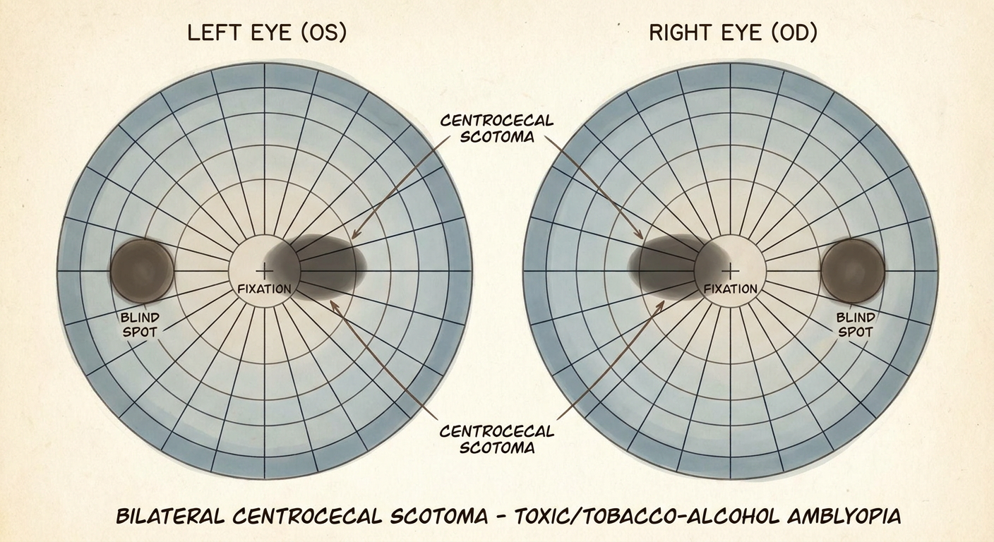
A medical ophthalmology diagram showing bilateral centrocecal scotoma visual field defects - two round visual field charts (right eye and left eye) each showing an oval-shaped dark scotoma area connecting the central fixation point to the blind spot, labeled diagram typical of toxic/tobacco-alcohol amblyopia
Toxic Amblyopia (Nutritional / Tobacco-Alcohol Amblyopia)
Definition
Toxic (nutritional) amblyopia is a bilateral, symmetrical, slowly progressive optic neuropathy caused by toxic and/or nutritional deficiency states, classically described in patients with heavy tobacco and alcohol use ("tobacco-alcohol amblyopia"). The term "amblyopia" here is used in its older sense of dimness of vision rather than the developmental squint-related amblyopia - it is really a bilateral optic neuropathy.
Etiopathogenesis
The underlying mechanism is thought to be deficient mitochondrial function in retinal ganglion cells, similar to the hereditary optic neuropathies (Leber's), which is why the papillomacular bundle (the tightly packed, metabolically demanding fibres serving central vision) is preferentially affected first.
Contributing factors:
- Vitamin B-complex deficiency - particularly B12 (cyanocobalamin) and B1 (thiamine); also B2 (riboflavin), B3 (niacin), B6 (pyridoxine)
- Direct toxic effect of tobacco (cyanide/thiocyanate) and alcohol
- Deficiency of copper, folic acid, and protein
- Strict vegan diets, poor nutrition in the elderly
- Pernicious anaemia (reduced B12 absorption)
- In resource-poor regions, pure dietary deficiency can occur epidemically (e.g., the Cuban optic/peripheral neuropathy epidemic)
- A similar picture can be produced by drug toxicity (e.g., ethambutol, isoniazid, linezolid, amiodarone, disulfiram) or environmental/occupational toxins - so "toxic optic neuropathy" is the broader umbrella term, of which tobacco-alcohol amblyopia is the classic nutritional/toxic subtype.
Clinical Features
Symptoms
- Insidious, painless, bilateral blurring of central vision
- Difficulty with colour vision, often noticed before acuity loss
- History of heavy smoking/alcohol intake, poor diet, or exposure to an offending drug/toxin
- Ask about peripheral neuropathy symptoms (numbness, gait disturbance) - suggests combined optic + peripheral neuropathy
Signs
- Visual acuity variably reduced (can range from mild to severe)
- Optic discs are usually normal-looking at presentation; some show subtle pallor, most often temporal disc pallor (site of papillomacular bundle entry)
- Pupillary reactions often normal, may be sluggish in severe cases
- Colour vision loss disproportionate to acuity loss - red desaturation
- Look for other stigmata of deficiency: Korsakoff syndrome, Wernicke encephalopathy, peripheral neuropathy, glossitis, pernicious anaemia signs
Visual field defect - the classic diagnostic hallmark
Bilateral, symmetrical centrocecal scotomas - an oval defect joining the point of fixation to the physiological blind spot. This is best demonstrated with a red target (the defect edges are poorly defined with a white target but sharply outlined with red).
Investigations
- Colour vision testing (Ishihara) - reduced disproportionately to acuity
- Perimetry - to demonstrate the centrocecal scotoma
- OCT - peripapillary RNFL thickening (early), helps exclude macular disease
- Fundus autofluorescence (FAF) - excludes cone/cone-rod dystrophy mimicking a central scotoma with dyschromatopsia
- VEP - markedly reduced P100 amplitude with normal/near-normal latency (demyelination vs axonal distinction)
- Blood work: serum and RBC folate, vitamin B12, thiamine (B1), riboflavin (B2); FBC and film (macrocytic anaemia); serum methylmalonate/homocysteine (sensitive markers of functional B12 deficiency even if serum B12 is low-normal)
- MRI brain/orbits - to exclude intracranial/compressive pathology, especially if the picture is atypical (marked asymmetry, disc pallor with good vision)
- LHON mitochondrial DNA testing - to exclude Leber hereditary optic neuropathy, an important differential/mimicker in young male "tobacco-alcohol amblyopia" cases (Leber disease can be misdiagnosed as toxic amblyopia)
Differential Diagnosis
- Leber hereditary optic neuropathy (LHON)
- Compressive optic neuropathy (chiasmal/orbital mass)
- Ethambutol/isoniazid/other drug-induced toxic optic neuropathy
- Cone or cone-rod dystrophy (central scotoma + dyschromatopsia, but disc usually normal and OCT/FAF show macular changes)
- Bilateral optic neuritis / NMOSD / MOG-antibody disease
Management
- Multidisciplinary care - co-manage with physician/neurologist
- Stop the offending agent immediately - abstain from alcohol and tobacco (compliance is often poor); withdraw any causative drug/toxin
- Dietary correction - balanced diet rich in fruit and green leafy vegetables
- Vitamin supplementation:
- Daily multivitamin
- Thiamine 100 mg twice daily
- Folate 1 mg daily
- Intramuscular hydroxocobalamin (vitamin B12) in severe/unresponsive cases - possibly acts partly by reversing cyanide toxicity (thiocyanate detoxification pathway); regimens range from weekly to monthly injections, continued long-term/for life in some cases
- Important pearl: if a patient is deficient in both B12 and folate, correct B12 first to avoid precipitating subacute combined degeneration of the spinal cord
Prognosis
Good if caught early and the patient complies with treatment, though recovery is slow - acuity recovers faster than colour perception. In advanced/neglected cases there can be substantial permanent visual deficit, but complete loss of useful vision is unusual; peripheral field is typically preserved even in severe disease.
Sources: Kanski's Clinical Ophthalmology - A Systematic Approach, 10th ed. (Nutritional optic neuropathy); Bradley and Daroff's Neurology in Clinical Practice (Tobacco-Alcohol or Nutritional Amblyopia); Adams and Victor's Principles of Neurology, 12th ed.
Exam tip: In a "toxic amblyopia" short-note question, examiners specifically want the centrocecal scotoma diagram, the B12/thiamine mechanism, and the treatment sequence (stop toxin → diet → thiamine/folate → B12, correcting B12 before folate). Also always mention LHON as a key differential since it's a classic "don't miss" comparison.
This is a shared conversation. Sign in to Orris to start your own chat.