
In case of VSD, do we have S2 wide splitting?
"ventricular septal defect" AND auscultation
ventricular septal defect S2 splitting auscultation
- Small VSD: normal S2 split.
- Moderate VSD with increased pulmonary flow: S2 may be narrowly split, often with a louder P2.
- Pulmonary hypertension/Eisenmenger physiology: P2 becomes loud and S2 splitting may become very narrow or single.
Why don't we have a two-wide split in case of VSD, as there is also blood flowing from left ventricle to right ventricle is present, right? So more amount of blood is entering into l- l- right ventricle. So a pulmonary valve has to be closed lately, whereas in l- left ventricle, the aortic valve has to close early, as it is having less amount of blood. Why isn't there?
"ventricular septal defect" AND auscultation
VSD why S2 not wide split left-to-right shunt ventricular systole physiology
Valve-closure timing is not determined simply by how much blood passes through a ventricle. It depends mainly on the timing of ventricular pressure fall relative to the great-artery pressure, plus the duration of ventricular ejection and pulmonary vascular “hangout” time.
Why VSD does not cause wide, fixed S2
What happens to A2?
Contrast with ASD
| Defect | Main time of shunt | Primary volume-loaded chamber | Typical S2 |
|---|---|---|---|
| ASD | Diastole | RV | Wide, fixed split |
| VSD | Systole | Pulmonary circulation, then LA/LV after recirculation | Usually normal or narrowly split; P2 may be loud |
ASD causes wide fixed splitting because it causes continuous RV diastolic volume overload.
VSD shunting is systolic, so it does not create the same persistent RV preload and delayed P2.
Why in VSD there is no left to right shunt in diastole?
"ventricular septal defect" AND hemodynamics
ventricular septal defect left to right shunt occurs systole not diastole ventricular pressure explanation
Pressure difference drives shunt flow
| Phase | LV pressure | RV pressure | LV to RV gradient | Result |
|---|---|---|---|---|
| Systole | about 120 mmHg | about 25 mmHg | Large | Large, fast LV-to-RV shunt |
| Diastole | low | low | Very small | Minimal, slow L-to-R flow |
Why this does not significantly preload the RV
VSD may have a small diastolic LV-to-RV flow, but the large, clinically important shunt is systolic because that is when the LV-RV pressure gradient is large.
In case of congenital cyanotic heart diseases, what do you mean by increased or decreased blood flow? What do you want to do with this?
"cyanotic congenital heart disease" AND "pulmonary blood flow"
cyanotic congenital heart disease increased decreased pulmonary blood flow classification radiograph
The amount of blood reaching the lungs through the pulmonary arteries for oxygenation.
Why do we care?
- How severe the cyanosis is
- Chest X-ray pulmonary vascular markings
- Whether the child tends toward cyanotic spells versus heart failure
- The likely diagnosis and immediate treatment
1. Decreased pulmonary blood flow
- More marked cyanosis
- Often cyanotic spells in TOF
- Chest X-ray: oligemic lung fields, meaning reduced pulmonary vascular markings or “dark” lungs
- Usually no pulmonary overcirculation or early heart failure
2. Increased pulmonary blood flow
- Cyanosis may be less dramatic than in low-pulmonary-flow lesions, though it can still be dangerous
- Chest X-ray: plethoric lungs, meaning prominent pulmonary vascular markings
- Pulmonary congestion and heart failure are more likely because the lungs receive too much flow
High-yield contrast
| Feature | Decreased pulmonary blood flow | Increased pulmonary blood flow |
|---|---|---|
| Blood reaching lungs | Too little | Too much |
| Lung X-ray | Oligemia, dark lung fields | Plethora, prominent vascular markings |
| Cyanosis | Often marked | Often milder, but variable |
| Heart failure | Less common initially | Commoner |
| Typical example | TOF | TGA with VSD / truncus |
The baby is blue partly because too little blood is being sent to the lungs to become oxygenated.
Plenty of blood reaches the lungs, but because of abnormal mixing or abnormal connections, the blood delivered to the body is still inadequately oxygenated.
How can we differentiate the lesions of increased blood flow in cynotic congential heart disease
"cyanotic congenital heart disease" AND diagnosis
cyanotic congenital heart disease increased pulmonary blood flow differentiate TGA TAPVC truncus single ventricle chest x ray
- Prominent hilar vessels extending to lung periphery
- Often cardiomegaly
- Sometimes pulmonary edema or heart-failure signs
High-yield approach
| Lesion with increased pulmonary blood flow | Typical clue | Chest X-ray | Other useful clue |
|---|---|---|---|
| TGA | Severe cyanosis in first hours to days of life, often little murmur | Narrow superior mediastinum with oval cardiac shadow: “egg on a string”; pulmonary plethora, especially if VSD/PDA | Usually RVH in neonates. Urgent echo confirms ventriculoarterial discordance. |
| TAPVC, unobstructed | Cyanosis plus heart failure, usually after a few weeks | Pulmonary plethora and cardiomegaly; “snowman/figure-of-8” occurs in supracardiac TAPVC, usually later, not in a newborn | Fixed split S2 and flow murmur may occur. If obstructed, there is severe respiratory distress and pulmonary edema, and the classic snowman is absent. |
| Truncus arteriosus | Mild cyanosis with early, prominent heart failure | Cardiomegaly with pulmonary plethora; a right aortic arch supports the diagnosis | Single loud S2, bounding pulses/wide pulse pressure, ejection click or truncal regurgitation murmur. |
| Single ventricle, unrestricted pulmonary flow | Mild cyanosis plus heart failure | Cardiomegaly and pulmonary plethora | Anatomy is variable. Echo defines the single-ventricle anatomy and outflow obstruction, if any. |
| DORV without pulmonary stenosis | Cyanosis severity depends on VSD position and physiology | Usually pulmonary plethora, often cardiomegaly | May resemble TGA physiologically. Echo is required to define the relation of great arteries and VSD. |
| Tricuspid atresia without pulmonary stenosis | Cyanosis with variable heart failure | Pulmonary plethora if there is no PS | ECG clue: left-axis deviation is classic. |
| HLHS or other left-sided obstructive lesions | Shock, poor pulses, acidosis as ductus closes, sometimes differential saturation | Often cardiomegaly and pulmonary venous congestion | Weak lower-body pulses and systemic hypoperfusion are more striking than cyanosis. |
Fast bedside distinctions
1. Very early, intense cyanosis with relatively quiet chest
- A baby may look very blue but not have major respiratory distress initially.
- The murmur may be absent or soft.
- CXR may show “egg on a string,” although this is not always present early.
2. Cyanosis plus marked respiratory distress and pulmonary edema
- This can resemble severe lung disease or sepsis.
- The child is very sick, tachypneic, and hypoxemic.
- Unlike unobstructed TAPVC, pulmonary venous obstruction causes pulmonary edema rather than simple pulmonary plethora.
3. Mild cyanosis + bounding pulses + early heart failure
- One arterial trunk supplies systemic and pulmonary circulations, so pulmonary flow is excessive as pulmonary vascular resistance falls after birth.
- A single loud S2 is a useful clue.
4. Cyanosis + heart failure, but no characteristic silhouette
A practical examination mnemonic
T-T-T-S-D
TGA
TAPVC
Truncus arteriosus
Single ventricle
DORV
Important limitation
- TGA with pulmonary stenosis may have decreased pulmonary flow.
- TAPVC with venous obstruction produces pulmonary edema and may not look simply plethoric.
- Single ventricle, DORV, and tricuspid atresia may have either increased or decreased pulmonary flow depending mainly on pulmonary stenosis/outflow obstruction.

Explain this
"cyanotic congenital heart disease" AND "pulmonary blood flow"
cyanotic CHD increased pulmonary blood flow TGA TAPVC chest xray egg on string snowman
Read the note as this flowchart
- Chest X-ray shows increased pulmonary vascular markings or pulmonary plethora
- The infant may develop congestive cardiac failure (CCF/CHF): tachypnea, poor feeding, sweating during feeds, hepatomegaly, poor weight gain
- Cyanosis is often less deep than in lesions with decreased PBF, but this is not always true
What “vascular marking/plethora” means
- Pulmonary arteries are enlarged
- Vessels are more visible even in outer lung fields
- Lungs look “busy” or plethoric
The two branches shown in the note
1. TAPVC with an ASD/PFO for obligatory mixing
TAPVC
“ASD ↓ TAPVC”
TAPVC requires an atrial-level communication, ASD/PFO, for survival.
Chest X-ray
- Cardiomegaly
- Increased pulmonary vascular markings
- The classic snowman sign or figure-of-8 sign
Important exception
2. TGA with a VSD, ASD/PFO, or PDA for mixing
TGA
- Deoxygenated systemic venous blood returns to RA → RV → aorta → body
- Oxygenated pulmonary venous blood returns to LA → LV → pulmonary artery → lungs
- ASD/PFO
- VSD
- PDA
“VSD ↓ TGA”
A VSD is one route for mixing in TGA, and TGA with VSD often has increased pulmonary flow.
Why TGA with VSD has increased PBF
Chest X-ray
- Egg-on-a-string or egg-on-side
- Narrow superior mediastinum, the “string”
- Increased pulmonary vascular markings
Corrected one-page summary
| Disease | Why cyanosis occurs | Why PBF is increased | Necessary/associated communication | Classic X-ray |
|---|---|---|---|---|
| TAPVC, unobstructed | Oxygenated pulmonary venous blood mixes with systemic venous blood on right side | All pulmonary venous return recirculates through right heart and lungs | ASD/PFO required for systemic output | Pulmonary plethora, cardiomegaly; snowman sign in supracardiac type later in infancy |
| TGA with VSD | Parallel circulations, with mixing of oxygenated and deoxygenated blood | VSD allows increased flow to pulmonary circuit | VSD, ASD/PFO, or PDA allows mixing | Egg-on-a-string, pulmonary plethora |
| Truncus arteriosus | One common arterial trunk receives mixed blood | Pulmonary arteries arise from common trunk without protective pulmonary stenosis | Usually associated VSD | Cardiomegaly, pulmonary plethora, often right aortic arch |
| Single ventricle/DORV without PS | Complete or substantial mixing | No significant obstruction to pulmonary outflow | Anatomy varies | Pulmonary plethora, often cardiomegaly |
One correction to the handwritten sentence
“Due to mixing they have mild cyanosis”
- Unobstructed TAPVC and truncus arteriosus may have mild to moderate cyanosis initially, with early CHF.
- TGA can cause profound cyanosis from birth, especially if there is poor mixing through ASD, VSD, or PDA.
- The degree of cyanosis depends on how much mixing occurs, pulmonary blood flow, and pulmonary venous obstruction.
Cyanosis + pulmonary plethora = think mixing lesion.
The main exam examples are TGA, unobstructed TAPVC, truncus arteriosus, single ventricle, and DORV without pulmonary stenosis.
What is mean by overriding of iota?
Quick answer
The aorta is “straddling” the VSD instead of being connected only to the LV.
- Mild overriding: aorta receives mostly LV blood.
- Marked overriding: aorta receives substantial RV blood.
- If the aorta arises almost entirely from the RV, it may be classified as double-outlet right ventricle (DORV).
, Overriding of Aorta.
- oxygenated blood from the LV, and
- deoxygenated blood from the RV.
Pathology of tof
anatomical diagram Tetralogy of Fallot VSD overriding aorta pulmonary stenosis right ventricular hypertrophy
"tetralogy of Fallot" AND pathophysiology
Tetralogy of Fallot pathology embryology anterior deviation infundibular septum authoritative
Tetralogy of Fallot: pathology and pathophysiology
Primary embryological defect
- Narrowing of the right ventricular outflow tract (RVOT)
- Failure of normal ventricular septum alignment, producing a large malalignment VSD
- The aorta shifts rightward and lies over the VSD, causing an overriding aorta
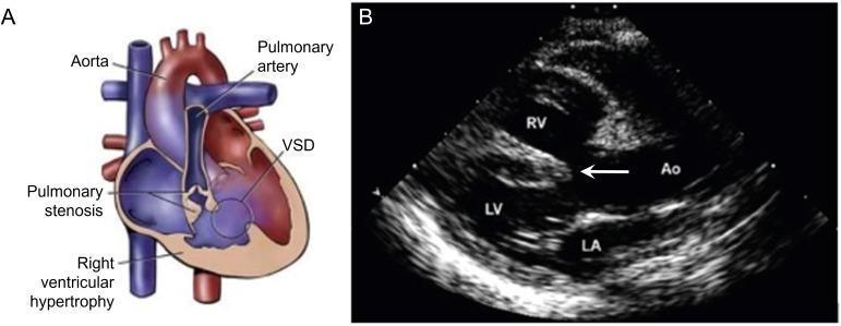
The four components
1. Pulmonary stenosis / RV outflow tract obstruction
2. Large VSD
- If RVOT obstruction is mild: blood can go into pulmonary artery, often left-to-right shunt, little or no cyanosis. This is called pink TOF.
- If RVOT obstruction is severe: blood is diverted through VSD into the aorta, right-to-left shunt, causing cyanosis.
3. Overriding aorta
4. Right ventricular hypertrophy
Central mechanism of cyanosis
Why “tet spells” occur
- Sudden infundibular muscle spasm, worsening RVOT obstruction
- Fall in systemic vascular resistance
Associations
- 22q11.2 deletion syndrome (DiGeorge syndrome)
- Right-sided aortic arch
- Coronary artery anomalies
- Pulmonary atresia in severe forms
Reason for ejection systolic murmur in tof
"tetralogy of Fallot" AND auscultation
Tetralogy of Fallot ejection systolic murmur RV outflow obstruction explanation
- Systolic ejection, because flow across the RVOT occurs during ventricular ejection
- Harsh, crescendo-decrescendo
- Best heard at the left upper or mid-sternal border
- May have a systolic thrill
Is the murmur from the VSD?
In TOF, the murmur is generally from pulmonary stenosis/RVOT obstruction, not from the VSD.
Important clinical correlation
A very cyanotic TOF child with a soft or disappearing murmur is more severe, not better.