
Sudden onset dysphagia
dysphagia sudden onset etiology diagnosis
dysphagia causes classification algorithm oropharyngeal esophageal
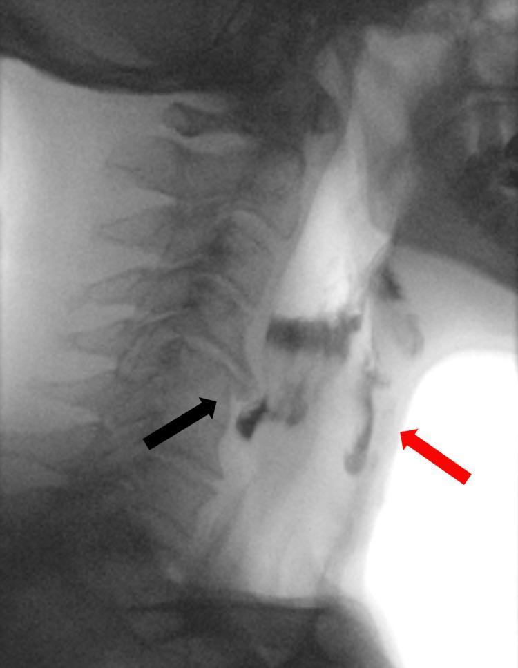
A lateral view videofluoroscopic swallowing study (VFSS) image demonstrating oropharyngeal dysphagia. The image captures the cervical spine and the pharyngeal/esophageal soft tissue structures during the transit of a radiopaque contrast bolus. A prominent anterior osteophyte at the C5 vertebral body level (indicated by a black arrow) causes focal extrinsic compression and narrowing of the proximal cervical esophagus. This mechanical obstruction leads to the stasis and pooling of the contrast material above the upper esophageal sphincter. Significantly, contrast is seen misdirected into the laryngeal vestibule and trachea (indicated by a red arrow), confirming laryngeal penetration and tracheal aspiration. This diagnostic image illustrates the clinical relationship between cervical spondylosis (DISH or severe osteophytosis) and mechanical dysphagia, highlighting the risk of respiratory complications due to impaired bolus clearance and subsequent airway entry.
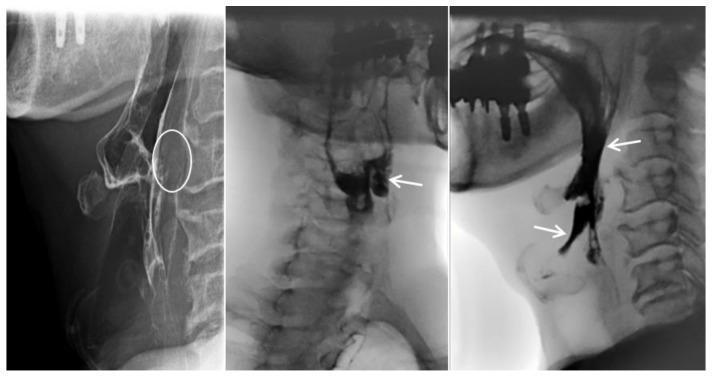
This diagnostic image consists of three lateral-view cervical esophagrams (fluoroscopic barium swallow study) demonstrating oropharyngeal dysphagia and mechanical obstruction. The first panel highlights prevertebral soft tissue swelling at the C3–C4 level (white circle), which causes extrinsic compression and anterior displacement of the esophagus. The subsequent panels illustrate functional swallowing impairment: radiopaque contrast material is seen abnormally pooling within the laryngeal vestibule and epiglottic space. White arrows indicate laryngeal penetration and aspiration, where contrast moves anteriorly into the airway rather than proceeding through the upper esophageal sphincter. These findings are clinically significant for diagnosing mechanical dysphagia, in this case secondary to an anteriorly extruded cervical disc. Key educational concepts include the radiographic identification of aspiration, the assessment of prevertebral soft tissue thickness, and the recognition of extrinsic esophageal compression in the context of cervical spine pathology.
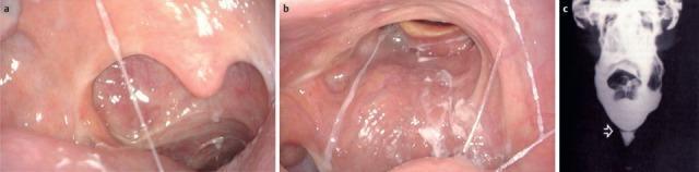
This composite educational clinical figure illustrates oropharyngeal and esophageal pathologies contributing to dysphagia. Image (a) is a transoral endoscopic view of an 87-year-old patient showcasing post-surgical changes following an extended partial resection of the right oropharynx and neck dissection. Visible features include a disrupted pharyngeal wall structure on the right and distorted mucosa consistent with radiotherapy and chemotherapy effects. Image (b) presents a transnasal endoscopic view of the velum and posterior oropharyngeal wall. It demonstrates significant scarring and asymmetrical contraction of the right-sided velum, with visible scar tissue extending across the posterior pharyngeal wall. Prominent white, viscous saliva is seen spanning the lumen, indicating impaired clearance and a transportation disorder. Image (c) is a contrast radiography (barium swallow) from a different patient, identifying tissue formations (indicated by an arrow) in the pharynx or esophagus that create mechanical obstacles. Together, these images teach how oncological treatments, scarring, and structural abnormalities disrupt the swallowing mechanism.
Sudden Onset Dysphagia
Step 1: Localize - Oropharyngeal vs. Esophageal
| Feature | Oropharyngeal (Transfer) | Esophageal |
|---|---|---|
| Timing | Difficulty within 1 second of swallowing | Delay several seconds after initiation |
| Location felt | Cervical / suprasternal notch | Mid-chest or lower |
| Associated symptoms | Coughing, choking, nasal regurgitation, drooling | Food "sticks," weight loss, heartburn |
| Type of bolus | Liquids often worse than solids | Solids typically worse initially |
- Sleisenger & Fordtran's Gastrointestinal and Liver Disease, p. 189-190
- Rosen's Emergency Medicine, p. 1217
Step 2: Causes of Sudden Onset Dysphagia
A. Oropharyngeal Causes
Neuromuscular (sudden-onset culprits)
- Stroke (CVA) - most common; pharyngeal weakness with failure of cricopharyngeus relaxation; onset is abrupt
- Myasthenia gravis - dysphagia is the presenting symptom in ~15%; worsens with repeated swallowing
- Botulism - infectious cause; descending paralysis
- Polymyositis / Dermatomyositis - inflammatory myopathy affecting palate, pharynx, upper esophagus
- Multiple sclerosis - demyelinating; sudden flares possible
- ALS (Lou Gehrig disease), Parkinson disease, Postpolio syndrome
- Thyroid dysfunction, Muscular dystrophy
Structural
- Zenker diverticulum - gurgling on swallowing, regurgitation of undigested food
- Carcinoma of the pharynx or proximal esophagus
- Proximal esophageal web (e.g., Plummer-Vinson syndrome)
- Osteophytes / cervical spondylosis - extrinsic compression (see image below)
- Infections of the pharynx or neck, thyromegaly, prior radiation/surgery
B. Esophageal Causes
Structural - Intrinsic
| Condition | Key feature |
|---|---|
| Food bolus impaction / foreign body | True sudden onset; often at a meal |
| Esophageal carcinoma | Age >50, progressive, weight loss |
| Peptic stricture (GERD) | Long history of heartburn; progressive solids |
| Schatzki ring | Episodic, nonprogressive; first episode often at a hurried meal with alcohol |
| Eosinophilic esophagitis (EoE) | Intermittent, young males; impaction risk |
| Medication-induced stricture | e.g., doxycycline, NSAIDs, bisphosphonates |
| Diverticula | Regurgitation of old undigested food |
Structural - Extrinsic
- Mediastinal mass, thoracic aortic aneurysm
- Dysphagia lusoria - aberrant subclavian artery compressing the esophagus
- Spinal osteophytes
Motility / Neuromuscular
| Condition | Key feature |
|---|---|
| Achalasia | Insidious progressive; solids AND liquids; bland regurgitation; weight loss |
| Distal esophageal spasm | Intermittent; chest pain; sensitive to hot/cold liquids |
| Hypercontractile (Jackhammer) esophagus | Chest pain + dysphagia |
| Scleroderma | Raynaud phenomenon; severe dysphagia signals peptic stricture |
| Chagas disease | Achalasia-like; relevant travel/endemic history |
| Reflux-related dysmotility | Background GERD |
- Sleisenger & Fordtran's, p. 190 (Boxes 13.1 and 13.2)
- Rosen's Emergency Medicine, p. 1218
Diagnostic Algorithm
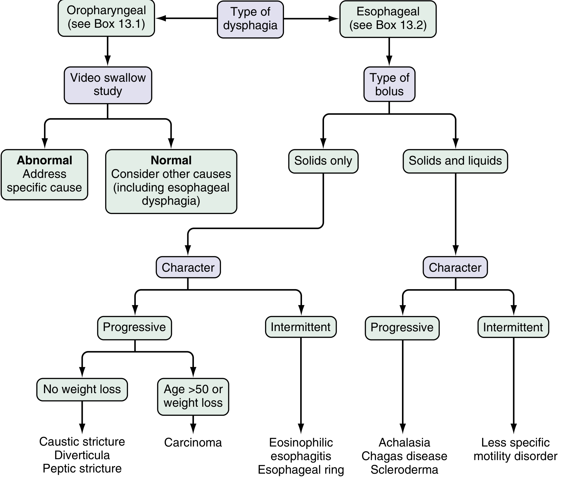
Three Key Diagnostic Questions for Esophageal Dysphagia
-
Solids only, or solids AND liquids?
- Solids only → suspect mechanical obstruction (ring, stricture, carcinoma)
- Both → suspect motility disorder (achalasia, spasm, scleroderma)
-
Intermittent or progressive?
- Intermittent → Schatzki ring, eosinophilic esophagitis, spasm
- Progressive → peptic stricture, carcinoma
-
Is there heartburn / weight loss?
- Heartburn + no weight loss → peptic stricture
- Age >50 + weight loss + rapidly progressive → carcinoma until proven otherwise
Emergency Red Flags (Sudden Onset)
| Feature | Concern |
|---|---|
| Associated new neurological deficits | Stroke / brainstem infarction |
| Fever + odynophagia | Retropharyngeal abscess, epiglottitis, esophagitis |
| Complete inability to swallow (sialorrhea) | High-grade obstruction, foreign body |
| Chest pain + dysphagia after vomiting/procedure | Esophageal perforation (Boerhaave) |
| Stridor / respiratory compromise | Airway emergency - foreign body |
| Recent choking episode at a meal | Food bolus impaction |
Investigations
| Investigation | Use |
|---|---|
| Barium swallow | First-line for structural and motility assessment |
| Upper GI endoscopy (EGD) | Visualize + biopsy + therapeutic (foreign body, dilation) |
| Videofluoroscopic swallowing study (VFSS) | Gold standard for oropharyngeal dysphagia |
| High-resolution manometry | Motility disorders (achalasia, spasm) |
| CT chest/neck | Extrinsic compression, mediastinal mass |
| Neurological workup | If stroke, MG, or MS suspected |
Summary Point
- Sleisenger & Fordtran's Gastrointestinal and Liver Disease, p. 189-192
- Rosen's Emergency Medicine, p. 1217-1218
- Yamada's Textbook of Gastroenterology, 7th ed., p. 628