
Hemodynamic disorders. Thrombosis. Etiopathogenesis and morphology. in detail
Reading File
Reading File
Finding Sources
Finding Sources
Searching PubMed
"thrombosis"[MeSH Terms] AND "pathogenesis"
Reading File
Reading File
Reading File
Reading File
Reading File
Reading File
Reading File
Searching Images
thrombus lines of Zahn microscopy
Reading File
query_images_db
Thrombosis: Etiopathogenesis and Morphology
1. Where thrombosis fits among hemodynamic disorders
Hemodynamic disorders cover edema, hyperemia and congestion, hemorrhage, hemostasis and thrombosis, embolism, infarction and shock. Hemostasis is the normal, localized clot that stops bleeding. Thrombosis is the inappropriate formation of a clot (thrombus) from the blood's own components within the intact cardiovascular system. Thrombosis underlies the most common and serious forms of cardiovascular disease, and it feeds embolism and infarction (Robbins, Cotran & Kumar Pathologic Basis of Disease, Ch. 4).
2. Etiopathogenesis: Virchow triad
The primary abnormalities that lead to thrombosis are (1) endothelial injury, (2) stasis or turbulent blood flow, and (3) hypercoagulability. Endothelial integrity is the most important factor. The three factors interact: endothelial injury alters flow and coagulability, and abnormal flow in turn injures endothelium.
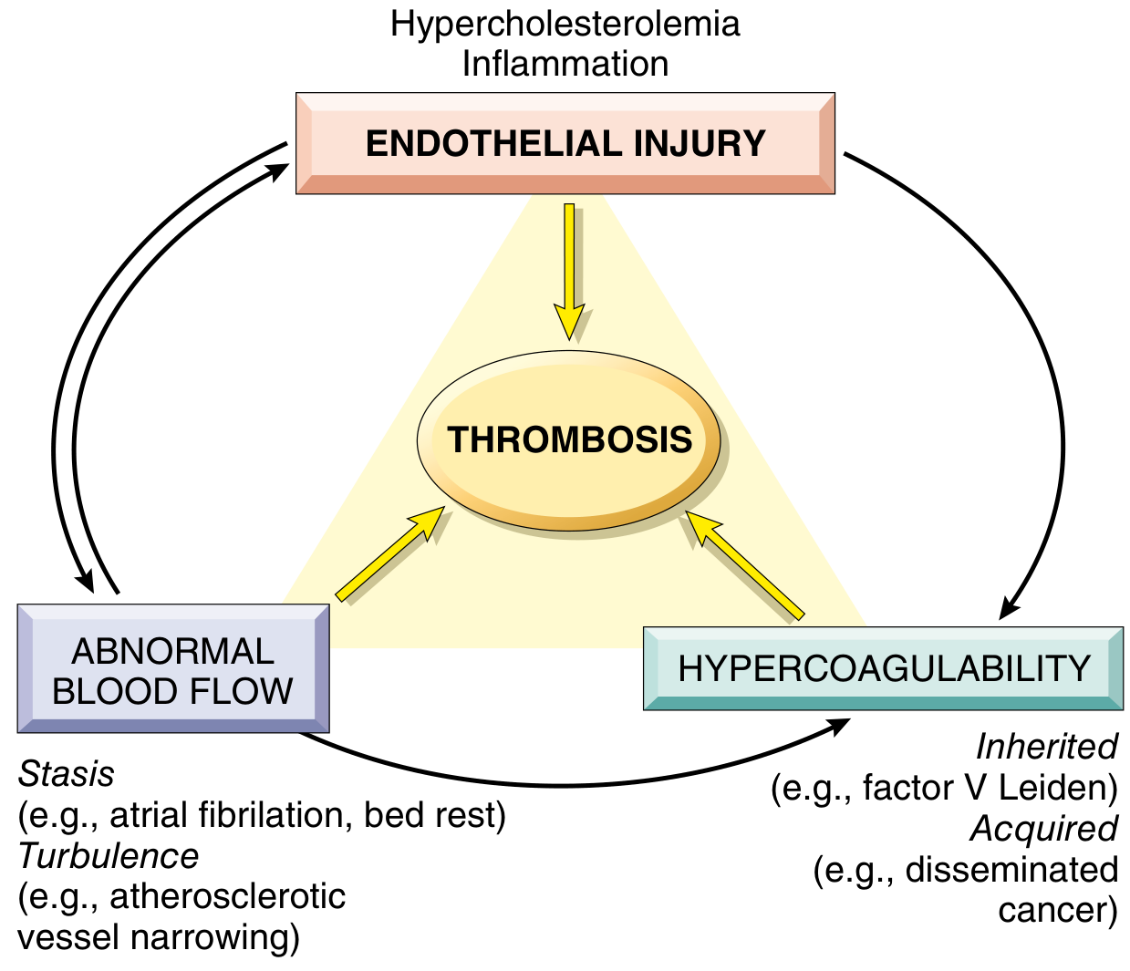
A. Endothelial injury
- Endothelial injury and platelet activation almost inevitably underlie thrombus formation in the heart and arteries. Blood flow is fast there, which impedes clotting.
- Platelet adhesion and activation is a prerequisite for thrombosis under high shear stress. This is why aspirin and other antiplatelet drugs are used in coronary disease and acute MI.
- Severe injury exposes subendothelial von Willebrand factor (vWF), collagen and tissue factor.
- Endothelial activation or dysfunction does not need gross denudation. Its causes include:
- physical injury
- infection
- abnormal flow
- inflammatory mediators (cytokines)
- hypercholesterolemia and homocystinemia
- cigarette smoke toxins
- Activated endothelium becomes prothrombotic in three ways:
- Procoagulant changes: thrombomodulin, protein C and tissue factor pathway inhibitor are downregulated, so thrombin activity is enhanced.
- Antifibrinolytic effects: plasminogen activator inhibitors (PAIs) are secreted and t-PA is downregulated.
- Platelet and leukocyte adhesion increases.
- Typical clinical settings are ulcerated atherosclerotic plaque, myocardial infarction with endocardial injury, vasculitis, trauma, and hemodynamic stress such as hypertension.
B. Alterations in normal blood flow (stasis and turbulence)
Normally, flow is laminar. Platelets run centrally, separated from the endothelium by a slower plasma layer. Turbulence and stasis cause three problems:
- They activate endothelium, which increases procoagulant activity and leukocyte adhesion.
- They disrupt laminar flow and bring platelets into contact with the endothelium.
- They prevent washout and dilution of activated clotting factors and the inflow of clotting factor inhibitors.
Turbulence is more important in arterial and cardiac thrombosis. It injures endothelium and forms countercurrents and local pockets of stasis. Stasis is the major contributor to venous thrombi.
Clinical examples:
- Ulcerated atherosclerotic plaques expose vWF and tissue factor and also cause turbulence.
- Aneurysms of the aorta and arteries cause local stasis.
- MI with non-contractile myocardium or ventricular aneurysm promotes mural thrombi.
- Rheumatic mitral stenosis with a dilated left atrium and atrial fibrillation gives profound stasis.
- Hyperviscosity (polycythemia vera) increases resistance to flow and causes small vessel stasis.
- Sickle cell anemia: deformed red cells impede flow in small vessels.
C. Hypercoagulability (thrombophilia)
It is particularly important in venous thrombosis. It is divided into primary (genetic) and secondary (acquired) causes.
Primary (inherited):
| Disorder | Key points |
|---|---|
| Factor V Leiden (Arg506Gln) | Factor V is resistant to cleavage by activated protein C, so a key anticoagulant pathway is lost. Present in 2 to 15% of people of European ancestry and in up to ~60% of patients with recurrent DVT. Autosomal dominant. Relative risk of venous thrombosis is 4 to 8 times higher in heterozygotes and ~12 times higher in homozygotes. |
| Prothrombin G20210A | Noncoding 3' UTR variant, found in 1 to 2% of people. Prothrombin levels are raised. Risk is 2 to 4 times higher in heterozygotes and ~7 times higher in homozygotes. |
| Antithrombin III, protein C, protein S deficiency | Rare. Venous thrombosis and recurrent thromboembolism begin in adolescence or early adulthood. |
| Homocysteinemia | Inherited (cystathionine β-synthase deficiency) or acquired (B6, B12 or folate deficiency). The prothrombotic effect is inconsistent across studies. |
| Raised factors VIII, IX, XI or fibrinogen; fibrinolysis defects | Common or very rare, with unknown genetics in many cases. |
Heterozygous factor V Leiden and prothrombin mutations alone give only a moderately increased risk. They become important when combined with acquired risk factors such as pregnancy, immobilization or long-haul flights. Always consider inherited causes in patients younger than 50 with thrombosis, even if acquired risk factors are present.
Secondary (acquired):
- Strong risk factors:
- prolonged bed rest or immobilization
- MI
- atrial fibrillation
- tissue injury (surgery, fracture, burn)
- cancer
- prosthetic cardiac valves
- DIC
- heparin-induced thrombocytopenia (HIT)
- antiphospholipid syndrome
- Other risk factors:
- cardiomyopathy
- nephrotic syndrome (loss of antithrombin)
- pregnancy and postpartum
- oral contraceptives
- sickle cell anemia
- smoking
- obesity
- advancing age (possibly from reduced endothelial PGI2)
Mechanisms differ by cause:
- Oral contraceptives and the hyperestrogenic state of pregnancy increase hepatic synthesis of coagulation factors and reduce anticoagulant synthesis.
- Disseminated cancers release procoagulants (Trousseau syndrome, migratory thrombophlebitis).
- In heart failure and trauma, stasis or vascular injury dominates.
Heparin-induced thrombocytopenia (HIT)
- IgG antibodies form against heparin-platelet factor 4 (PF4) complexes. Heparin displaces PF4 from endothelium and induces a conformational change that creates a neoantigen.
- The antibody-PF4 complexes bind platelet Fc receptors and activate the platelets, which become procoagulant. Macrophages also remove antibody-coated platelets, causing thrombocytopenia.
- Thrombosis occurs in about 50% of cases, in both veins and arteries. Sequelae include skin necrosis, limb gangrene, stroke and MI.
- Low molecular weight heparin carries a lower risk. Direct factor Xa or thrombin inhibitors avoid it.
- Vaccine-induced thrombotic thrombocytopenia (VITT) with adenoviral vector COVID-19 vaccines is a similar anti-PF4 mechanism. It can cause cerebral venous sinus thrombosis.
Antiphospholipid antibody syndrome (APS)
- Features: autoantibodies, venous or arterial thromboses, and pregnancy complications (recurrent miscarriage, fetal death, preterm birth, preeclampsia), plus thrombocytopenia and valve vegetations.
- Antibodies target anionic phospholipid-binding proteins, especially β2-glycoprotein I and cardiolipin. They activate endothelium, platelets, monocytes and neutrophils, activate complement and inhibit fibrinolysis.
- Early fetal loss mainly reflects antibody interference with trophoblast growth and differentiation (failed placentation), not thrombosis.
- About 50% of cases are primary. The rest are secondary, most often to SLE. Antibodies are found in 5 to 15% of healthy people, so a "second hit" (infection, smoking, pregnancy) is probably needed.
- Lupus anticoagulant paradox: in vitro the antibodies inhibit phospholipid-dependent tests and prolong the PTT, but in vivo they cause thrombosis. A false-positive VDRL/syphilis test occurs because the test antigen contains cardiolipin.
D. Link to normal hemostasis (mechanistic summary)
- Exposed vWF and collagen cause platelet adhesion (GPIb), then activation, ADP and TxA2 release, and aggregation (GPIIb/IIIa-fibrinogen).
- Tissue factor activates the extrinsic pathway and generates thrombin. Thrombin cleaves fibrinogen to fibrin and further activates platelets.
- The counter-regulatory systems are:
- antithrombin III
- the protein C/S-thrombomodulin pathway
- tissue factor pathway inhibitor
- t-PA/plasmin fibrinolysis
- endothelial PGI2, NO and adenosine diphosphatase
- Thrombosis results when the balance tips toward procoagulant activity.
3. Morphology
General features
- Thrombi can arise anywhere in the cardiovascular system. Size and shape depend on the site and cause.
- Arterial and cardiac thrombi begin at sites of turbulence or endothelial injury. Venous thrombi occur at sites of stasis.
- Thrombi are focally attached to the vessel surface at the point of origin.
- Arterial thrombi grow retrograde. Venous thrombi extend in the direction of blood flow. Both propagate toward the heart.
- The propagating tail is poorly attached and prone to fragmentation and embolization.
- Lines of Zahn are visible grossly and microscopically. They are laminations of pale platelet-fibrin layers alternating with darker red cell-rich layers. They show the thrombus formed in flowing blood, so they distinguish antemortem thrombi from bland, nonlaminated postmortem clots.
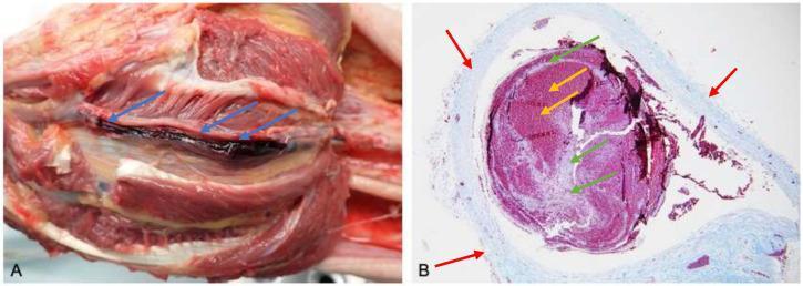
Posterior tibial vein thrombus (A) and a section showing lines of Zahn (B): red cell-rich dark layers alternating with pale fibrin-leukocyte layers.
Types by site
| Type | Features |
|---|---|
| Mural thrombi (heart chambers, aortic lumen) | Promoted by abnormal contraction (arrhythmia, dilated cardiomyopathy, MI) or endomyocardial injury (myocarditis, catheter trauma). Aortic mural thrombi lie on ulcerated atheroma or aneurysms. |
| Arterial thrombi | Frequently occlusive. Most common sites, in decreasing order, are coronary, cerebral and femoral arteries. They are friable meshworks of platelets, fibrin, red cells and degenerating leukocytes (pale, "white" or mixed thrombi). They usually overlie a ruptured atherosclerotic plaque. Vasculitis or trauma are other causes. |
| Venous thrombi (phlebothrombosis) | Almost invariably occlusive, forming a long luminal cast. They contain many entrapped red cells and relatively few platelets, so they are called red or stasis thrombi. They are firm, focally attached and show lines of Zahn. About 90% involve the lower limb veins. Other sites are upper limb veins, periprostatic and ovarian/periuterine plexuses, dural sinuses, portal vein and hepatic vein. |
| Valvular vegetations | Thrombi on heart valves. Infective (endocarditis on damaged valves) or sterile: nonbacterial thrombotic endocarditis in hypercoagulable states (e.g., cancer), and Libman-Sacks endocarditis in SLE. |
| Capillary / microvascular (hyaline) thrombi | Fibrin thrombi in DIC, seen only microscopically. |
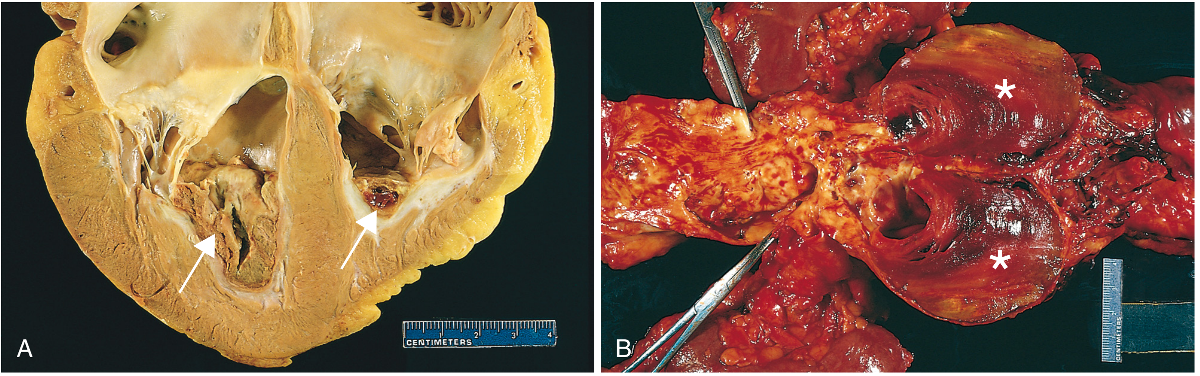
(A) Thrombus in the ventricular apices over a fibrous scar. (B) Laminated thrombus in an abdominal aortic aneurysm with friable mural thrombi on atheroma.
Antemortem thrombus versus postmortem clot
| Feature | Thrombus (antemortem) | Postmortem clot |
|---|---|---|
| Consistency | Firm, dry, friable | Gelatinous, rubbery |
| Attachment | Attached to the wall | Not attached |
| Surface/cut face | Rough, dull; lines of Zahn | Smooth, shiny; no lamination |
| Appearance | Mixed red and gray-white layers | Dark red dependent "currant jelly" portion with a yellow "chicken fat" supernatant layer |
4. Fate of the thrombus
- Propagation: more platelets and fibrin accumulate and the thrombus enlarges, possibly to total occlusion.
- Embolization: fragments travel to distant sites.
- Dissolution: fibrinolysis shrinks recent thrombi. Older thrombi have extensive cross-linked fibrin and resist lysis. This is why t-PA works only in the first few hours of a coronary thrombosis.
- Organization and recanalization: ingrowth of endothelial cells, smooth muscle cells and fibroblasts. New capillary channels partly restore the lumen. Eventually only a fibrous lump may remain, incorporated into the vessel wall.
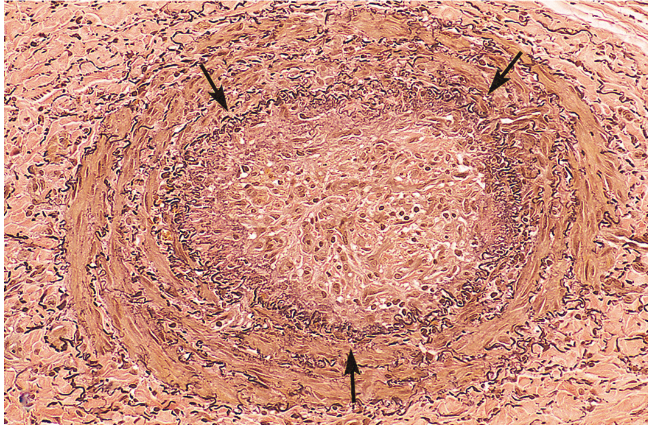
Elastic stain: the original lumen, bounded by the internal elastic lamina, is completely filled with organized thrombus.
Other outcomes:
- The center may undergo enzymatic digestion (lysosomal enzymes from trapped leukocytes and platelets).
- Bacteremia can infect the thrombus. It then erodes the wall and may form a mycotic aneurysm.
5. Clinical consequences
- Venous thrombosis
- Superficial thrombi (saphenous veins, varicosities) cause local congestion, swelling and pain. They rarely embolize, but the edema predisposes to infection and varicose ulcers.
- Deep vein thrombosis (DVT) at or above the knee (popliteal, femoral, iliac veins) is serious because of embolization to the lungs and pulmonary infarction.
- Local pain and edema are often absent because collaterals open. About 50% of DVTs are asymptomatic and are found only after embolization.
- Predisposing factors: bed rest, heart failure, trauma, surgery, burns, pregnancy, cancer and others.
- Related terms are thrombophlebitis, migratory thrombophlebitis (Trousseau sign) and phlegmasia alba dolens.
- Arterial thrombosis: the main problem is occlusion of a critical vessel, causing MI, stroke or limb ischemia and gangrene. Embolization causes downstream infarcts.
- Cardiac mural thrombi: systemic embolization, e.g., to brain, kidney or spleen.
- DIC: widespread microthrombi cause consumption of platelets and clotting factors, with ischemic injury and bleeding.
6. High-yield points
- Virchow triad: endothelial injury, stasis or turbulence, and hypercoagulability.
- Endothelial injury is the main factor for arterial and cardiac thrombi. Stasis and hypercoagulability are the main factors for venous thrombi.
- Factor V Leiden is the commonest inherited thrombophilia. The mutation is Arg506Gln and it causes protein C resistance.
- Lines of Zahn are the hallmark of an antemortem thrombus.
- Venous thrombi are red and occlusive. Arterial thrombi are pale and mixed, and often only partly occlusive.
- Four fates: propagation, embolization, dissolution, and organization/recanalization.
Recent literature check
I ran a quick PubMed search for recent reviews on thrombosis pathogenesis. Two recent systematic reviews are relevant. Red blood cells in thrombosis (PMID 40154506) argues that red cells are active participants in clot formation and stability. A review of thrombophilia (PMID 40157494) covers pathophysiology, risk factors and molecular diagnosis. Neither contradicts the textbook account above.
Source: Robbins, Cotran & Kumar Pathologic Basis of Disease, Ch. 4 (Thrombosis, Hypercoagulability, Morphology, Fate of the Thrombus). The post-mortem clot comparison table and the DIC and migratory thrombophlebitis notes also draw on standard pathology teaching beyond the retrieved text.
I can go into detail on embolism, infarction, shock or edema next, which are the other parts of hemodynamic disorders.
This is a shared conversation. Sign in to Orris to start your own chat.