
Differentiate between monosynaptic reflex and polysynaptic reflex
Reading File
Finding Sources
Finding Sources
Reading File
Reading File
Reading File
Reading File
Reading File
Loading Image
Loading Image
Reading File
Reading File
Loading Image
Reading File
I now have comprehensive content from multiple authoritative sources. Let me compile a thorough, well-structured answer.
Monosynaptic Reflex vs. Polysynaptic Reflex
Definition
| Feature | Monosynaptic Reflex | Polysynaptic Reflex |
|---|---|---|
| Synapses involved | Only one synapse between afferent and efferent neuron | Two or more synapses; one or more interneurons interposed between afferent and efferent neurons |
| Interneurons | Absent - the Ia afferent synapses directly on the alpha motor neuron | Present - can be 2 to hundreds of synaptic relays |
| Classic example | Knee jerk (stretch reflex/myotatic reflex) | Withdrawal reflex (flexion reflex); crossed extensor reflex |
| Receptor type | Muscle spindle (proprioceptor) | Cutaneous nociceptors, exteroceptors (skin, pain receptors) |
| Afferent fiber | Group Ia (large-diameter, fast myelinated) | Group II, III, IV; Aδ and C fibers |
| Neurotransmitter at central synapse | Glutamate (excitatory) | Glutamate (excitatory) + GABA/Glycine at inhibitory interneurons |
| Response speed (latency) | Very short (~20-30 ms) - fastest possible | Longer latency due to additional synaptic delays |
| Complexity of response | Simple, stereotyped: same muscle contracts | Complex: involves flexors, extensors, contralateral limb |
| Scope | Ipsilateral, single-muscle group | Can be bilateral (crossed extensor) and multi-segmental |
| Susceptibility to fatigue | Less susceptible | More susceptible (more synapses = more fatigue) |
| Modifiability | Less modifiable | Highly modifiable by descending inputs and experience |
| Frequency | Rare in practice | Most reflexes in the body are polysynaptic |
Monosynaptic Reflex: The Stretch Reflex
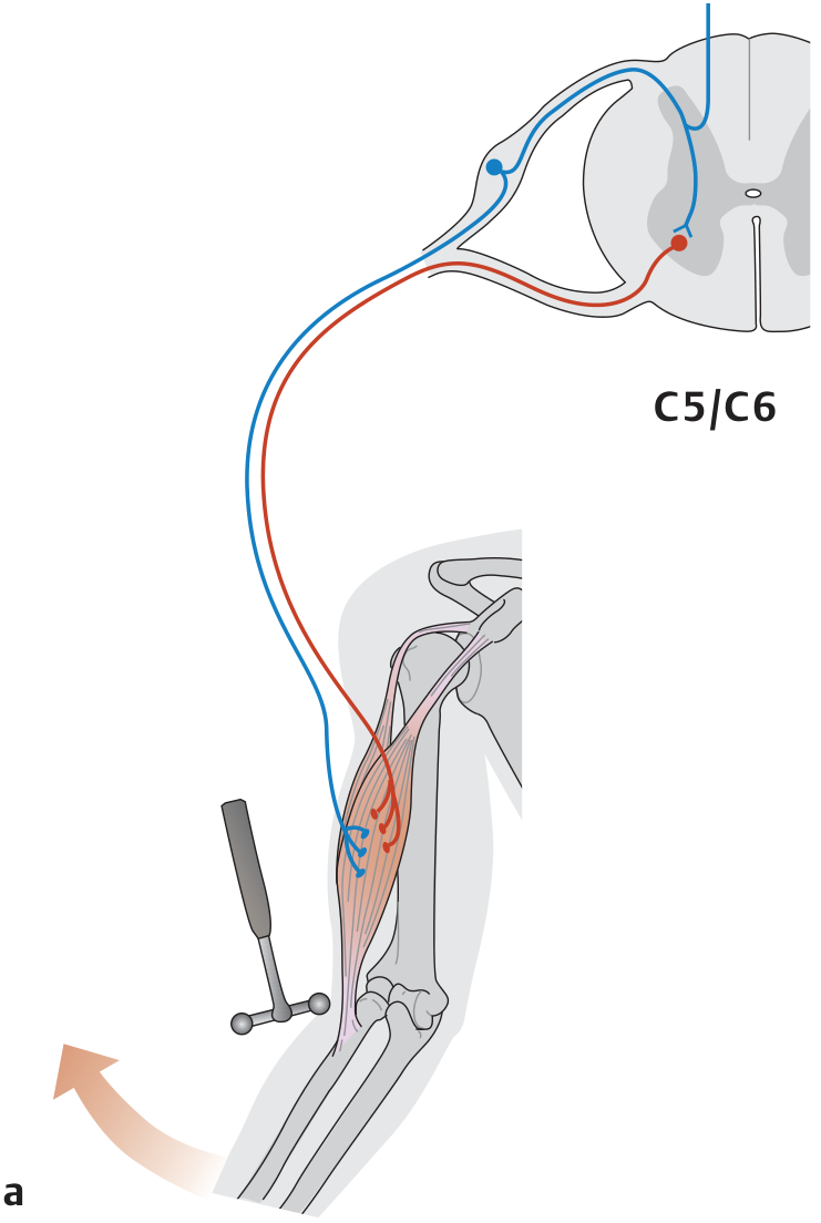
The stretch reflex (myotatic reflex) is the only truly monosynaptic reflex in the human body. When a skeletal muscle with an intact nerve supply is stretched, it contracts reflexively.
Circuit:
Muscle spindle (receptor) → Ia afferent fiber → dorsal root ganglion → direct synapse on alpha motor neuron in ventral horn → same (homonymous) muscle contracts
The Ia sensory axon also contacts interneurons that inhibit antagonist muscle motor neurons (reciprocal innervation), so while the agonist contracts, the antagonist relaxes. This inhibitory arm itself involves an interneuron and is therefore disynaptic - only the excitatory arm to the agonist is monosynaptic.
Key points (from Ganong's and Kandel's):
- Stimulus is muscle stretch; receptor is the muscle spindle
- Afferents are large, fast, myelinated Group Ia fibers
- Neurotransmitter at the central synapse is glutamate
- The knee jerk (patellar tendon - L4), ankle jerk (Achilles - S1), biceps reflex (C5/C6), and triceps reflex (C7) are all examples
Clinical grading of stretch reflexes (DTRs):
- 0 = absent, 1+ = hypoactive, 2+ = normal, 3+ = hyperactive without clonus, 4+ = hyperactive with mild clonus, 5+ = sustained clonus
- Absent DTR suggests a lesion anywhere in the reflex arc (muscle spindle, Ia afferent, or motor neuron) - e.g., peripheral neuropathy
- Hyperactive DTR suggests an upper motor neuron lesion (loss of descending inhibitory control)
Polysynaptic Reflex: The Withdrawal (Flexion) Reflex
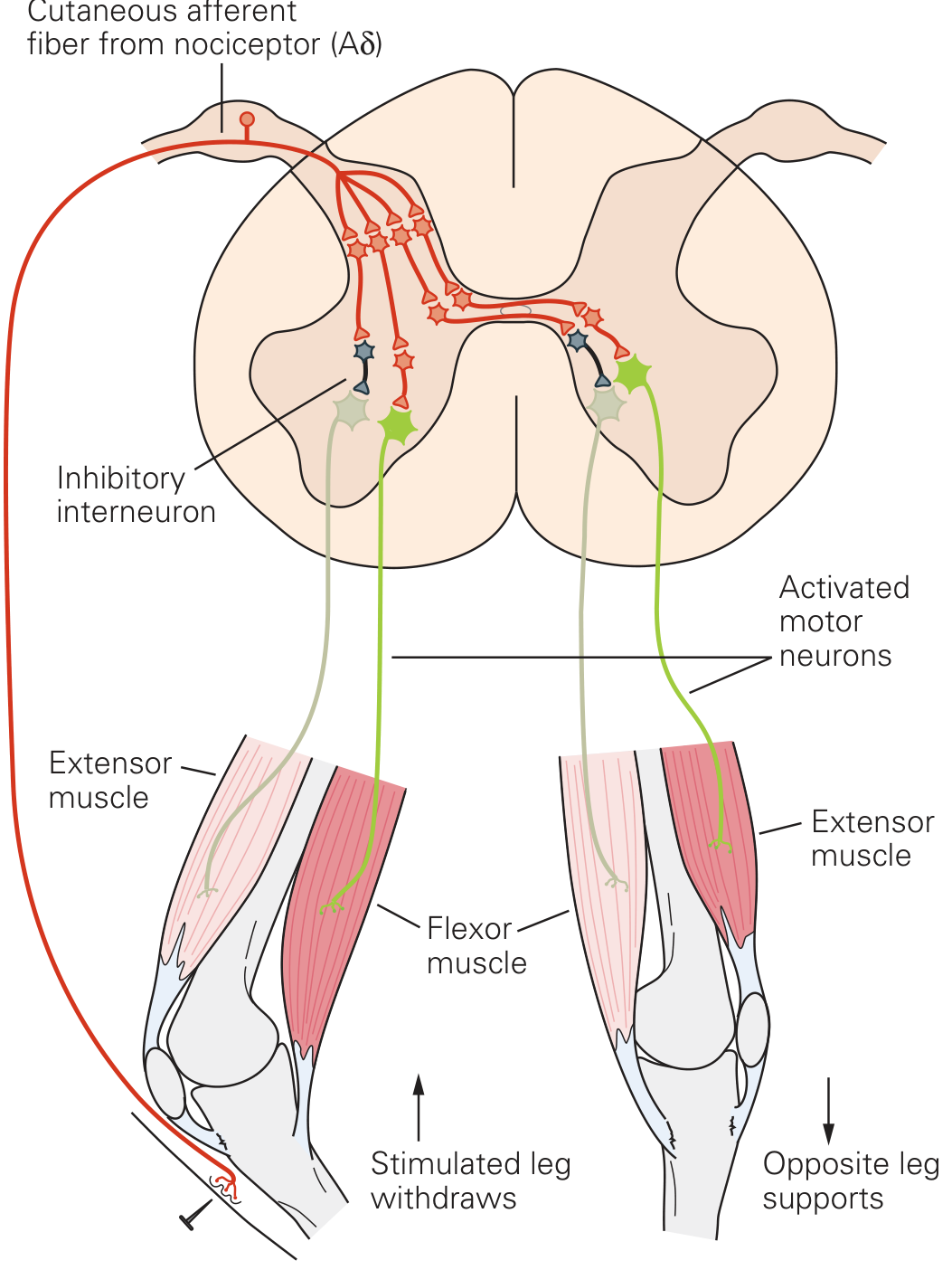
The withdrawal reflex is the prototype polysynaptic reflex. A noxious stimulus to a limb causes that limb to withdraw while the contralateral limb extends to support the body (crossed extensor reflex).
Circuit:
Nociceptor in skin (receptor) → Aδ afferent fiber → excitatory interneurons → ipsilateral flexor motor neurons (flexion/withdrawal) AND → inhibitory interneurons → ipsilateral extensor motor neurons (relaxed) AND crossed excitatory interneurons → contralateral extensor motor neurons (contract to support weight)
Key points:
- Multiple interneurons allow convergence of many sensory inputs
- The response is bilateral and multi-segmental
- Interneurons allow the reflex to be modified by descending cortical and brainstem inputs
- Inhibitory interneurons (using GABA and glycine) ensure antagonist muscles relax during the response
- The tonic vibration reflex also has a long latency implying a polysynaptic pathway
Key Differentiating Features - Summary
Monosynaptic reflex (stretch reflex):
- One synapse only
- Receptor = muscle spindle (proprioceptor), Ia fiber
- Same-muscle contraction (agonist activation + reciprocal inhibition of antagonist via interneuron)
- Short latency, ipsilateral, simple
- Only example in humans: the deep tendon reflexes
Polysynaptic reflex (withdrawal reflex):
- Multiple synapses (interneurons)
- Receptor = cutaneous exteroceptors (nociceptors), Aδ/C fibers
- Complex response: flexion ipsilateral + extension contralateral
- Longer latency, bilateral, multi-segmental
- Highly modifiable by higher centers; most reflexes in the body are polysynaptic
- Subject to temporal and spatial summation, facilitation, and occlusion
Sources:
- Ganong's Review of Medical Physiology, 26th Ed., pp. 235-240
- Kandel's Principles of Neural Science, 6th Ed., pp. 808-811 (Chapter 32)
- THIEME Atlas of General Anatomy and Musculoskeletal System, p. 109
- Miller's Review of Orthopaedics, 9th Ed., p. 76
Classification and properties of receptors
Finding Sources
Finding Sources
Finding Sources
Finding Sources
Reading File
Reading File
Reading File
Reading File
Reading File
Loading Image
Reading File
Reading File
Loading Image
Reading File
I now have everything needed for a comprehensive, well-illustrated answer. Let me compile it.
Classification and Properties of Sensory Receptors
What is a Sensory Receptor?
A sensory receptor is a specialized structure that detects environmental stimuli (mechanical, thermal, chemical, photic, noxious) and transduces them into electrical signals (receptor potentials), which then trigger action potentials in the afferent sensory nerve. All sensory systems encode four elementary attributes of a stimulus: modality, location, intensity, and duration (Ganong's, p. 169).
Classification of Receptors
Receptors can be classified by multiple criteria:
1. By Type of Stimulus (Modality) - Most important classification
| Receptor Type | Stimulus | Examples | Location |
|---|---|---|---|
| Mechanoreceptors | Pressure, vibration, stretch, touch | Pacinian corpuscle, Meissner corpuscle, Merkel cells, Ruffini endings, hair cells, baroreceptors | Skin, vessels, cochlea, joints |
| Thermoreceptors | Temperature change | Cold receptors (TRPM8), warm receptors (TRPV3/TRPV4) | Skin |
| Nociceptors | Extremes of pressure, temperature, noxious chemicals | Mechanical nociceptors, thermal nociceptors, polymodal nociceptors | Skin, deep tissue |
| Photoreceptors | Light (photons) | Rods, cones | Retina |
| Chemoreceptors | Chemical changes | Olfactory receptors, taste buds, carotid bodies (PO₂), ventrolateral medulla (pH of CSF) | Nose, tongue, vasculature |
| Osmoreceptors | Osmolality | Hypothalamic osmoreceptors | Hypothalamus |
| Proprioceptors | Body position / muscle length / tension | Muscle spindles (Ia), Golgi tendon organs, joint receptors | Muscle, tendons, joints |
(Costanzo Physiology, Table 3.2)
2. By Location
| Category | Description | Examples |
|---|---|---|
| Exteroceptors | Respond to stimuli from the external environment | Cutaneous touch, pain, temperature, photoreceptors, chemoreceptors for smell/taste |
| Interoceptors (Visceroceptors) | Respond to stimuli from internal organs | Baroreceptors, chemoreceptors (carotid body), gut stretch receptors |
| Proprioceptors | Respond to stimuli from within muscles, tendons, and joints; sense body position and movement | Muscle spindles, Golgi tendon organs, semicircular canal hair cells |
3. By Structure (Morphology)
| Type | Description | Examples |
|---|---|---|
| Free (bare) nerve endings | Unmyelinated or thinly myelinated, unencapsulated | Nociceptors, thermoreceptors |
| Encapsulated endings | Nerve terminal surrounded by connective tissue capsule | Meissner, Pacinian, Ruffini corpuscles |
| Specialized sensory cells | Distinct receptor cells synapsing onto sensory neurons | Hair cells (cochlea, vestibular), rods and cones, taste cells |
4. By Adaptation Speed
This is one of the most physiologically important classifications:
| Type | Also Called | Behavior | Examples |
|---|---|---|---|
| Rapidly adapting (RA) | Phasic receptors | Fire only at onset (and offset) of a stimulus; detect changes and movement | Pacinian corpuscle (fast vibration), Meissner corpuscle (slow vibration/light touch) |
| Slowly adapting (SA) | Tonic receptors | Continue firing throughout the duration of a maintained stimulus | Merkel cells (sustained pressure), Ruffini endings (skin stretch), muscle spindles, nociceptors |
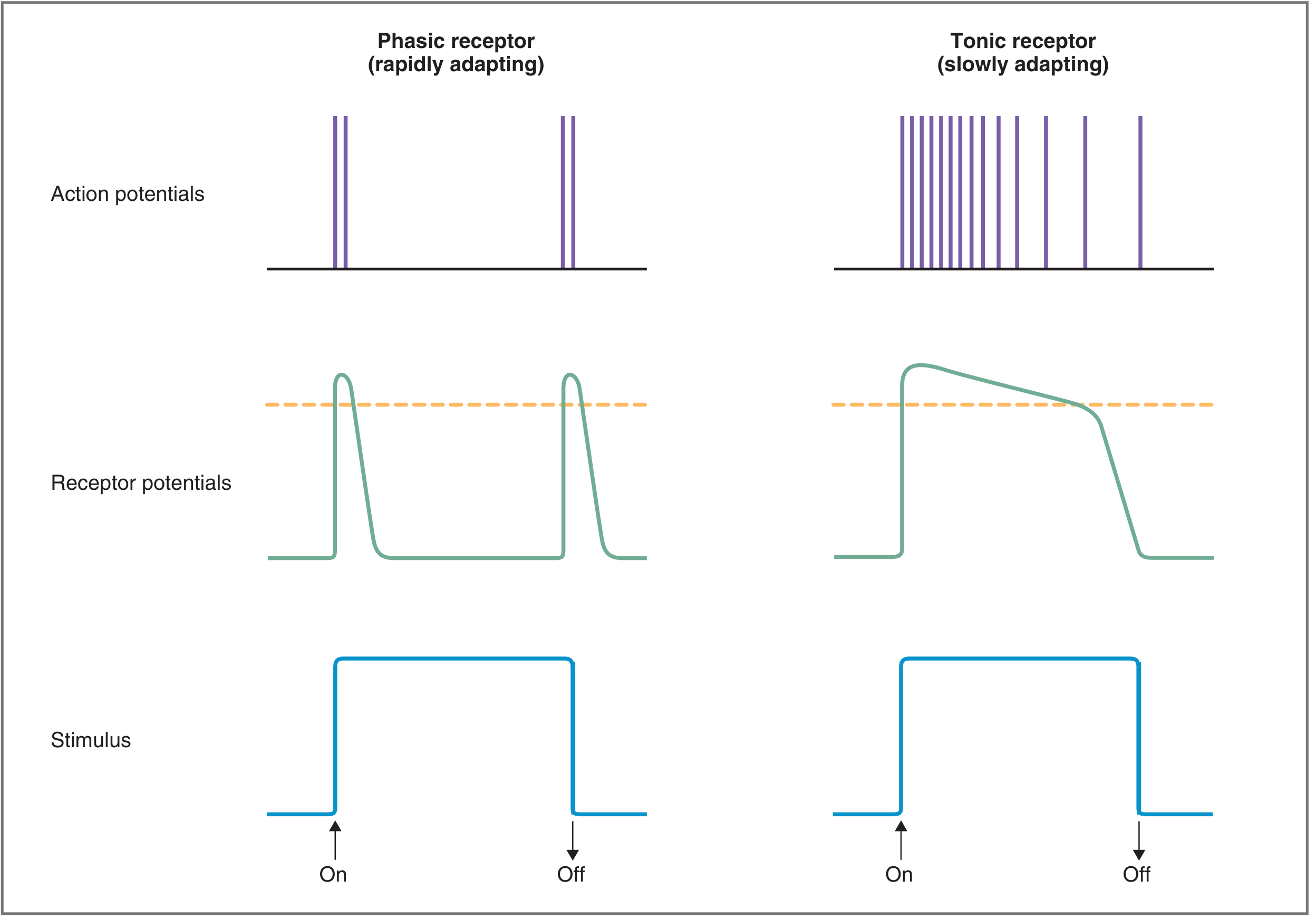
Phasic (rapidly adapting) receptors respond at onset and offset only; tonic (slowly adapting) receptors fire continuously throughout the stimulus. - Costanzo Physiology, Fig. 3.8
5. By Adequate Stimulus (Principle of Specific Nerve Energies)
Each receptor has an "adequate stimulus" - the particular form of energy to which it is most sensitive. For example:
- Photoreceptors respond to light, not to touch
- Nociceptors respond to noxious stimuli, not to light
This concept is called the law of specific nerve energies (Johannes Müller) - the sensation perceived depends on which nerve is activated, not the nature of the stimulus.
The Four Cutaneous Mechanoreceptors (most tested)
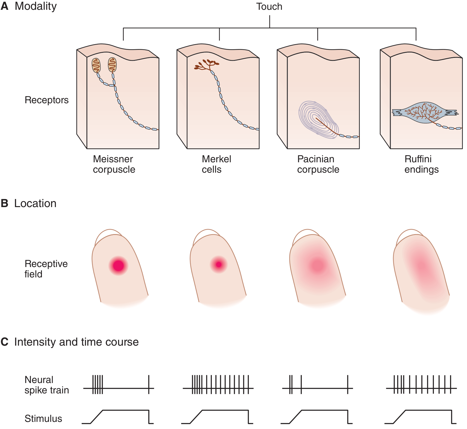
| Receptor | Adaptation | Receptive Field | Stimulus Detected | Skin Type | Fiber |
|---|---|---|---|---|---|
| Meissner corpuscle | Rapidly adapting (RA1) | Small, sharp | Slow vibration, light touch, texture | Glabrous only | Aβ |
| Merkel cells | Slowly adapting (SA1) | Small, sharp | Sustained pressure, fine detail, edges | Glabrous | Aβ |
| Ruffini endings | Slowly adapting (SA2) | Large, diffuse | Skin stretch, finger position, fluttering | Glabrous + hairy | Aβ |
| Pacinian corpuscle | Rapidly adapting (RA2) | Large, diffuse | Fast vibration, deep pressure | Glabrous + hairy | Aα/Aβ |
Properties of Sensory Receptors
1. Receptor (Generator) Potential
- When a stimulus activates a receptor, ion channels open/close → current flow → change in membrane potential called the receptor potential (also called generator potential)
- The receptor potential is a graded potential - its amplitude is proportional to stimulus intensity
- It is NOT an action potential; it increases or decreases the probability that an action potential will occur
- If the receptor potential reaches threshold → action potentials fire in the afferent nerve
- The frequency of action potentials encodes stimulus intensity (Costanzo, pp. 82-83)
2. Adequate Stimulus
- Each receptor has a specific form of energy (its "adequate stimulus") to which it has the lowest threshold
- Stimuli of other modalities can activate the receptor, but only at much higher intensities
3. Threshold
- The minimum stimulus intensity required to activate a receptor
- Small subthreshold stimuli produce receptor potentials but no action potentials
- Summation of subthreshold stimuli can reach threshold (spatial and temporal summation)
4. Adaptation
- If a constant-strength stimulus is maintained, firing frequency declines over time - this is adaptation
- Phasic/rapidly adapting receptors: detect the onset and offset of stimuli (changes); useful for detecting motion, vibration (e.g., Pacinian corpuscles - you stop noticing clothing on your skin)
- Tonic/slowly adapting receptors: continue signaling during sustained stimuli; useful for maintaining postural tone, sustained pain warning (e.g., muscle spindles, nociceptors)
5. Receptive Field
- The area of the body surface (or space) that, when stimulated, changes the firing rate of a sensory neuron
- Small receptive fields = high spatial acuity (fingertips - Merkel, Meissner)
- Large receptive fields = low spatial acuity (back - Pacinian, Ruffini)
- Two-point discrimination is directly related to receptive field size
6. Sensory Coding
Four attributes are always encoded (Ganong's, p. 172):
- Modality - type of stimulus (labeled-line principle: each modality has dedicated pathways)
- Location - encoded by receptive fields and enhanced by lateral inhibition
- Intensity - encoded by firing frequency, number of receptors activated, and type of receptor activated
- Duration - encoded by duration of firing; shaped by adaptation
7. Law of Projection
Sensory information is always perceived as coming from the receptor site (peripheral end), not from anywhere along the nerve pathway. Stimulating a sensory nerve anywhere along its length is perceived as a stimulus at its receptor end (e.g., tingling in the fingers when the ulnar nerve at the elbow is struck - "funny bone" phenomenon).
8. Convergence and Divergence
- Convergence: multiple afferent fibers synapse on a single second-order neuron → amplification
- Divergence: a single afferent fiber contacts multiple second-order neurons → wider broadcast of information
Nociceptors - Special Properties
Nociceptors carry pain signals via two types of fibers:
- Aδ fibers (thin myelinated, 2-5 μm, 12-35 m/s) → First (fast) pain - sharp, well-localized
- C fibers (unmyelinated, 0.4-1.2 μm, 0.5-2 m/s) → Second (slow) pain - dull, burning, diffuse
Many nociceptors are TRP channels (Transient Receptor Potential):
- TRPV1 - activated by heat (>45°C), acids, and capsaicin
- TRPM8 - activated by cold (<24°C) and menthol
- TRPV3/TRPV4 - warm temperatures (33-39°C / 25-34°C)
Nociceptors are slowly adapting - they do not fatigue, which serves as a persistent warning of ongoing tissue damage.
Sources:
- Ganong's Review of Medical Physiology, 26th Ed., pp. 169-177
- Costanzo Physiology, 7th Ed., pp. 82-86
- Medical Physiology (Boron & Boulpaep), pp. 340-343
This is a shared conversation. Sign in to Orris to start your own chat.