
What is lupus pernio
lupus pernio sarcoidosis skin lesion face
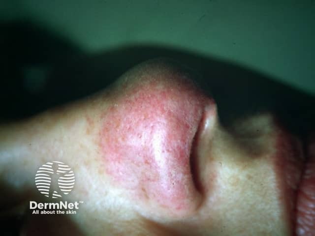
This high-resolution clinical photography shows cutaneous lupus pernio, the characteristic violaceous, indurated plaques of sarcoidosis involving the nasal dorsum and surrounding integument. Acquisition modality: bedside clinical photography, full-face anterior view, with close-up focus on the nose (nasal pyramid, alar rims) to document contour irregularities and color. The lesion appears as erythematous-violaceous plaques with firm, raised induration, subtle scaling, and blending erythema into adjacent normal skin. Dermal granulomatous inflammation in lupus pernio typically yields palpable plaques and nodules; in this image, the lesion is non-ulcerated, symmetric along the nasal region, with mild edema and vascular prominence. In the clinical context, lupus pernio strongly suggests sarcoidosis and often indicates chronic systemic involvement, particularly of the lungs and upper airways; dermatologic findings may precede or accompany pulmonary disease. Differential diagnoses include granulomatous rosacea, tuberculoid granulomatous lesions, and other granulomatous dermatoses; biopsy would show noncaseating granulomas with tight epithelioid granulomas and multinucleated giant cells. This image supports diagnostic correlation, educational case illustration, and differential diagnosis discussion for dermatology, rheumatology, pulmonology, and pathology. It has clinical relevance for prognosis and monitoring response to therapy, including corticosteroids or steroid-sparing agents, and for documenting treatment outcomes. Correlation with biopsy and systemic evaluation remains essential for definitive diagnosis.
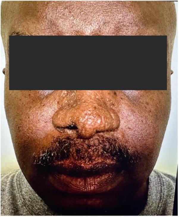
This clinical photograph shows the frontal view of a patient with cutaneous sarcoidosis, specifically lupus pernio, following systemic treatment. The image focuses on the mid-face region, including the nose, cheeks, and upper lip. While there is evidence of post-inflammatory texture changes and some subtle residual thickening, there is a marked absence of the highly characteristic indurated, violaceous, or reddish-brown shiny nodules and plaques typically seen in active lupus pernio. The nasal bridge and alae appear relatively smooth and the previous bulbous distortion has largely resolved. The skin tone on the cheeks and perioral area is more uniform, though some mild follicular prominence and chronic scarring may persist. This image serves as an educational example of the therapeutic response of cutaneous sarcoidosis to Janus kinase (JAK) inhibitor therapy, illustrating the significant reduction in granulomatous infiltration and the restoration of normal facial contours.
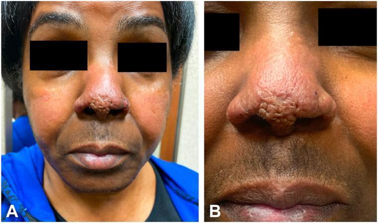
Clinical photograph of a 63-year-old African American woman displaying characteristic cutaneous manifestations of lupus pernio, a form of sarcoidosis. Figure A shows a frontal view of the face with indurated, violaceous-to-hyperpigmented papules that have coalesced into a plaque on the nasal tip and bilateral alar rims, creating a distinctive 'cobblestone' texture. Erythematous to hypopigmented patches are visible on the medial cheeks, notably sparing the nasolabial folds. An erosion on the left alar rim indicates a prior biopsy site. Figure B provides a close-up view highlighting the atrophic and firm papules along the nasal tip and left alar rim. These findings are highly suggestive of chronic sarcoidosis, often associated with upper respiratory tract involvement and systemic disease. The educational focus is on the morphology and distribution of sarcoidal skin lesions on the central face.
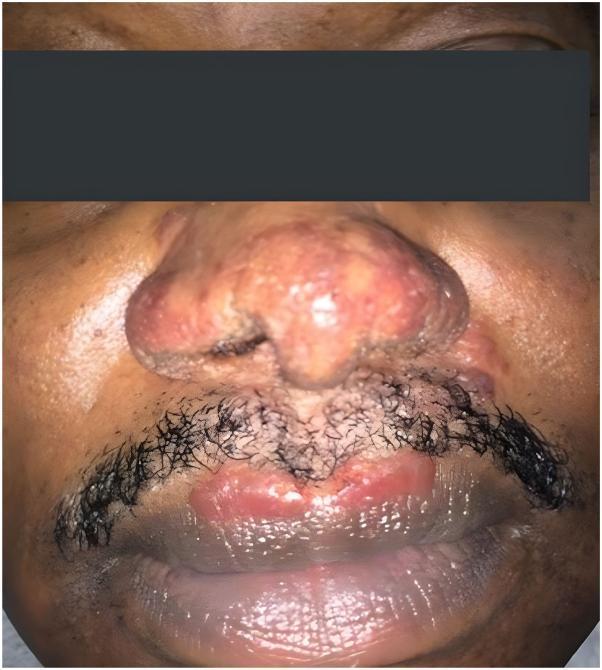
This clinical photograph displays a front-on view of the mid-face of a 56-year-old male, highlighting prominent dermatological manifestations of cutaneous sarcoidosis, specifically lupus pernio. The nasal region is significantly involved, showing diffuse, pinkish-red to violaceous infiltration with multiple confluent, shiny nodules that distort the nasal contour. The surface appears irregular and indurated. In the perioral area, reddish-brown, shiny, well-demarcated plaques are visible along the vermilion border of the upper lip and extending into the philtrum and mustache region. The affected skin shows a characteristic waxy or translucent texture. This image serves as a classic educational representation of chronic sarcoidosis, where granulomatous infiltration typically affects the 'central face' (nose, lips, and cheeks). Clinical significance lies in recognizing these specific morphologies, which are often refractory to treatment and can be associated with underlying systemic sarcoidosis or upper respiratory tract involvement.
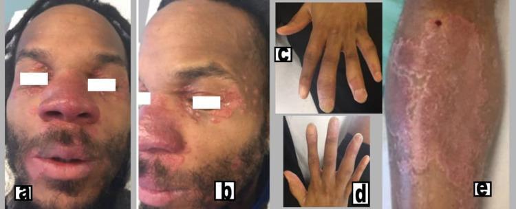
This composite clinical photograph displays the multi-systemic cutaneous and musculoskeletal manifestations of chronic sarcoidosis. Figures (a) and (b) show the patient's face with characteristic lupus pernio, presenting as indurated, violaceous (purplish) plaques and nodules affecting the nose, cheeks, and periorbital regions. Figure (e) illustrates a large, well-demarcated, erythematous to purplish indurated plaque on the medial aspect of the right leg. Figures (c) and (d) demonstrate clinical dactylitis ('sausage digits') of the hands, characterized by diffuse swelling of the fingers involving the proximal interphalangeal (PIP) and distal interphalangeal (DIP) joints. The image serves as an educational resource for identifying the classic visual signs of sarcoidosis, specifically the association between lupus pernio skin lesions and sarcoid dactylitis or inflammatory arthritis. It is intended for medical students and clinicians specializing in dermatology and rheumatology.
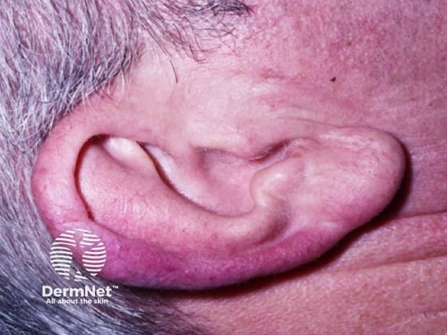
This is a high-resolution clinical photograph of a cutaneous lesion in the auricular region, captured in a lateral view of the external ear. The focal area is consistent with lupus pernio, the chronic, violaceous plaque-form of cutaneous sarcoidosis that commonly involves the nose, cheeks, ears, and lips, with the pinna often affected. The lesion appears as a well-defined, violaceous to reddish-purple plaque on the helix/pinna, with subtle surface sclerosis and mild edema; surrounding skin shows faint erythema without obvious ulceration. In cutaneous sarcoidosis, such plaques reflect dermal granulomatous infiltration; histology (if obtained) would typically show noncaseating granulomas composed of epithelioid histiocytes and Langhans-type giant cells, often with sparse lymphocytic rim. Clinically, lupus pernio is a marker of chronic sarcoidosis and may correlate with systemic involvement, including pulmonary, ocular, and cardiac disease; therefore, thorough evaluation (chest imaging, ACE levels, calcium metabolism, hepatic function) is recommended. Differential considerations include granulomatous rosacea, actinic granuloma, other granulomatous dermatitis, and infectious etiologies in the appropriate context. This image is valuable for dermatology education, sarcoidosis characterization, and monitoring therapeutic response to corticosteroids, immunomodulators, or biologic agents. This photographic depiction supports clinical diagnosis and aids multidisciplinary management decisions.
"lupus pernio" sarcoidosis
Lupus Pernio
Clinical Features
- Nose (tip and alar rims - most characteristic)
- Cheeks, lips, forehead
- Earlobes
- Digits
- Brown to violaceous, smooth, shiny, indurated plaques
- Often symmetric
- May be deeply disfiguring
- Rarely resolve spontaneously
- Can resemble rhinophyma when on the nose
Systemic Associations
| Systemic Involvement | Frequency |
|---|---|
| Chronic fibrotic respiratory tract disease | ~75% of lupus pernio patients |
| Granulomas in bones (punched-out cysts, typically fingers) | ~43% |
| Chronic ocular lesions | ~37% |
| Sinus involvement | ~50% |
| Upper respiratory tract involvement | Very common |
Complications
- Nasal mucosal and underlying bone involvement can lead to nasal perforation and collapse of the nasal bridge (mimicking granulomatosis with polyangiitis)
- Upper aerodigestive tract involvement
- Lesions may ulcerate if treated with laser (including pulsed-dye laser) - important clinical warning
- Chronic, recalcitrant course
Variants
- Angiolupoid sarcoidosis: Pink and violaceous papules/plaques with prominent telangiectasias on the face; considered a variant of lupus pernio
Histology
Differential Diagnosis
| Condition | Distinguishing features |
|---|---|
| Lupus vulgaris (cutaneous TB) | Caseating granulomas; diascopy shows "apple jelly" nodules |
| Granulomatous rosacea | No systemic involvement |
| Rhinophyma | Pure sebaceous hypertrophy; no violaceous color |
| Lymphoma cutis | Violaceous plaques; biopsy shows lymphoid infiltrate |
| Cutaneous lupus erythematosus | ANA/dsDNA positive; different histology |
| Granulomatosis with polyangiitis | ANCA positive; necrotizing vasculitis on biopsy |
Management
- Topical or intralesional corticosteroids - for localized/mild disease
- Systemic corticosteroids - first-line for systemic disease
- Steroid-sparing agents: Hydroxychloroquine, methotrexate, azathioprine
- Biologics (TNF inhibitors): Infliximab, adalimumab for refractory disease
- JAK inhibitors: Emerging therapy (as seen in post-treatment images showing significant resolution)