
I have pain in opening of stomach
Reading File
Pain in the "opening of the stomach" likely refers to pain in the epigastric region - the upper middle part of the abdomen, or possibly at the gastroesophageal junction (where the esophagus meets the stomach). The most common causes include gastritis, peptic ulcer disease, GERD, or gastric outlet issues.
Searching sources...
Finding Sources
Finding Sources
Searching the Web
pain in epigastric region stomach opening causes symptoms treatment
Reading File
Reading File
Primary and secondary ossification
Primary ossification occurs in the diaphysis (shaft) of long bones during fetal development, forming the primary ossification center. Secondary ossification occurs in the epiphyses (ends) after birth, forming secondary ossification centers. Together they are responsible for bone growth in length via the growth plate (epiphyseal plate) between them.
Searching sources...
Finding Sources
Searching the Web
primary and secondary ossification centers bone development anatomy
Finding Sources
Finding Sources
Reading File
Reading File
Reading File
Loading Image
Loading Image
Here is a complete overview of primary and secondary ossification, drawn from authoritative textbooks:
Primary and Secondary Ossification Centers
Background: Endochondral Ossification
Almost all long bones (limbs, vertebrae, ribs) develop through endochondral ossification - bone forms on a pre-existing hyaline cartilage model (anlage). Flat bones of the skull, mandible, and clavicle form by intramembranous ossification (no cartilage model needed). - Histology: A Text and Atlas, p. 640
Primary Ossification Center (Diaphyseal Center)
| Feature | Detail |
|---|---|
| Location | Diaphysis (shaft of the long bone) |
| Timing | Appears ~8th week of gestation; all long bone primary centers present by 12th week |
| Process | Chondrocytes hypertrophy → matrix calcifies → vascular invasion → osteoblasts deposit bone on cartilage spicules |
| Result | Forms the shaft of the bone; creates the medullary canal |
Steps in sequence:
- Central cartilage in the diaphysis is resorbed, forming the medullary canal
- Periosteal capillaries invade the diaphysis
- Osteoblasts (from periosteum) deposit a periosteal bony collar on the cartilage surface
- This penetrates inward to form the primary ossification center
- Bone grows toward both epiphyses
At birth, the diaphyses are largely ossified - The Developing Human (Langman's Embryology), p. 908
Secondary Ossification Center (Epiphyseal Center)
| Feature | Detail |
|---|---|
| Location | Epiphyses (ends of the long bone) |
| Timing | Most appear after birth (first few years of life); a few are present at birth (e.g., distal femur, proximal tibia) |
| Process | Epiphyseal cartilage cells hypertrophy → vascular connective tissue invasion → ossification spreads radially outward |
| Result | Forms the bony epiphysis; leaves behind articular cartilage and the growth plate |
After the secondary center forms, only two cartilaginous structures remain:
-
Articular cartilage (persists lifelong)
-
Epiphyseal cartilage plate / growth plate (persists until skeletal maturity)
-
The Developing Human, p. 909; Robbins Basic Pathology, p. 771
The Growth Plate (Physis)
The growth plate is the plate of cartilage trapped between the primary and secondary ossification centers. It has 5 sequential zones:
| Zone | Activity |
|---|---|
| 1. Reserve zone | Resting chondrocytes, stem cells |
| 2. Proliferative zone | Chondrocytes divide rapidly, form columns |
| 3. Hypertrophic zone | Chondrocytes enlarge significantly |
| 4. Zone of apoptosis and mineralization | Chondrocytes die, matrix calcifies |
| 5. Primary spongiosa | Osteoblasts deposit bone on calcified cartilage spicules |
This drives longitudinal bone growth. - Robbins Basic Pathology, p. 771
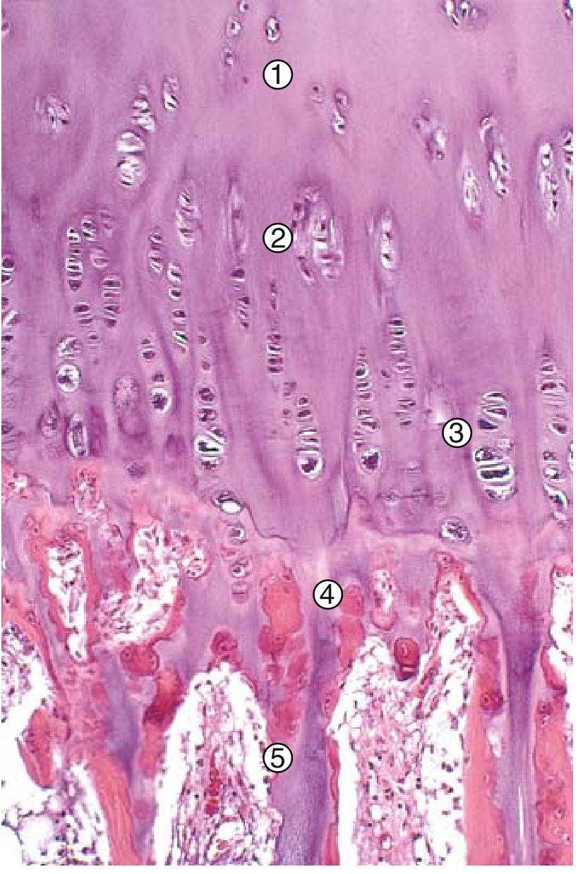
Endochondral Ossification - Full Diagram
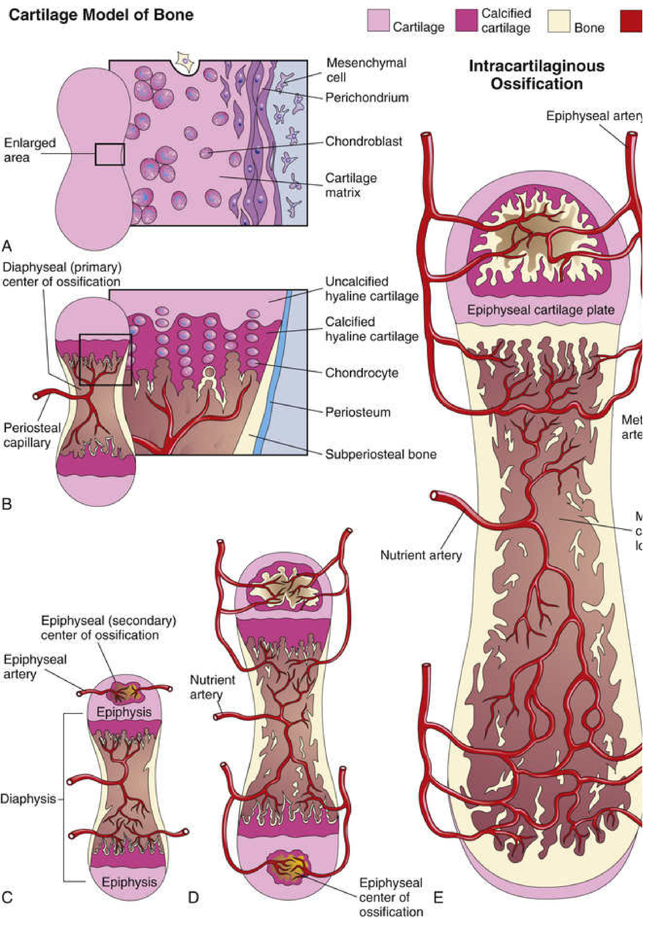
The diagram above shows:
- (A) Cartilage model with mesenchymal cells and perichondrium
- (B) Primary (diaphyseal) ossification center forming with periosteal capillary invasion
- (C) Epiphyseal (secondary) ossification center appearing in both epiphyses
- (D-E) Mature bone with epiphyseal cartilage plate, nutrient artery, and metaphyseal blood supply
Key Comparison Table
| Feature | Primary Center | Secondary Center |
|---|---|---|
| Location | Diaphysis (shaft) | Epiphysis (end) |
| Timing | Fetal life (~8th week) | After birth (mostly) |
| Ossification spreads | Toward epiphyses | Centrifugally (outward) |
| What remains cartilage | Growth plate + articular cartilage | Articular cartilage only (eventually) |
| Blood supply | Nutrient artery | Epiphyseal artery |
Epiphyseal Fusion
When growth is complete (most bones by ~20 years), the growth plate is entirely replaced by spongy bone - the epiphysis and diaphysis fuse permanently and no further elongation is possible. - The Developing Human Clinically Oriented Embryology, p. 909
Bone width increases separately via appositional (periosteal) growth, not the ossification centers.
This is a shared conversation. Sign in to Orris to start your own chat.