
Give an account of sources, functions and deficiency manifestations of retinol
Reading File
Finding Sources
Finding Sources
Finding Sources
Finding Sources
Reading File
Reading File
Reading File
Reading File
Reading File
Loading Image
Reading File
Loading Image
I now have excellent, comprehensive material from multiple authoritative sources. Here is a complete, well-structured account of retinol.
Retinol (Vitamin A) - Sources, Functions, and Deficiency Manifestations
Overview and Chemical Forms
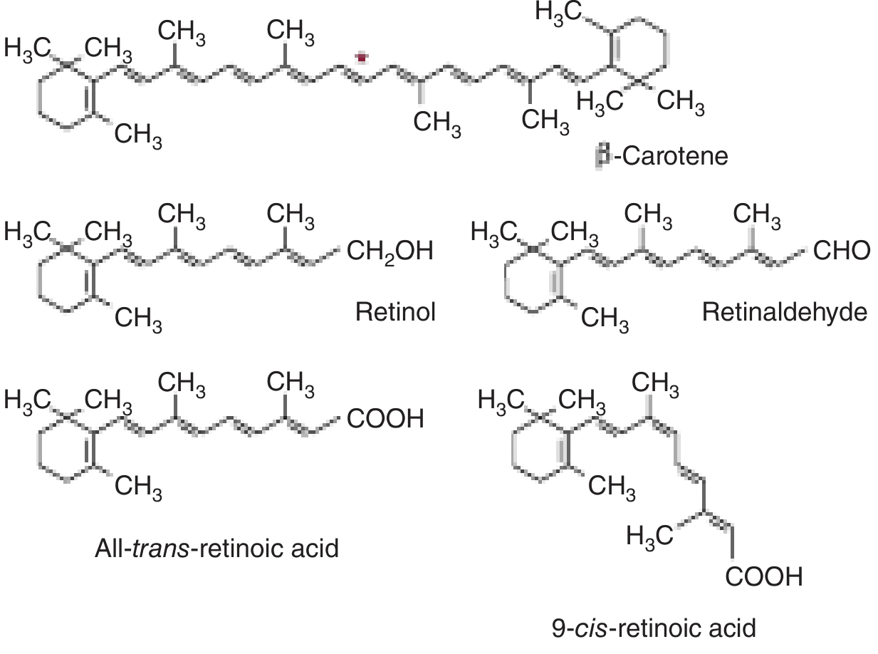
Retinol is a fat-soluble vitamin belonging to the retinoid family. The term "Vitamin A" is generic and encompasses several biologically active compounds:
- Retinol - the transport and storage form (as retinyl ester)
- Retinaldehyde (retinal) - the aldehyde form, active in the visual cycle
- Retinoic acid - the acid form, active in gene regulation
- Provitamin A carotenoids (e.g., β-carotene) - plant-derived precursors
Sources
Preformed Vitamin A (Animal Sources)
Found exclusively in foods of animal origin as retinyl esters:
- Liver (especially fish liver oils such as cod liver oil) - richest source
- Egg yolk
- Milk, butter, cheese
- Fish (fatty fish, oily fish)
- Fortified foods (margarine, cereals in some countries)
Provitamin A Carotenoids (Plant Sources)
Found in plants as carotenoids that are converted to retinol in intestinal mucosa:
- β-Carotene - most important; cleaved by intestinal carotene dioxygenase to yield retinaldehyde, then reduced to retinol
- Also: α-carotene, γ-carotene, β-cryptoxanthin
- Yellow/orange vegetables: carrots, squash, sweet potato, pumpkin
- Dark leafy greens: spinach, kale, broccoli (carotenoids masked by chlorophyll)
Carotenoids contribute approximately 30% of dietary vitamin A. Since absorption and conversion are inefficient, 6 μg β-carotene = 1 μg preformed retinol (1 retinol activity equivalent, RAE). Carotenoids also circulate unchanged in the blood due to low intestinal carotene dioxygenase activity.
(Robbins & Kumar Basic Pathology; Harper's Illustrated Biochemistry, 32nd Ed.)
Absorption, Transport, and Storage
- Absorption is fat-dependent and requires bile salts and pancreatic enzymes
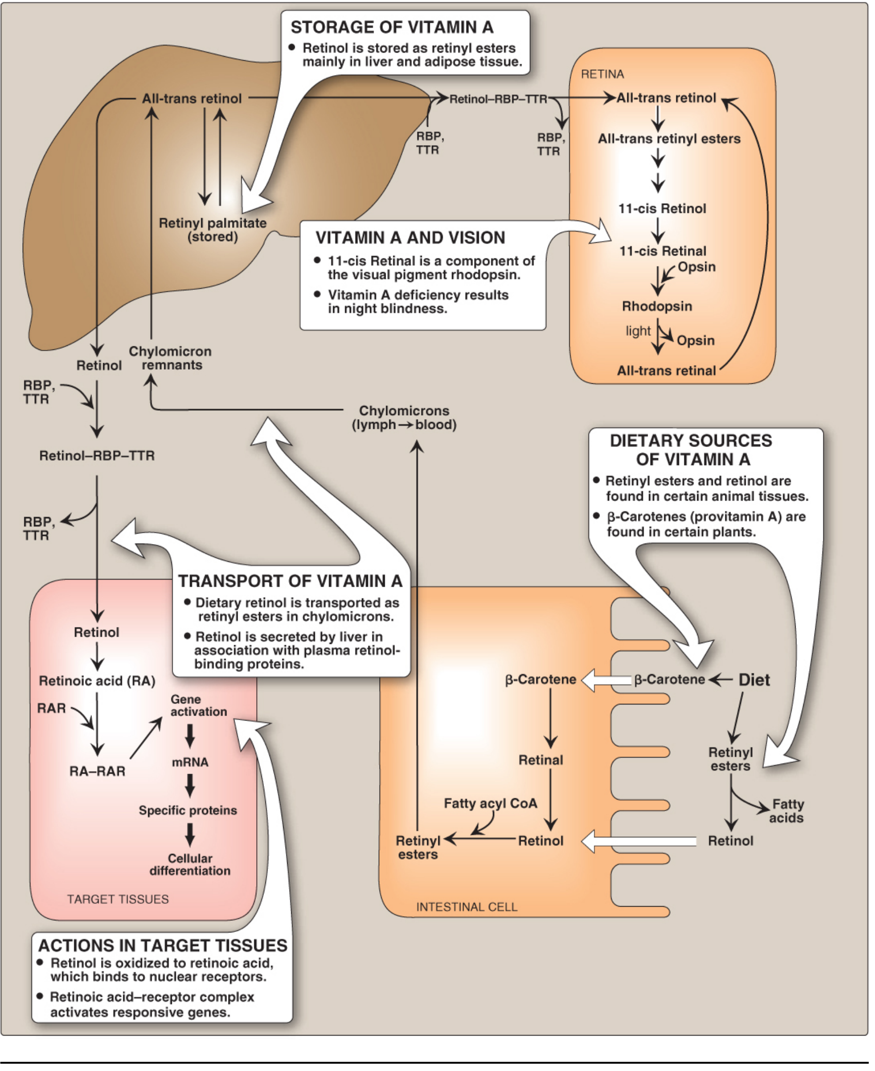
- Retinyl esters and β-carotene are absorbed in the intestinal wall; β-carotene is converted to retinol there
- Retinol is packaged into chylomicrons and transported via lymph to the bloodstream
- Taken up by the liver (via apolipoprotein E receptor); >90% of body stores reside in hepatic perisinusoidal stellate (Ito) cells as retinyl palmitate - sufficient for at least 6 months on an adequate diet
- Released from liver bound to retinol-binding protein (RBP), which circulates complexed with transthyretin (TTR)
- Peripheral cells take up retinol via cell-surface RBP receptors; RBP is then recycled back into blood
(Lippincott Illustrated Reviews Biochemistry, 8th Ed.; Robbins & Kumar Basic Pathology)
Functions
1. Vision (Visual Cycle)
Vitamin A (as 11-cis retinal) is the prosthetic group of the light-sensitive opsin proteins:
- Rhodopsin = 11-cis retinal + opsin (in rod cells; for dim/scotopic vision)
- Iodopsins (3 types) = 11-cis retinal + cone opsins (for color/photopic vision in bright light)
Mechanism: Light isomerizes 11-cis retinal → all-trans retinal, bleaching rhodopsin and releasing opsin. This activates the G protein transducin, generating a nerve impulse to the brain. Regeneration requires isomerization of all-trans retinal back to 11-cis retinal. Deficiency reduces synthesis of all four visual pigments.
(Lippincott Illustrated Reviews Biochemistry, 8th Ed.; Harper's Illustrated Biochemistry, 32nd Ed.)
2. Epithelial Cell Differentiation and Maintenance
- Retinol is oxidized intracellularly to retinoic acid, which binds nuclear retinoic acid receptors (RARs)
- The retinoic acid-RAR complex forms heterodimers with RXR (retinoic X receptor) that bind to retinoic acid response elements on DNA, regulating transcription of genes for keratin, growth factor receptors, tumor suppressor genes, and other proteins
- Promotes the differentiation of mucus-secreting columnar epithelium throughout the body
- Without vitamin A, the epithelium undergoes squamous metaplasia and keratinization
(Robbins & Kumar Basic Pathology; Lippincott Illustrated Reviews Biochemistry, 8th Ed.)
3. Reproduction
- Retinol and retinal are essential for normal reproduction
- Supports spermatogenesis in males
- Prevents fetal resorption in females (retinoic acid alone cannot substitute for these functions)
4. Immune Function
- Regulates differentiation of immune system cells
- Vitamin A supplementation reduces mortality from diarrhea by ~30% and morbidity by ~15%
- Maintains mucosal barrier integrity against pathogens
- Note: RBP is a negative acute-phase protein - its synthesis falls during infection, reducing circulating vitamin A and further impairing immune defense during illness
(Robbins & Kumar Basic Pathology; Harper's Illustrated Biochemistry, 32nd Ed.)
5. Bone Growth and Development
- Required for normal osteoblast and osteoclast activity
- Deficiency leads to impaired bone growth and remodeling
6. Metabolic Effects (via RXR)
- Retinoids inhibit adipogenesis and stimulate lipid breakdown
- RXR (activated by 9-cis retinoic acid) heterodimerizes with PPARs (peroxisome proliferator-activated receptors), regulating fatty acid oxidation, adipogenesis, and lipoprotein metabolism
(Robbins & Kumar Basic Pathology)
Deficiency Manifestations
Vitamin A deficiency is the most important preventable cause of blindness worldwide. It may result from poor nutrition, fat malabsorption (celiac disease, Crohn's disease, colitis), bariatric surgery, or prolonged use of mineral oil laxatives.
Ocular Manifestations (Xerophthalmia - Progressive Sequence)
| Stage | Manifestation |
|---|---|
| 1 | Loss of sensitivity to green light - earliest sign |
| 2 | Night blindness (nyctalopia) - impaired dark adaptation, inability to see in dim light; visual threshold is increased |
| 3 | Xerosis conjunctivae - dryness of conjunctiva; replacement of lacrimal/mucus-secreting epithelium by keratinized epithelium |
| 4 | Bitot's spots - buildup of keratin debris forming small, foamy, opaque plaques on the conjunctiva (pathognomonic) |
| 5 | Xerosis cornae - corneal dryness and erosion of the roughened corneal surface |
| 6 | Keratomalacia - softening and ulceration/destruction of the cornea |
| 7 | Corneal perforation and blindness - irreversible |
(Robbins & Kumar Basic Pathology; Goodman & Gilman's Pharmacological Basis of Therapeutics; Andrews' Diseases of the Skin)
Epithelial and Skin Manifestations
- Squamous metaplasia of mucus-secreting epithelia throughout the body
- Respiratory tract: loss of mucociliary epithelium predisposes to secondary pulmonary infections (pneumonia)
- Urinary tract: desquamation of keratin debris → renal and urinary bladder stones
- Skin: hyperplasia and hyperkeratinization of the epidermis; plugging of adnexal gland ducts → follicular or papular dermatosis (phrynoderma) - "toad skin" appearance with follicular hyperkeratosis
- Salivary glands, pancreas, and other glandular epithelia: metaplasia and dysfunction
Immune System Deficiency
- Increased susceptibility to infectious diseases even with mild deficiency
- Impaired T-lymphocyte and natural killer cell function
- Suppressed antibody responses
- Reduced regeneration of damaged epithelia
Growth and Development
- Impaired skeletal growth (bone remodeling defects)
- In children: growth retardation
- Reproductive failure: defective spermatogenesis, fetal resorption
Summary of Deficiency Manifestations by System
| System | Manifestation |
|---|---|
| Eyes | Night blindness → xerophthalmia → keratomalacia → blindness |
| Conjunctiva | Bitot's spots |
| Skin | Phrynoderma (follicular hyperkeratosis) |
| Respiratory | Squamous metaplasia → recurrent infections |
| Urinary | Squamous metaplasia → calculi |
| Immune | Increased susceptibility to infection, increased child mortality |
| Reproductive | Infertility, fetal resorption, defective spermatogenesis |
| Bone | Growth retardation in children |
Recommended Dietary Allowance
- Expressed as Retinol Activity Equivalents (RAE):
- 1 RAE = 1 μg all-trans retinol = 12 μg β-carotene = 24 μg α-carotene or β-cryptoxanthin
- Old unit: 1 IU = 0.3 μg retinol (3.33 IU = 1 μg retinol)
Sources:
- Robbins & Kumar Basic Pathology, p. 292-293
- Lippincott Illustrated Reviews: Biochemistry, 8th Ed., p. 1077-1078
- Harper's Illustrated Biochemistry, 32nd Ed., p. 548-549
- Goodman & Gilman's Pharmacological Basis of Therapeutics
- Andrews' Diseases of the Skin
This is a shared conversation. Sign in to Orris to start your own chat.