
Cellulitis opd management
cellulitis outpatient management antibiotics
cellulitis skin infection clinical appearance
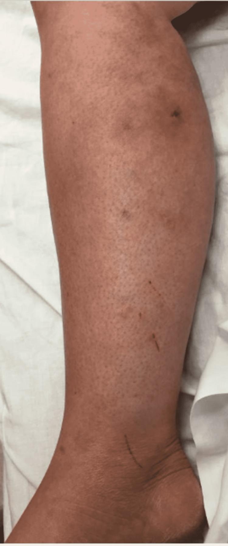
This clinical photograph displays the right lower extremity (RLE) of a patient, showing the lateral and posterior aspects of the lower leg and ankle. The skin exhibits diffuse, patchy erythema with a somewhat mottled appearance, characteristic of resolving cellulitis. Several small, discrete, hyperpigmented macules or crusting lesions are scattered across the mid-calf region. Linear abrasions or excoriations are visible near the lateral malleolus and lower shin. The image illustrates the clinical progression of an atypical mycobacterial infection (specifically Mycobacterium chelonae) undergoing treatment. The lack of severe edema or fluctuance suggests improvement from an acute infectious state. This visual is relevant for dermatology and infectious disease specialties, particularly in identifying the cutaneous manifestations of non-tuberculous mycobacteria (NTM) which can mimic standard bacterial cellulitis.
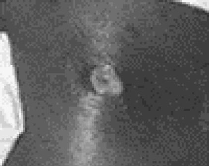
Clinical photograph of an acute skin and soft-tissue infection (SSTI) featuring a necrotic pustule. The image shows a raised, central lesion with a non-uniform appearance, including a core consistent with tissue necrosis, ulceration, and the presence of inflammatory exudate. This primary lesion is surrounded by a significant area of cellulitis, characterized by diffuse cutaneous erythema and apparent edema with ill-defined borders. Such presentations are classically associated with community-associated methicillin-resistant Staphylococcus aureus (CA-MRSA), specifically strains containing the Panton-Valentine leukocidin (PVL) gene, which promotes tissue destruction and abscess formation. The clinical significance of this visual is to demonstrate the progressive nature of CA-MRSA infections, which may initially present as painful pustules resembling spider bites before rapidly evolving into necrotizing abscesses with spreading secondary cellulitis.
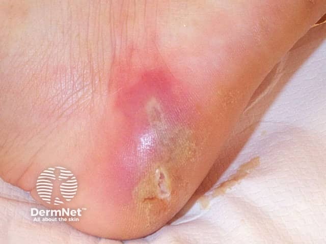
Clinical photograph of acute cellulitis involving the soft tissues of the foot/ankle. Acquisition: high-magnification, close-up dermatologist’s view, not a radiologic modality. The image shows a localized erythematous, warm, and tender plaque with surrounding edema on the plantar-lateral aspect of the foot near the ankle. The skin surface is glossy with mild desquamation and a small crust or erosion at the lesion margin, consistent with superficial disruption. No obvious purulence is visible in this frame, but the erythema extends beyond the edge of the lesion, suggesting active inflammatory infiltration of the dermis and subcutaneous tissue. This appearance aligns with cellulitis rather than dermatitis, erysipelas, or venous stasis changes, though mimics may include contact dermatitis. Key features include abrupt onset, focal swelling, heat, and tenderness, supporting a bacterial etiology typically caused by streptococci or staphylococci. Clinically, this finding warrants prompt antibiotic therapy and evaluation for systemic involvement. When differentiating from abscess, necrotizing fasciitis, or deep-seated infection, consider imaging or ultrasound if signs of abscess or rapid progression arise. Document exposure history, diabetes or edema, and immunocompromise status. This image is suited for dermatology education, infectious disease training, and clinical decision support databases, illustrating surface dermatologic signs that guide triage and therapeutic choices.
cellulitis treatment antibiotics
PMID: 39240378
Cellulitis: OPD Management
Definition & Pathophysiology
- Non-purulent cellulitis: Beta-hemolytic streptococci (S. pyogenes) - responsible for ~75% of cases
- Purulent cellulitis: S. aureus, including CA-MRSA
- Special contexts: Vibrio vulnificus (saltwater exposure), Pasteurella multocida (cat/dog bite), Pseudomonas spp.
Who Can Be Managed as an OPD Patient?
- Hemodynamically stable, no sepsis or SIRS criteria
- No systemic toxicity (no high fever/rigors suggesting bacteremia)
- Immunocompetent host
- No rapid progression
- No involvement of face/orbit (periorbital/orbital cellulitis often needs IV therapy)
- Able to tolerate oral medications
- Reliable follow-up within 48-72 hours possible
- Systemic toxicity (fever >38.5°C, hypotension, tachycardia)
- Immunocompromised (HIV, transplant, chemotherapy, diabetes with poor control)
- Facial cellulitis with orbital involvement
- Rapidly spreading despite 48h oral therapy
- Severe comorbidities (cardiac, renal, hepatic failure)
- Unable to tolerate oral medications
Severity Classification & Antibiotic Choice
Mild (No systemic signs, no MRSA risk) - FIRST LINE
| Drug | Dose | Duration |
|---|---|---|
| Cephalexin | 500 mg PO QID | 5-7 days |
| Dicloxacillin | 500 mg PO QID (on empty stomach) | 5-7 days |
| Amoxicillin-clavulanate | 625 mg PO TDS | 5-7 days |
- Andrews' Diseases of the Skin and Rosen's Emergency Medicine both recommend cephalexin or dicloxacillin as initial empiric therapy for mild outpatient cellulitis.
- A 2024 network meta-analysis (Shu Z et al., Arch Dermatol Res 2024) found no significant difference in cure rates among antibiotics for cellulitis, with cefaclor showing favorable outcomes and ceftriaxone having fewest GI side effects.
Mild - ALTERNATIVES (if penicillin allergy or MRSA risk factors)
| Drug | Dose | Notes |
|---|---|---|
| Clindamycin | 300-450 mg PO TID | Covers MRSA + streptococci; first choice if MRSA suspected |
| TMP-SMX (Co-trimoxazole) | 1-2 DS tabs PO BD | MRSA active but poor strep coverage - add cephalexin if non-purulent |
| Doxycycline | 100 mg PO BD | MRSA active but limited strep coverage |
| Azithromycin/Clarithromycin | 500 mg OD | Second-line; macrolide resistance limits use |
MRSA risk factors: Prior MRSA infection/colonization, antibiotic use in past 90 days, healthcare worker contacts, recurrent SSTIs, failure of beta-lactam therapy.
Purulent Cellulitis (with draining pus but no abscess requiring I&D)
- First line: Clindamycin or TMP-SMX - both have efficacy against CA-MRSA
- If abscess is also present: Incision & Drainage (I&D) is the definitive treatment; adjunct antibiotics with TMP-SMX or clindamycin improve cure rates
Antibiotic Duration
- Standard uncomplicated cellulitis: 5 days (evidence supports this is as effective as 10 days for mild cases)
- If no improvement at 48-72 hours: reassess and consider escalating to IV or changing antibiotic spectrum
- Symptoms may transiently worsen in the first 24-48h despite appropriate antibiotics
General OPD Management Measures
- Mark the border of erythema with a skin marker at the first visit - monitor for progression or regression at follow-up
- Elevate the affected limb - significantly aids in reducing swelling and accelerating resolution (Textbook of Family Medicine 9e)
- Analgesia: NSAIDs or paracetamol for pain
- Treat the portal of entry: tinea pedis (use antifungal cream), wound care, drain any abscess
- Warm soaks/compresses - soothing, may help with local inflammation
- Hydration and rest
Follow-Up
- Review at 48-72 hours - mandatory for OPD cases
- If worsening (extension beyond marked border, increasing fever, increased pain): step up to IV antibiotics (admit or give IV outpatient with teicoplanin/ceftriaxone as appropriate)
- Full resolution may take 2-4 weeks; residual induration is common
Differentials to Exclude Before Diagnosing Cellulitis
- Stasis/gravitational dermatitis - bilateral, no fever, centered on medial malleoli, itchy
- Allergic contact dermatitis - pruritic, geometric borders
- Deep venous thrombosis - unilateral edema, pain, no erythema center
- Gout/pseudogout - overlies joint, responds to NSAIDs
- Erythema nodosum - nodular, multifocal
- Erythema migrans (Lyme) - expanding annular lesion, tick exposure history
- Necrotizing fasciitis - must exclude; disproportionate pain, rapid spread, crepitus, skin necrosis - requires emergency surgery
Special Situations in OPD
| Situation | Modification |
|---|---|
| Recurrent cellulitis | Investigate and treat predisposing factors (tinea, lymphedema, venous stasis); prophylactic penicillin V or erythromycin if >2 episodes/year |
| Diabetic foot | Broader coverage (amox-clav), podiatry referral; lower threshold to admit |
| Facial cellulitis | Consider CT orbit; lower threshold to admit for IV |
| Bite-associated cellulitis | Amoxicillin-clavulanate covers Pasteurella; co-amoxiclav 5-7 days |
| Penicillin anaphylaxis | Clindamycin or TMP-SMX |
Key Points
- Most uncomplicated cellulitis in immunocompetent patients responds well to 5-7 days of oral cephalexin or dicloxacillin
- CA-MRSA is the main cause of purulent SSTIs but is an uncommon cause of non-purulent cellulitis
- Always mark the borders and ensure follow-up in 48-72 hours
- Limb elevation is a simple but effective adjunct
- A 2024 network meta-analysis confirms no single antibiotic is clearly superior; choose based on purulence, MRSA risk, and local resistance patterns