
I want microscopic image of dry yeast and wet yeast.
Searching the Web
microscopic image dry yeast cells
Searching the Web
microscopic image wet yeast cells active
Loading Image
Loading Image
Loading Image
Loading Image
Loading Image
Loading Image
Here are microscopic images of dry yeast and wet (active/hydrated) yeast side by side, with descriptions of what you're seeing:
Dry Yeast - Under the Microscope
Dry yeast cells are dormant and densely packed. Under the microscope they appear as small, granular, mostly featureless specks with little internal detail visible - the cells are desiccated and in a state of suspended animation.
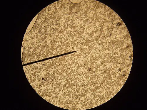
Dry yeast (baker's yeast) viewed under microscope - the cells appear as a dense, granular mass with minimal individual cell definition.
Wet / Active (Hydrated) Yeast - Under the Microscope
Once rehydrated, yeast cells swell, become turgid, and show clear oval-to-round shapes. Active cells often show budding (asexual reproduction), internal granules, vacuoles, and a visible cell wall.
1. Active wet yeast cells - light microscope (x400-1000)

Saccharomyces cerevisiae (baker's/brewer's yeast) hydrated and active - clearly defined oval cells, many showing daughter buds attached. Scale bar visible.
2. High-magnification single yeast cell (10 microns scale)

A single fully hydrated yeast cell at high magnification (~10 µm scale). Internal structures are visible: vacuoles (large clear bubbles), lipid droplets, and cytoplasm granules - all signs of a metabolically active cell.
3. Yeast cells, pseudohyphae, and true hyphae comparison

Wet yeast morphology: left panel shows individual oval yeast cells, middle shows pseudohyphae (budded chains), right shows true hyphae - all forms seen in active/wet conditions.
4. Dense active yeast colony (S. cerevisiae, phase contrast)
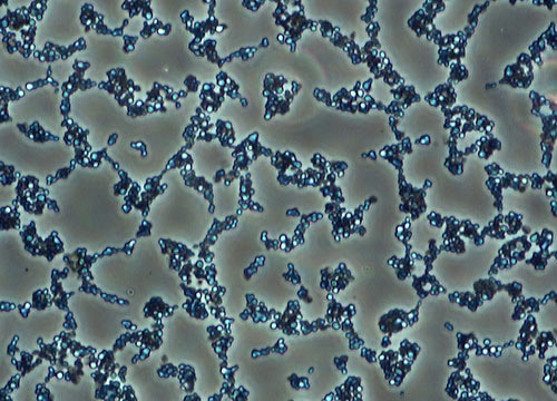
Active hydrated Saccharomyces cerevisiae at high density - the dark, clumped oval cells are visibly distinct and turgid compared to the flat granular appearance of dry yeast.
Key Differences at a Glance
| Feature | Dry Yeast | Wet / Active Yeast |
|---|---|---|
| Cell shape | Shrunken, irregular | Round to oval, turgid |
| Internal structure | Not visible | Vacuoles, granules visible |
| Budding | Absent | Often present |
| Movement | None | Slight Brownian motion |
| Cell boundary | Indistinct | Clear, defined wall |
| Appearance | Dense granular mass | Distinct individual cells |
When dry yeast is rehydrated with warm water (~38-40°C), the cells absorb water, their membranes repair, and they resume metabolism - typically visible as budding and CO₂ production within 5-10 minutes under a microscope.
This is a shared conversation. Sign in to Orris to start your own chat.