
Detailed symptomatology of respiratory medicine
respiratory symptoms chest diagram dyspnea cough hemoptysis
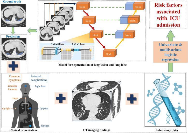
This infographic illustrates a multifaceted deep learning and statistical workflow for predicting ICU admission in patients with COVID-19 pneumonia. The diagram integrates three primary data sources: Clinical presentation (annotating symptoms like cough, dyspnea, myalgia, and high fever), CT imaging findings (showing a 3D volumetric reconstruction of lung parenchyma with ground-glass opacities and consolidation), and Laboratory data (represented by genomic and biochemical icons). A central component features a 'Model for segmentation of lung lesion and lung lobe' utilizing a U-Net based architecture with pseudo 3D convolutions. This model generates a 'Prediction' of infected regions, which is compared against the 'Ground truth' manual segmentation on axial CT slices. These segments, combined with clinical and lab metrics, feed into 'Univariate & multivariate logistic regression' to identify significant 'Risk factors associated with ICU admission.' The visual highlights the synergy between computer-aided diagnostic systems and traditional clinical data in assessing disease severity and predicting adverse hospital outcomes in respiratory infections.
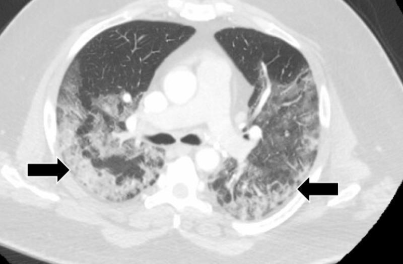
This diagnostic image is an axial contrast-enhanced CT scan of the chest in a lung window setting. It demonstrates bilateral, diffuse ground-glass opacities (GGO) characterized by a hazy increase in pulmonary density that does not obscure the underlying bronchial and vascular structures. The abnormalities are most prominent in the peripheral and posterior aspects of the lower lobes (indicated by black arrows), showing a patchy, confluent distribution. While the overall lung architecture is preserved, the opacities indicate interstitial thickening or partial alveolar filling. Central mediastinal structures, including the heart, bifurcation of the trachea (carina), and main stem bronchi, are clearly visible. These radiological findings are commonly associated with viral pneumonia (such as COVID-19), pulmonary edema, or diffuse alveolar hemorrhage, and are used here to evaluate respiratory symptoms like dyspnea and cough in the clinical context of a 47-year-old male.
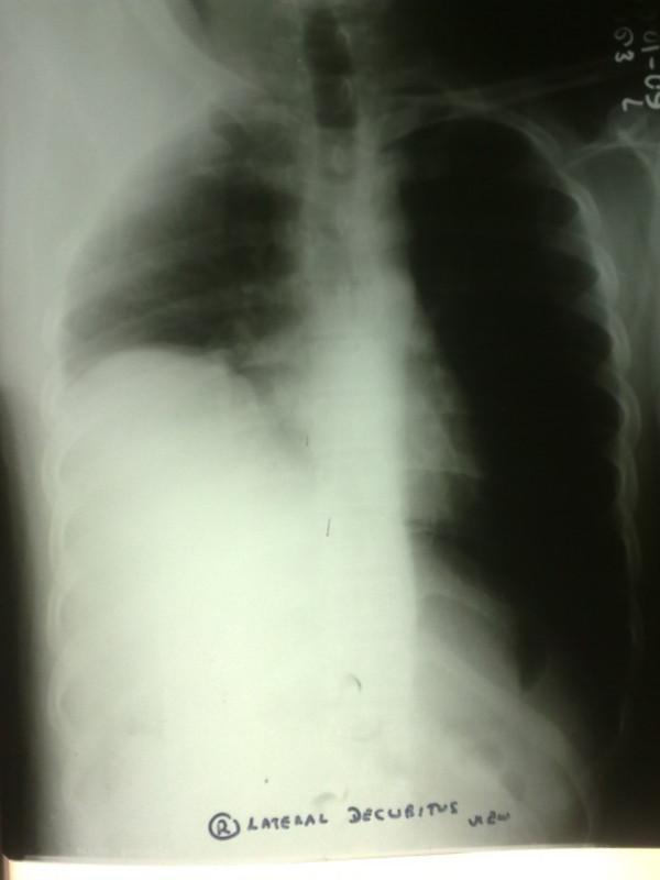
This diagnostic image is a posterior-anterior (PA) chest radiograph demonstrating a significant abnormality of the diaphragm. The primary finding is a markedly elevated right hemidiaphragm, which occupies a substantial portion of the right hemithorax, leading to reduced volume of the right lung field. The lung parenchyma above the elevated diaphragm shows relatively normal lucency, although there is increased basal opacity corresponding to the upward displacement of abdominal contents. In contrast, the left lung field appears hyperlucent, likely representing compensatory expansion. The mediastinal structures remain relatively midline. Clinically, this radiographic appearance is characteristic of phrenic nerve palsy or diaphragmatic eventration, resulting in respiratory symptoms such as dyspnea and cough. This material is suitable for medical students and clinicians focusing on pulmonology and radiology to understand the visual presentation of diaphragmatic dysfunction.
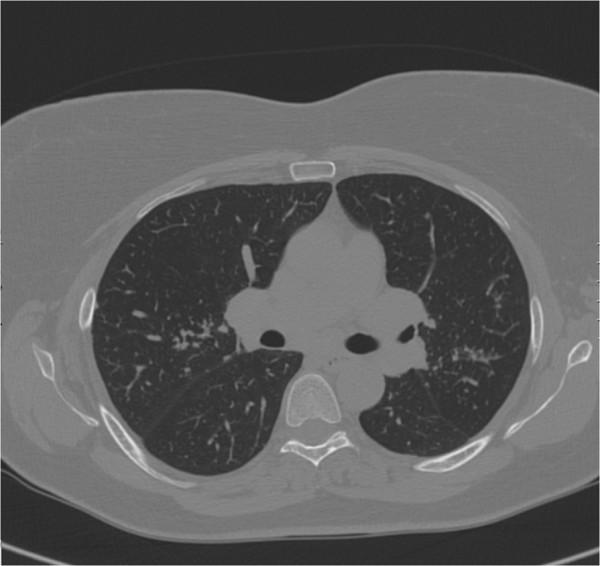
This diagnostic image is an axial Computed Tomography (CT) scan of the chest, displayed in a lung window setting. The image reveals diffuse, bilateral pulmonary pathology characterized by numerous small, well-defined, round nodules distributed throughout the visible lung parenchyma in all lobes. The distribution appears random or perilymphatic, consistent with granulomatous disease or metastatic processes. The mediastinal window view captures the level of the carina, showing the bifurcation of the primary bronchi and the descending aorta, which appear within normal limits. There is no evident pleural effusion. The surrounding thoracic cage, including the sternum, ribs, and vertebral body, shows normal bony density and integrity. This visual is significant for medical education in pulmonology and radiology, illustrating the presentation of multiple pulmonary nodules and adenomegaly in the context of chronic respiratory symptoms like dry cough and dyspnea.
Detailed Symptomatology of Respiratory Medicine
1. COUGH
Definition and Significance
Duration-Based Classification
| Category | Duration | Common Causes |
|---|---|---|
| Acute | < 3 weeks | Viral URTI, bacterial pneumonia, exacerbations of asthma/COPD, pulmonary embolism, inhaled irritants |
| Subacute | 3 - 8 weeks | Post-infectious cough, upper airway cough syndrome (rhinosinus disease), exacerbations of underlying illness |
| Chronic | > 8 weeks | Upper airway cough syndrome, asthma, gastroesophageal reflux disease (GERD), non-asthmatic eosinophilic bronchitis, ACE inhibitor use |
Red Flag Features (Warrant Urgent Investigation)
- Hemoptysis
- New cough in an active smoker older than 45 years
- Change in character of an existing cough or change in voice
- Any cough in a 55-80-year-old with >30 pack-years of smoking history
- Cough associated with: prominent dyspnea at rest or at night, hoarseness, fever, weight loss, dysphagia, vomiting, or recurrent pneumonia
Cough Variant Presentations
- Cough-variant asthma: Cough may be the sole presenting feature of asthma
- GERD-related cough: Cough can be the only symptom of gastroesophageal reflux, without any heartburn
- Drug-induced: ACE inhibitors and stagliptin (a DPP-4 inhibitor) are recognised causes. DPP-4 inhibition in mucosa promotes inflammation, manifesting as rhinorrhoea, post-nasal cough, and fatigue - easily mistaken for a seasonal cold
- Psychogenic cough: Nearly all patients labelled "psychogenic" were found, on careful 3-month evaluation, to have one of the organic causes listed above. Behavioural modification is effective even when a pulmonary cause exists.
Complications of Persistent Cough
- Tussive syncope (cough syncope)
- Retinal vessel rupture
- Persistent headache
- Chest wall and abdominal muscle pain/trauma
- Rib fractures
- Disrupted sleep and quality of life
2. DYSPNEA (Breathlessness / Shortness of Breath)
Definition
Pathophysiologic Mechanisms
-
Sensory input to the cerebral cortex from specialised receptors:
- Mechanoreceptors in the upper airways and face
- Airways and lung receptors (via vagus nerves)
- Respiratory muscle and chest wall receptors
- Chemoreceptors (central and peripheral)
- All signals process through spinal cord to supraspinal regions en route to the sensorimotor cortex
-
Perception - how the cortex interprets that input, modulated by cognitive and affective factors
- Air hunger - linked to hypercapnia, hypoxia, and breath-holding mechanisms
- Excessive effort - linked to breathing against resistance (e.g., airway obstruction, respiratory muscle weakness)
Key Clinical Features to Assess
- Onset: Sudden vs. gradual
- Temporal pattern: Exertional vs. rest, nocturnal (orthopnoea/PND), episodic
- Severity: Quantified with MRC scale (Grade 0 to 4) or Borg scale
- Triggers: Exercise, allergens, cold air, posture
- Accompanying features: Wheeze, cough, chest tightness, oedema, cyanosis, fever
Differential Diagnoses by Pattern
| Pattern | Likely Cause |
|---|---|
| Exertional dyspnea, progressive | COPD, ILD, pulmonary hypertension, heart failure |
| Orthopnoea (lying flat) | Cardiogenic pulmonary oedema, bilateral diaphragm weakness |
| Paroxysmal nocturnal dyspnea | Left heart failure |
| Episodic dyspnea + wheeze | Asthma |
| Acute onset, pleuritic pain | Pulmonary embolism, pneumothorax |
| Dyspnea + fever + productive cough | Pneumonia |
| Chronic progressive + dry cough | Interstitial lung disease |
3. HEMOPTYSIS
Definition and Classification
- Massive hemoptysis: variously defined as >200-600 mL blood in 24 hours - but any hemoptysis threatening life should be treated as massive. - Harrison's Principles of Internal Medicine, p. 635
Distinguishing Hemoptysis from Hematemesis
| Feature | Hemoptysis | Hematemesis |
|---|---|---|
| Colour | Bright red | Dark/coffee-ground |
| pH | Alkaline | Acid |
| Admixture | Frothy sputum, alveolar macrophages laden with hemosiderin | Food particles |
| History | Lung/airway disease | Gastric disease |
Differential Diagnosis by Age and Setting
- Before middle age: Infections predominate (TB, bronchiectasis, pneumonia)
- After 40-45 years or in smokers: Bronchogenic carcinoma heads the list
- Residual pulmonary cavity (post-TB, fungal disease): Aspergilloma or reactivation
- Predisposing vascular factors (oral contraceptives, heart failure): Pulmonary embolism
Common Causes (Fishman's classification)
- Bronchitis, bronchiectasis (most common, especially massive hemoptysis from bronchial arteries)
- Bronchogenic carcinoma, endobronchial metastases (breast, colon, kidney, melanoma, carcinoid)
- Dieulafoy's disease of the bronchus (rare - submucosal broncho-pulmonary arterial fistula)
- Infections: TB, pneumonia, lung abscess, invasive aspergillosis (especially in immunocompromised after granulocytopenia resolves - cavitation causes massive hemoptysis)
- Granulomatosis with polyangiitis (GPA/Wegener's)
- Goodpasture syndrome (diffuse alveolar haemorrhage)
- Drug-related: Anti-VEGF agents (bevacizumab, ramucirumab) in squamous NSCLC
- Pulmonary embolism
- Pulmonary AVM (arteriovenous malformation)
- Mitral stenosis
- Post-lung biopsy
- Chemotherapy-induced diffuse alveolar haemorrhage (bone marrow transplant)
Management Approach
- Lateral decubitus position with bleeding side down to prevent aspiration into unaffected lung
- If large-volume bleeding persists: intubation + emergency bronchoscopy
- Stable patients: CT angiography to identify bleeding source
- Bronchial artery embolization is the first-line definitive procedure - controls bleeding in 75-90% of patients
- Rebleeding after embolization: 20-50%; re-embolization usually effective
- Surgery (lobectomy) is a salvage strategy - better outcomes in non-urgent settings
4. CHEST PAIN
Types of Respiratory Chest Pain
Pleuritic Pain
- Localised and unilateral
- Distributed along intercostal nerve zones
- Diaphragmatic pleurisy refers to the ipsilateral shoulder and neck
- Described as "sharp," "burning," or "a catch"
- Defining feature: clearly related to respiratory movements - worsens on deep inspiration; coughing or sneezing causes intense distress
- Patients often experience secondary dyspnea because inspiratory pain makes them aware of every breath
- Sudden onset: Spontaneous pneumothorax, pulmonary embolism, bacterial pneumonia
- Gradual onset (days): Tuberculosis
- Slow development: Primary or secondary malignancies
- Chronic pleuritic pain: Characteristic of mesothelioma
Pericardial Pain
- Sharp, retrosternal
- Relieved by sitting up and leaning forward
Intercostal Neuritis / Radiculitis
- Superficial, knifelike quality
- Worsened by vigorous respiratory movements, but unlike pleurisy, not by ordinary breathing
- Lancinating or electric shock-like sensations unrelated to movement
- Hyperalgesia over intercostal nerve distribution = confirmatory
- Vesicular rash appearing 1-2 days later = herpes zoster
Musculoskeletal Chest Pain
- Tietze syndrome, costochondritis, rib fractures
- Point tenderness is characteristic
Cardiac Chest Pain (must be excluded)
-
Angina: Substernal "pressure," "constriction," or "squeezing"; induced by exercise, heavy meals, emotion; radiation to neck or ulnar aspect of arms
-
Variant (Prinzmetal) angina: Same quality as typical angina but at rest
-
Acute MI: Greater intensity and duration; profuse sweating, nausea, hypotension, arrhythmias
-
Murray & Nadel's Textbook of Respiratory Medicine, p. 394-396; Fishman's Pulmonary Diseases and Disorders, p. 426-430
5. WHEEZE AND STRIDOR
Wheeze
- Expiratory wheeze: Most common; associated with lower airway obstruction (asthma, COPD, acute infective tracheobronchitis)
- Biphasic wheeze: Both inspiratory and expiratory; suggests mid-airway obstruction
- Key distinction: Not all that wheezes is asthma - cardiac wheeze (cardiac asthma from LVF), anaphylaxis, foreign body, and vocal cord dysfunction can all produce wheeze
Stridor
- Inspiratory stridor: Obstruction above the glottis
- Expiratory stridor: Lower respiratory tract, prolonged expiratory phase (tracheobronchitis)
- Biphasic stridor: Particularly ominous - suggests severe airway compromise
6. OTHER IMPORTANT RESPIRATORY SYMPTOMS
Sputum
- Volume, colour, consistency are all clinically significant
- Purulent (yellow/green): Bacterial infection (pneumonia, COPD exacerbation)
- Rusty / "prune juice" appearance: Classical pneumococcal pneumonia
- Pink frothy: Pulmonary oedema
- Foul-smelling, copious: Lung abscess, bronchiectasis
- "Currant jelly": Klebsiella pneumonia
Hoarseness
- New hoarseness + chronic cough = red flag
- Indicates laryngeal involvement (recurrent laryngeal nerve palsy from mediastinal mass/lung apex tumour), laryngeal cancer, or vocal cord dysfunction
Clubbing
- Peripheral soft tissue swelling of terminal digits
- Associated with: Lung cancer (especially squamous cell), bronchiectasis, ILD (IPF), empyema, mesothelioma, and congenital cyanotic heart disease
Cyanosis
- Central cyanosis: Blue discolouration of lips, tongue, mucous membranes; implies SpO₂ < ~85% or PaO₂ < 60 mmHg
- Peripheral cyanosis: Affects extremities due to reduced peripheral circulation
Fatigue / Weight Loss
- Constitutional B symptoms in context of respiratory disease signal malignancy, TB, or connective tissue disease-associated ILD
- In COPD: Fatigue compounds exercise limitation significantly
7. ADVENTITIOUS BREATH SOUNDS - A Quick Symptom-Oriented Reference
| Sound | Mechanism | Clinical Association |
|---|---|---|
| Fine crackles | Explosive reopening of small collapsed airways | ILD/pulmonary fibrosis (late inspiratory, bilateral basal) |
| Coarse crackles | Secretions in larger airways | Pneumonia, COPD, bronchiectasis |
| Wheeze | Narrowed lower airways, turbulent flow | Asthma, COPD, foreign body |
| Stridor | Upper airway obstruction | Epiglottitis, croup, tracheal tumour |
| Pleural friction rub | Inflamed pleural surfaces rubbing | Pleuritis, pulmonary infarction, pleurisy |
| Bronchial breathing | Consolidated lung acting as acoustic conductor | Lobar pneumonia, lung abscess |
| Egophony ("ee" → "ay") | Enhanced sound transmission through consolidation + effusion | Lobar pneumonia + pleural effusion |
8. OCCUPATIONAL AND ENVIRONMENTAL HISTORY
-
Cigarette smoke: Quantified in pack-years (packs/day × years)
-
Workplace exposures: Silica, asbestos, coal dust, beryllium, organic antigens - symptoms improving on weekends ("Monday morning tightness") suggest occupational asthma
-
Geographic exposure: Cryptococcosis (southwestern USA), histoplasmosis (southern/midwestern USA), South American blastomycosis
-
Humidifiers/cooling towers: Hypersensitivity pneumonitis, Legionella
-
Drug history: Bleomycin, nitrofurantoin, methotrexate, amiodarone, beta-blockers (bronchospasm), aspirin (pulmonary oedema)
-
Family history: Cystic fibrosis, alpha-1 antitrypsin deficiency, hereditary haemorrhagic telangiectasia
-
Fishman's Pulmonary Diseases and Disorders, p. 425-430
Summary: Symptom-Disease Mapping
| Symptom Cluster | Priority Diagnoses |
|---|---|
| Chronic cough + wheeze + nocturnal symptoms | Asthma |
| Chronic dyspnea + productive cough + smoking history | COPD |
| Hemoptysis + weight loss + smoking >40y | Lung cancer |
| Hemoptysis + night sweats + immunosuppression | Tuberculosis |
| Pleuritic pain + sudden dyspnea + hypoxia | Pulmonary embolism / Pneumothorax |
| Progressive dyspnea + dry cough + fine crackles + clubbing | Interstitial lung disease (IPF) |
| Purulent sputum + copious volume + recurrent infections | Bronchiectasis |
| Cough + hoarseness + dysphagia | Mediastinal malignancy / Superior vena cava syndrome |
| Wheeze + urticaria + angioedema | Anaphylaxis |
- Murray & Nadel's Textbook of Respiratory Medicine (2-Volume, 9780323655873) - Chapters on Cough, Dyspnea, Chest Pain, Adventitious Sounds
- Fishman's Pulmonary Diseases and Disorders (2-Volume Set, 9781260473940) - "Approach to the Patient with Respiratory Symptoms" (Ch. 27)
- Harrison's Principles of Internal Medicine 22E (2025) (9781265977061) - Hemoptysis, Upper Respiratory Symptoms