
What is atropine indications contraindications how to give
atropine indications contraindications dose administration clinical
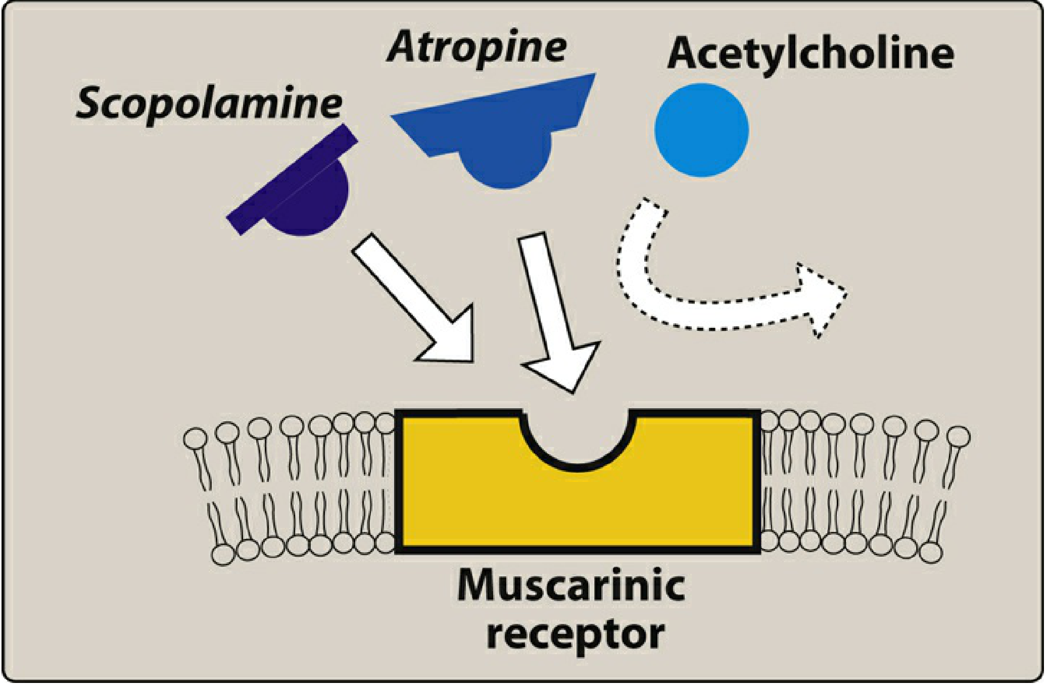
atropine mechanism muscarinic receptor pharmacology diagram
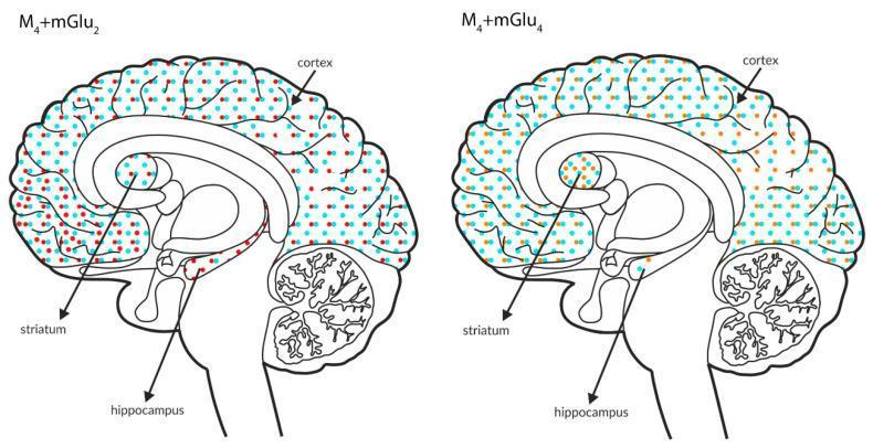
This medical illustration consists of two sagittal-view anatomical diagrams of the human brain, serving as a Comparison Chart for neurotransmitter receptor co-expression. The diagrams specifically map the distribution of muscarinic and metabotropic glutamate receptors across the cortex, striatum, and hippocampus. The left schematic illustrates the co-expression of M4 receptors (light blue dots) and mGlu2 receptors (red dots), showing dense, uniform coverage in the cortex and striatum, with a relatively lower density in the hippocampus. The right schematic illustrates the co-expression of M4 receptors (light blue dots) and mGlu4 receptors (orange dots); here, the distribution is uniform in the cortex, but notably less dense in the striatum and hippocampus compared to the first diagram. These diagrams facilitate a visual comparison of neuroreceptor localization, which is critical for understanding the pathophysiology of schizophrenia and the development of targeted antipsychotic therapies. The content is designed for intermediate to advanced neuroscience education, focusing on neuroanatomy and synaptic pharmacology.
A molecular pharmacology diagram illustrating the conformational switch of the Cannabinoid Receptor 2 (CB2R) between active (left) and inactive (right) states. The visual depicts a G protein-coupled receptor (GPCR) model featuring alpha-helical transmembrane domains. The central mechanism shown is the 'toggle switch' involving the Trp258 residue within a 'secondary site/toggle pocket'. On the left, the agonist HU-308 binds to the primary site, leaving Trp258 in an upright active conformation. On the right, a modified ligand stabilizes the inactive state through a stereogenic phenyl group that engages in an edge-to-face pi-interaction with Trp258, effectively 'switching off' receptor signaling. Key molecular modifications to the ligand for therapeutic development are highlighted: 1) fluorophore conjugation for imaging, 2) stereogenic phenyl group for functional inactivation (e.g., inhibiting beta-arrestin association and ERK1/2 phosphorylation), 3) azide incorporation for improved affinity, and 4) a novel resorcinol moiety. The diagram highlights structural biology concepts in drug design, specifically targeting the active/inactive equilibrium of GPCRs for pain modulation research.
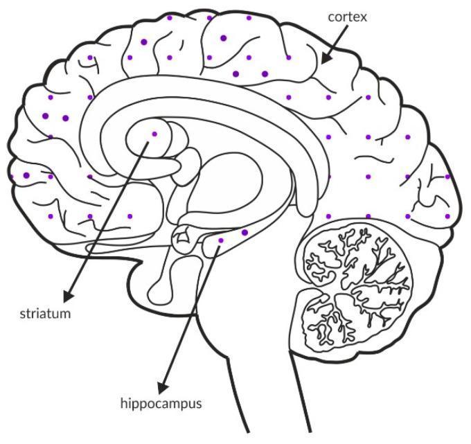
This medical illustration is an anatomical diagram showing a sagittal cross-section of a healthy human brain, used to visualize the distribution and expression intensity of M5 muscarinic receptors. The diagram employs a dotted pattern overlay where the density of purple dots serves as a semi-quantitative indicator of receptor concentration. Anatomical structures explicitly labeled with arrows include the cerebral cortex, striatum, and hippocampus. The cortex and hippocampus exhibit a moderate density of dots, representing significant receptor expression in these regions. In contrast, the striatum shows a lower density of dots, indicating a relatively lower concentration of M5 receptors. This schematic serves as an educational tool for neuroscience and neuropharmacology, illustrating specific regional receptor localization within the central nervous system and providing clinical context for potential drug targets in the treatment of cognitive or positive symptoms in neuropsychiatric disorders like schizophrenia.
Atropine - Complete Drug Reference
Mechanism of Action
Indications
| Clinical Use | Details |
|---|---|
| Symptomatic bradycardia | First-line therapy; increases heart rate by blocking M2 receptors at the SA node |
| Organophosphate / nerve agent poisoning | Reverses muscarinic symptoms (bronchospasm, hypersecretion, bradycardia, GI effects) |
| Pre-anesthetic/preoperative | Antisialagogue - reduces oral/bronchial secretions before intubation or surgery |
| Ophthalmic | Mydriasis and cycloplegia for refraction testing, uveitis, ophthalmic procedures |
| GI antispasmodic | Relaxes smooth muscle in irritable bowel, biliary/renal colic |
| Anticholinesterase antidote | Reverses muscarinic excess from physostigmine, neostigmine overdose, or mushroom poisoning (muscarine-containing species) |
| Pre-succinylcholine in children | Prevents profound bradycardia/sinus arrest caused by succinylcholine in pediatric patients |
No longer indicated: Atropine is NOT recommended for asystole or pulseless electrical activity (PEA) - removed from ACLS algorithms.
- Tintinalli's Emergency Medicine, p. 2782
- Lippincott Illustrated Reviews: Pharmacology, p. 178-179
Contraindications
| Contraindication | Reason |
|---|---|
| Acute angle-closure glaucoma | Mydriasis raises intraocular pressure dangerously |
| Tachycardia | Will further accelerate heart rate |
| Urinary tract obstruction / urinary retention | Bladder detrusor relaxation worsens retention |
| Ileus / GI obstruction | Reduces GI motility, worsens obstruction |
| Thyrotoxicosis | Risk of extreme tachycardia |
| Myasthenia Gravis | Worsens neuromuscular junction dysfunction |
| Pyloric stenosis | Reduces GI motility, worsens obstruction |
| Known hypersensitivity | Absolute contraindication |
Note: In severe or life-threatening muscarinic poisoning (organophosphates), the above contraindications may be overridden by clinical necessity.
- Harriet Lane Handbook (23rd ed.), p. 1027
- LHSC Critical Care Drug Reference
Dosing and Administration
Routes
- IV (preferred in emergencies) - give as rapid bolus; slow IV can cause paradoxical bradycardia
- IM - effective; use 1 mg/mL concentration
- IO (intraosseous) - if IV not available
- ET (endotracheal tube) - dilute in 1-2 mL NS; only if no IV/IO access; less predictable absorption
- Ophthalmic - topical drops/ointment
- Nebulized - for bronchospasm (rarely used now; ipratropium preferred)
Adult Dosing
| Indication | Dose | Notes |
|---|---|---|
| Symptomatic bradycardia | 0.5-1 mg IV Q3-5 min | Max total dose: 3 mg (0.04 mg/kg) |
| Organophosphate poisoning | 2-5 mg IV Q3-5 min | Titrate until secretions dry; may need massive doses |
| Pre-intubation antisialagogue | 0.5 mg IV/IM | Give 1-2 min before intubation |
| Ophthalmic (uveitis) | 1-2 drops 1% solution OD-BID | - |
| AtroPen (IM autoinjector) | 2 mg (green pen) | For nerve agent/organophosphate exposure |
Key rule: Doses <0.5 mg IV and slow injection are associated with paradoxical bradycardia (due to M1 pre-junctional blockade allowing more ACh release). Always give at least 0.5 mg fast.
Pediatric Dosing
| Indication | Dose | Notes |
|---|---|---|
| Bradycardia / CPR | 0.02 mg/kg IV/IO/IM Q5 min x 2-3 doses | Max single dose: 0.5 mg (child), 1 mg (adolescent); max total: 1 mg (child), 2 mg (adolescent) |
| Pre-intubation | 0.02 mg/kg IV/IM | Max 0.5 mg/dose |
| ET administration | 0.04-0.06 mg/kg | Dilute in NS (1-2 mL), flush with 1 mL NS |
| Organophosphate poisoning | 0.05-0.1 mg/kg Q5-10 min | Until bronchial/oral secretions stop |
| AtroPen (by weight) | See table below | - |
- <7 kg: 0.25 mg (yellow pen)
- 7-18 kg: 0.5 mg (blue pen)
- 18-41 kg: 1 mg (dark red pen)
-
41 kg: 2 mg (green pen)
Neonates: Atropine for neonatal bradycardia is no longer recommended. The minimum 0.1 mg rule no longer applies for pre-intubation in neonates.
- Harriet Lane Handbook (23rd ed.), p. 1026-1027
Dose-Dependent Effects
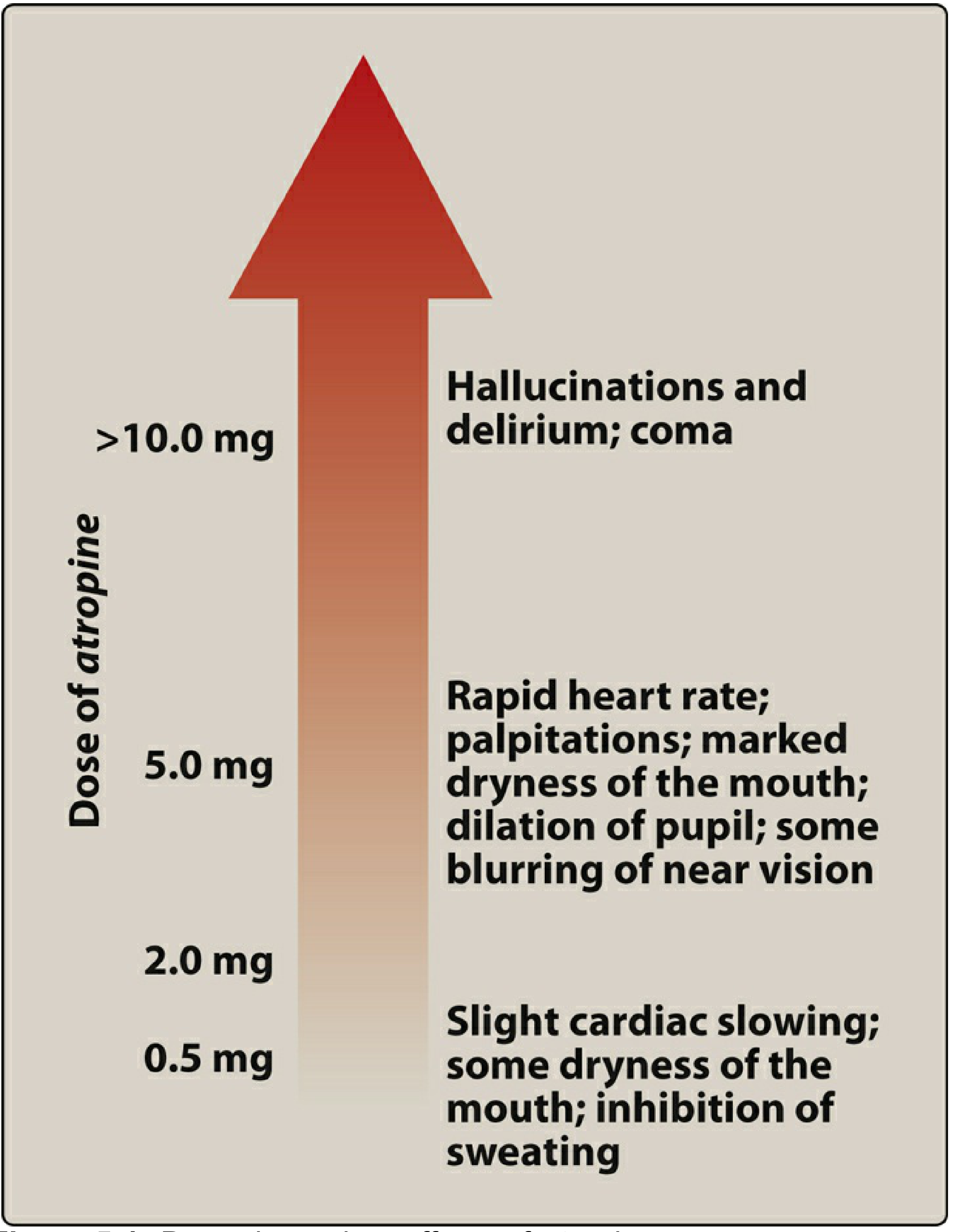
| Dose | Effects |
|---|---|
| 0.5 mg | Slight cardiac slowing (paradoxical), dry mouth, inhibited sweating |
| 2 mg | Tachycardia, marked dry mouth |
| 5 mg | Rapid HR, palpitations, marked dry mouth, pupil dilation, blurred vision |
| >10 mg | Hallucinations, delirium, coma |
Pharmacokinetics
| Parameter | Value |
|---|---|
| Onset (IV) | Immediate |
| Onset (IM) | 15-30 min |
| Half-life (adults) | 2.1-3.9 hours (~4 hours) |
| Metabolism | Hepatic (partial) |
| Elimination | Primarily renal (urine) |
Adverse Effects
- Cardiovascular: Tachycardia, palpitations (at higher doses)
- Eyes: Mydriasis, cycloplegia, blurred vision, photophobia
- GI: Dry mouth (xerostomia), constipation, reduced GI motility
- GU: Urinary retention
- Skin: Dry, hot, flushed skin; inhibited sweating - can cause hyperthermia, especially dangerous in children
- CNS: Restlessness, confusion, hallucinations, delirium (especially in elderly and children)
Key Drug Interactions
- Atropine + phenothiazines = increased anticholinergic effect
- Atropine + quinidine = increased anticholinergic effect
- Atropine + procainamide = increased anticholinergic effect