
POLYsomnography for ent from standard ent text books
Reading File
Now let me search the medical library for polysomnography content from ENT textbooks.
Finding Sources
Finding Sources
Excellent! I found polysomnography content in multiple ENT textbooks. Let me read all the relevant sections in parallel.
Reading File
Reading File
Reading File
Excellent content from all three ENT textbooks. Now let me read the remaining sections - Limitations of PSG from Cummings, the Scott-Brown's Vol 1 (overnight PSG section), and search for more content including adult OSA PSG parameters.
Reading File
Reading File
Finding Sources
Let me now fetch the PSG images and get more content from Scott-Brown's Vol 1 for adult criteria and from Cummings adult OSA section.
Loading Image
Loading Image
Reading File
I now have comprehensive content from all ENT textbooks. Let me also fetch the obstructive apnea PSG tracing image from Cummings.
Loading Image
Loading Image
I now have rich, comprehensive content from all major ENT textbooks. Let me compile the complete answer.
Polysomnography (PSG) - From Standard ENT Textbooks
1. Definition and Overview
Polysomnography (PSG) is the gold standard investigation for the diagnosis of sleep-disordered breathing (SDB), including obstructive sleep apnea (OSA). It involves simultaneous recording of multiple physiological parameters during sleep to characterize sleep architecture, respiratory events, and their consequences.
(Cummings Otolaryngology; K.J. Lee's Essential Otolaryngology; Scott-Brown's Otorhinolaryngology)
2. Parameters Recorded in a Full (Level 1) PSG
A complete polysomnogram records the following:
| Category | Parameters Recorded |
|---|---|
| Sleep staging | Electroencephalography (EEG), Electrooculography (EOG), Electromyography (EMG) |
| Respiratory effort | Chest and abdominal wall motion (strain gauges / respiratory inductive plethysmography) |
| Airflow | Nasal-oral thermistor, nasal pressure transducer |
| Gas exchange | Pulse oximetry (SpO2), end-tidal CO2 or transcutaneous CO2 |
| Cardiac | Electrocardiogram (ECG) |
| Other | Body position, snoring sound, video monitoring |
| Optional | Esophageal pressure (Pes) - for UARS detection |
Esophageal pressure monitoring (Pes) is the most sensitive method to detect Upper Airway Resistance Syndrome (UARS), but is not routinely performed. (Cummings Otolaryngology)
3. Levels of Sleep Studies (AASM Classification)
(K.J. Lee's Essential Otolaryngology)
Level 1 PSG - The standard, attended, hospital-based overnight study:
- Requires at least 6 hours of recording with sleep efficiency ≥70%
- A technician hooks up all leads and remains present overnight
- Records EEG, ECG, EOG, nasal airflow, SpO2, thoracic and abdominal movements, leg movements, body position, and snoring
- Can distinguish central from obstructive apnea
- Can be performed as a split-night study: diagnostic first half + CPAP titration second half
- Limitation: expensive, resource-intensive; patients often report atypical sleep in the lab
Level 2 PSG - Home-based but with identical recording channels to Level 1; no technician present. Rarely used due to high rate of lost data from displaced leads.
Level 3 PSG (Ambulatory Sleep Study) - Home-based with fewer channels, typically recording SpO2, body motion, snoring, and chest movement. Growing rapidly due to convenience, lower cost, and improved accuracy. Must be part of a comprehensive sleep program and interpreted by a qualified expert.
Level 4 PSG - Home oximetry alone. Simple but insufficient for therapeutic planning; prone to false positives (mild COPD) and false negatives (OSA without major desaturation). Rarely used.
4. Key Definitions Measured on PSG
Obstructive Apnea: Cessation of airflow with continued respiratory effort.
- Adults: duration ≥10 seconds
- Children: duration ≥2 times the typical breath interval (due to faster respiratory rates)
Obstructive Hypopnea: Partial reduction in airflow with continued respiratory effort (no universally agreed upon definition in children across centers).
Central Apnea: Cessation of airflow due to absent respiratory effort.
Apnea-Hypopnea Index (AHI): Number of apneas + hypopneas per hour of sleep. The primary diagnostic measure.
Respiratory Disturbance Index (RDI): Number of all respiratory events including respiratory effort-related arousals (RERAs) per hour of sleep. RERAs require esophageal pressure monitoring and are seldom routinely measured.
Obstructive Hypoventilation: End-tidal CO2 ≥50 mmHg for more than 8-10% of total sleep time.
5. PSG Tracings - Typical Patterns
Fig. A - Obstructive Apnea (Cummings, Fig. 186.4A): The nasal-oral thermistor and expired CO2 flatten during the apneic episode while chest and abdominal wall movements continue, indicating effort against a closed airway. SpO2 falls shortly after the event.
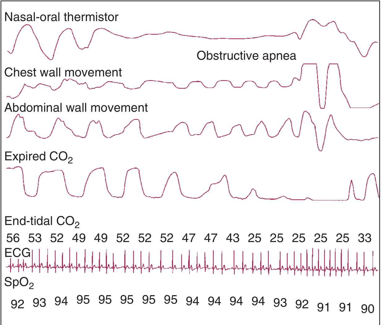
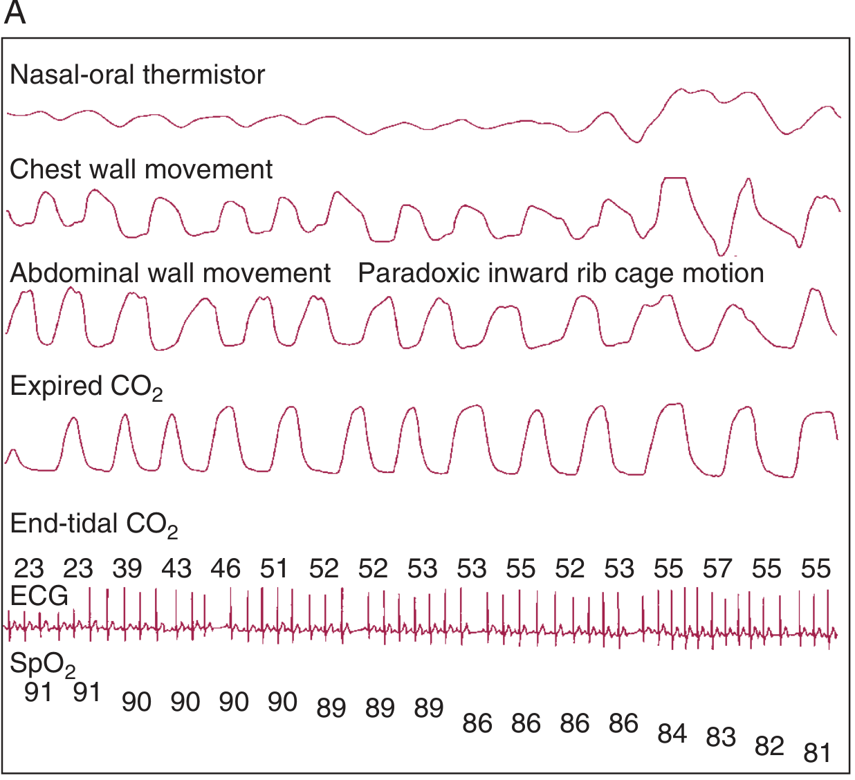
Fig. B - Obstructive Hypoventilation: Continuous partial airway obstruction results in elevated end-tidal CO2 and a progressive fall in SpO2, with paradoxic inward rib cage motion visible.
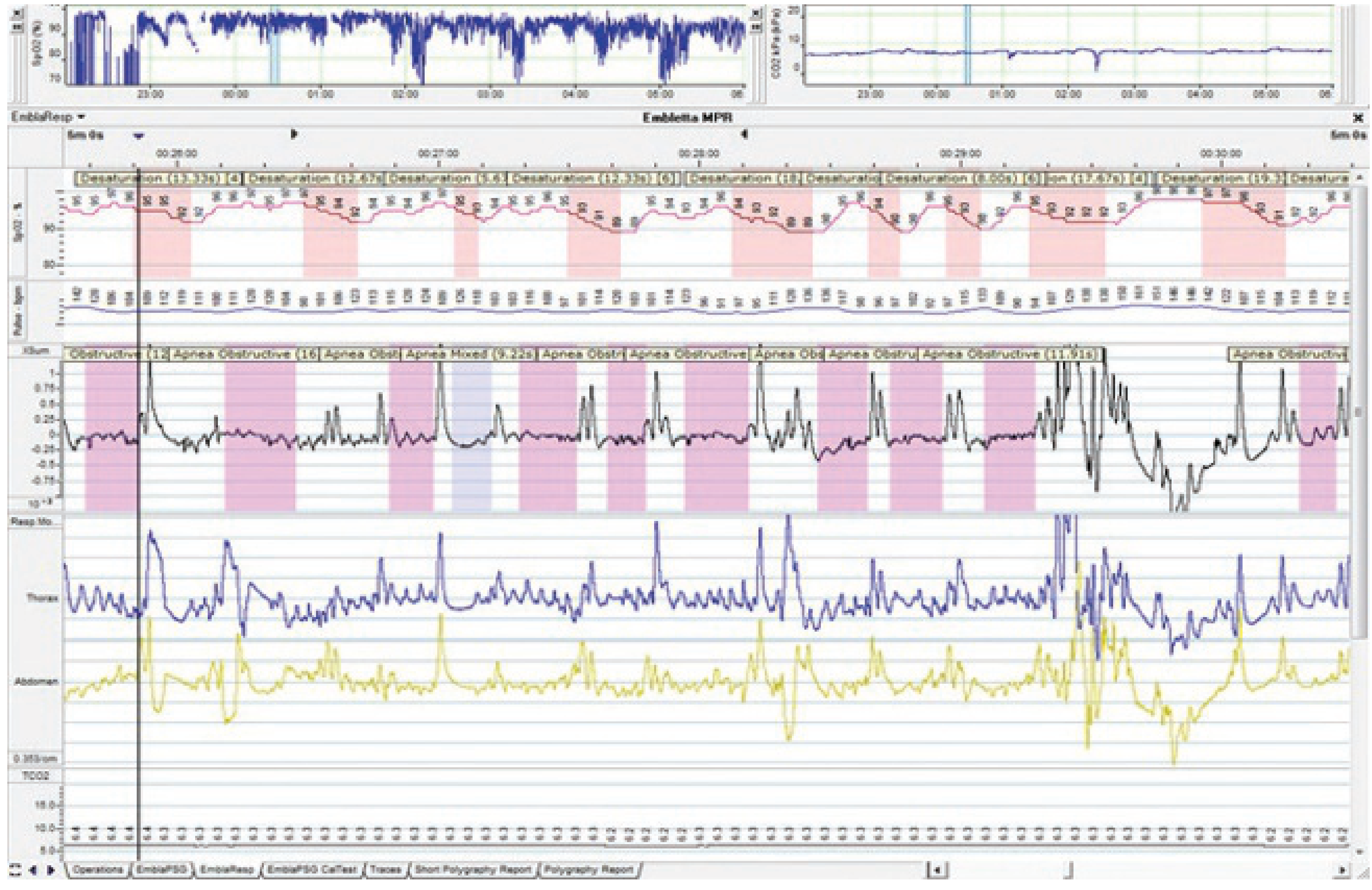
Real PSG from Scott-Brown's (Fig. 27.3): A multi-channel overnight study showing repeated desaturations (low SpO2) and decreased airflow despite ongoing thoracic and abdominal respiratory effort - confirming OSA.
6. Severity Classification of OSA on PSG
Adults (Scott-Brown's Otorhinolaryngology)
| Severity | AHI (events/hour) |
|---|---|
| No OSA | AHI <5 |
| Mild OSA | AHI ≥5 and <15 |
| Moderate OSA | AHI ≥15 and <30 |
| Severe OSA | AHI ≥30 |
Blood pressure is typically at risk when AHI >30. Note: these criteria do not account for desaturation index or duration of individual apneas.
Children (Pediatric PSG - Cummings, Box 186.2)
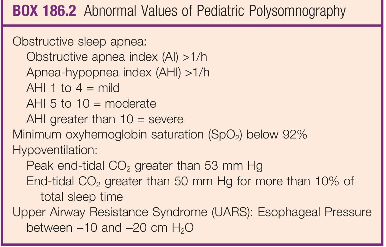
Abnormal Pediatric PSG Criteria:
- OSA: Obstructive apnea index (AI) >1/hour; AHI >1/hour
- Mild: AHI 1-4 | Moderate: AHI 5-10 | Severe: AHI >10
- Minimum SpO2 below 92%
- Hypoventilation: Peak end-tidal CO2 >53 mmHg; OR end-tidal CO2 >50 mmHg for >10% of total sleep time
- UARS: Esophageal pressure between -10 and -20 cm H2O
In normal children (Marcus et al.), average AI = 0.1 ± 0.5/hr; minimum SpO2 = 96 ± 2%, meaning AI >1/hr is abnormal.
7. When to Use PSG in ENT Practice
Indications
- Diagnosis and quantification of OSA (adults and children)
- Differentiating OSA from primary (simple) snoring
- Detecting UARS
- Pre-operative assessment before adenotonsillectomy in high-risk children (per AAO-HNS guidelines):
- Obesity, Down syndrome, craniofacial abnormalities, neuromuscular disorders, sickle cell disease, mucopolysaccharidoses
- Healthy children where clinical picture is uncertain or tonsil size is discordant with SDB severity
- Children who remain symptomatic after adenotonsillectomy
- Before initiating CPAP/BiPAP in high-risk children or surgical failures
- Complex cases: narcolepsy, parasomnias, periodic limb movement disorder (where EEG/EMG are essential)
- Neuromuscular disorders requiring nocturnal ventilation
- Suspected obesity-hypoventilation syndrome (transcutaneous CO2 monitoring needed)
(Cummings Otolaryngology; Scott-Brown's Vol 1)
8. When is Level 1 (In-Hospital) PSG Needed vs. Home Testing?
(Scott-Brown's Otorhinolaryngology, Vol 1)
In-hospital PSG required for:
- Complex OSA (suspected OHS - needs transcutaneous CO2 monitoring)
- Neuromuscular disorders (may need assisted ventilation during the study)
- Narcolepsy, parasomnias, periodic limb movement syndrome (require full EEG/EOG/EMG)
- CPAP titration in complex patients
- Unattended studies that previously failed (disconnected leads)
Home (Level 3) ambulatory testing acceptable for:
- Straightforward suspected OSA in otherwise healthy adults
- Improves diagnostic adherence (patients trust the diagnosis more in their own home)
- More convenient and lower cost
9. Limitations of PSG
(Cummings Otolaryngology)
- PSG has never been validated to predict risk of adverse outcomes or response to treatment.
- A dichotomy often exists between PSG severity and clinical symptom severity - very symptomatic children may have normal PSGs; asymptomatic snorers may have severe findings.
- No standardized monitoring method across centers; no universal consensus on what constitutes an "abnormal" study.
- First-night effect: Patients frequently report that sleep in the laboratory was atypical - especially in children under 3, where caution is needed in interpreting results (Scott-Brown's Vol 2).
- Scoring variability: How hypopneas are scored (AASM vs. Stanford criteria) dramatically changes results - in one study, OSA prevalence varied from 19% to 99% depending on the definition used.
- In children, sleep architecture differs with age: preschoolers have more central apneas and are less likely to awaken than school-age children with comparable SDB.
- The accuracy of clinical assessment alone vs. PSG in children ranges from 30-88% depending on criteria used - highlighting both the value and the pitfalls of PSG.
10. Alternatives to PSG
| Alternative | Method | Notes |
|---|---|---|
| Ambulatory device (e.g. ApneaLink Plus) | Nasal airflow + chest movement + SpO2 | Comparable to PSG for OSA detection in obese adolescents |
| Actigraphy | Wrist-worn accelerometer | Good for sleep-wake pattern estimation; no respiratory data |
| Home oximetry (Level 4) | SpO2 only | Insufficient alone; false positives (COPD) and false negatives (mild OSA) |
| Clinical scoring tools | PSQ-22 (sensitivity 78%, specificity 72%) | Can predict PSG positivity but does not replace it |
| CAS-15 | 77.3% sensitivity, 60.7% specificity for AHI >2 in children |
11. PSG in the Context of UARS
Upper Airway Resistance Syndrome (UARS) is a form of SDB that does not meet standard apnea/hypopnea criteria. On PSG, UARS is characterized by:
- A repetitive pattern of increasing negative esophageal pressure culminating in an arousal
- Awake esophageal pressure is normally -10 to -5 cm H2O
- UARS is diagnosed when pressure repeatedly reaches -10 to -20 cm H2O before arousal
This requires esophageal pressure monitoring (Pes) - not routinely available in most centers. (Cummings Otolaryngology)
Sources:
- Cummings Otolaryngology Head and Neck Surgery (9780323612173), Chapter 186 - Polysomnography & Pediatric Sleep
- K.J. Lee's Essential Otolaryngology (9781260122237) - Levels of PSG, OSA severity
- Scott-Brown's Otorhinolaryngology Head & Neck Surgery Vol 2 (9781138094634) - Pediatric PSG
- Scott-Brown's Otorhinolaryngology Head & Neck Surgery Vol 1 (9781138094642) - Overnight PSG, OSA criteria, CPAP indications
This is a shared conversation. Sign in to Orris to start your own chat.