
Limitation for pulse oximeter in simple way
pulse oximeter limitations simple explanation
Limitations of Pulse Oximeter (Simple Summary)
1. Cannot Detect CO2 (Hypoventilation)
2. Blind to Hypoventilation in Patients on Supplemental Oxygen
3. Cannot Detect Hyperoxia
4. Confused by Abnormal Hemoglobins
- Carboxyhemoglobin (CO poisoning): The oximeter reads CO-bound hemoglobin as oxygenated, giving a falsely normal reading. A patient poisoned by carbon monoxide may show SpO2 = 99% while actually starving for oxygen.
- Methemoglobin: The oximeter tends to read around 85% regardless of actual saturation.
5. Insensitive to Small Changes in PaO2 (Flat Part of Curve)
6. Interference from Physical Factors
- Nail polish or artificial nails - dark colours block the light
- Poor circulation / low cardiac output - weak pulse means unreliable reading
- Cold fingers - vasoconstriction reduces signal
- Shivering or movement - motion artifact causes false readings
- Venous pulsations (e.g., tricuspid regurgitation) - the device may pick up venous blood instead of arterial
7. Skin Pigmentation
8. Dyes and Substances
Quick Summary Table
| Limitation | What Goes Wrong |
|---|---|
| CO2/hypoventilation | Not detected at all |
| Supplemental O2 | Hides hypoventilation |
| CO poisoning | Falsely normal SpO2 |
| Methemoglobinemia | Reads ~85% regardless |
| Poor perfusion/cold | Unreliable or no signal |
| Nail polish/dark nails | Blocks light |
| Dark skin | May overestimate SpO2 |
| Motion/shivering | False readings |
oxyhemoglobin and carbooxyhemoglobin
oxyhemoglobin vs carboxyhemoglobin difference mechanism
oxyhemoglobin dissociation curve carboxyhemoglobin left shift
Oxyhemoglobin (HbO₂) vs Carboxyhemoglobin (COHb)
What Are They?
| Oxyhemoglobin (HbO₂) | Carboxyhemoglobin (COHb) | |
|---|---|---|
| Formed by | Hemoglobin + Oxygen (O₂) | Hemoglobin + Carbon Monoxide (CO) |
| Where formed | In the lungs (normal process) | When CO is inhaled (abnormal) |
| Color | Bright cherry red | Also cherry red (this causes confusion!) |
| Function | Carries O₂ to tissues | Cannot carry any oxygen - useless |
| Bond type | Reversible, loosely bound | Very tight, hard to reverse |
Why is Carboxyhemoglobin So Dangerous?
- CO "kicks out" oxygen and grabs the hemoglobin binding sites for itself. - Katzung's Pharmacology
- The remaining oxyhemoglobin (not yet taken by CO) also has trouble releasing its oxygen to tissues. This is called the Haldane effect - a left shift of the oxyhemoglobin dissociation curve, meaning oxygen clings tighter and tissues starve even more.
- CO binds to cytochrome oxidase in the mitochondria, blocking the cell's ability to use oxygen at all (even if some oxygen was delivered). - Tintinalli's Emergency Medicine
The Dissociation Curve - Key Visual
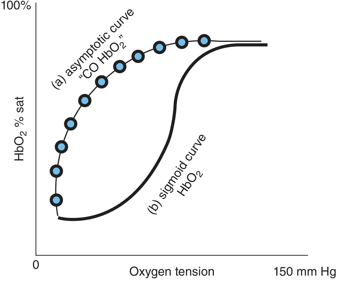
- Curve (b) = Normal sigmoid shape - oxygen releases well to tissues at lower oxygen tensions
- Curve (a) = CO-poisoned curve - becomes asymptotic, shifted far LEFT - oxygen barely releases to tissues even at very low oxygen tensions
Why the Pulse Oximeter Gets Fooled
Normal vs Dangerous COHb Levels
| Person | COHb Level |
|---|---|
| Healthy non-smoker | < 1% |
| Smoker | 5-10% |
| Headache, malaise | ~15% |
| Headache, fatigue, poor coordination | ~25% |
| Collapse, syncope | ~40% |
| Death possible | > 60% |
Treatment of CO Poisoning
co-oximetery
co-oximetry principle wavelengths hemoglobin species measurement
Co-oximetry - Simple Explanation
What is it?
The Key Principle: Beer-Lambert Law
"You need at least as many wavelengths as there are hemoglobin species you want to measure."
Pulse Oximeter vs Co-oximeter - Side by Side
| Feature | Pulse Oximeter | Co-oximeter |
|---|---|---|
| Wavelengths used | 2 (660 nm red + 940 nm infrared) | 4 to 256 wavelengths |
| Where used | Bedside / non-invasive | Laboratory (blood sample needed) |
| What it measures | Only HbO₂ and Hb (deoxyHb) | HbO₂, HbO, COHb, MetHb, SulfHb |
| Can detect CO poisoning? | NO - falsely normal | YES |
| Can detect MetHb? | Only approximate (~85%) | YES, accurately |
| Type of saturation reported | Functional SpO₂ | Fractional SaO₂ |
| Invasive? | No | Yes (arterial blood sample) |
Functional vs Fractional Saturation - The Critical Difference
SpO₂ = HbO₂ / (HbO₂ + Hb) × 100%
SaO₂ = HbO₂ / (HbO₂ + Hb + COHb + MetHb) × 100%
The Absorption Spectra - Why It Matters
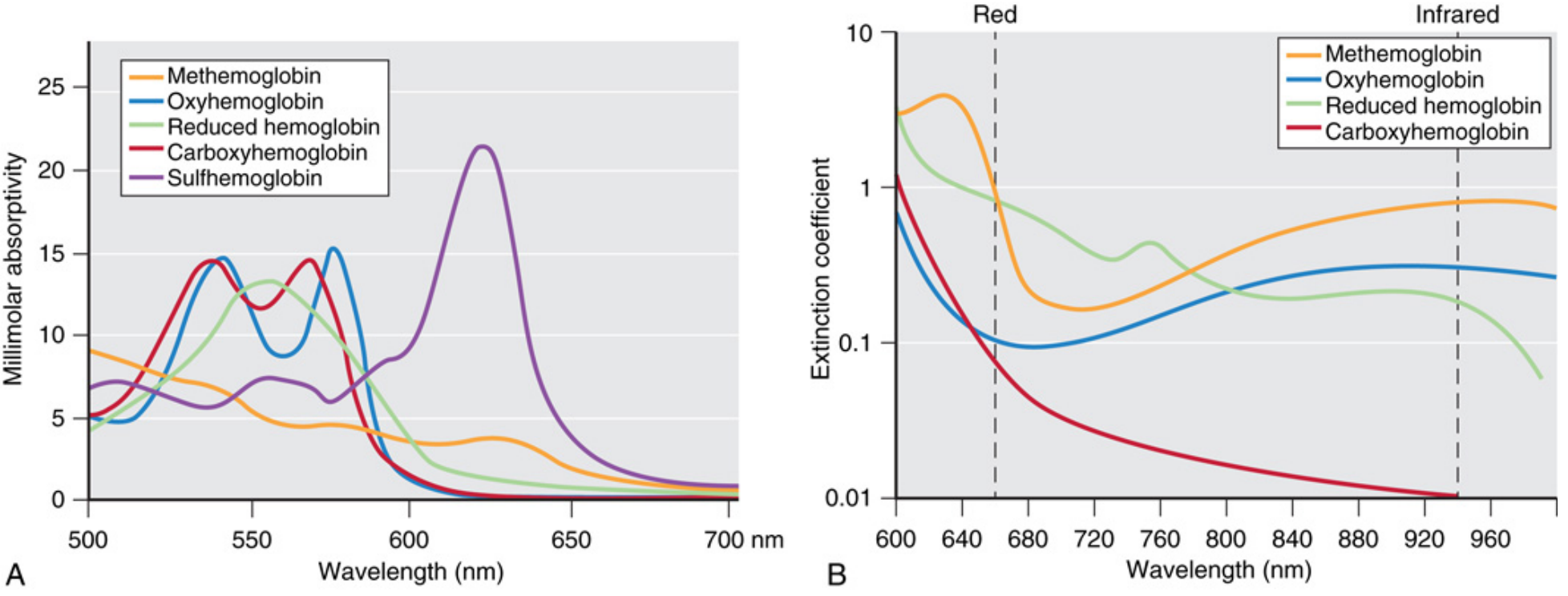
- Graph A (left): Shows how each hemoglobin species has a unique light absorption "fingerprint" across visible wavelengths (500-700 nm)
- Graph B (right): Shows the extinction coefficients at the red (660 nm) and infrared (940 nm) wavelengths used by a pulse oximeter. Notice: carboxyhemoglobin (red line) and oxyhemoglobin (blue) overlap near 660 nm - this is exactly why the pulse oximeter cannot tell them apart
- A co-oximeter uses many more wavelengths to separate all these curves
5 Hemoglobin Species a Co-oximeter Measures
| Hemoglobin | Meaning | Clinical Importance |
|---|---|---|
| HbO₂ (Oxyhemoglobin) | Hb carrying O₂ | Normal |
| Hb (Deoxyhemoglobin) | Hb without O₂ | Normal (venous blood) |
| COHb (Carboxyhemoglobin) | Hb bound to CO | CO poisoning |
| MetHb (Methemoglobin) | Hb with Fe³⁺ (oxidized) | Drug toxicity (dapsone, nitrates) |
| SulfHb (Sulfhemoglobin) | Hb bound to sulfur | Rare, from certain drugs |
When to Use a Co-oximeter (Instead of Relying on Pulse Ox)
- Suspected CO poisoning (house fire, smoke inhalation)
- Suspected methemoglobinemia (cyanosis with high SpO₂, after dapsone/nitrates)
- Patient on supplemental oxygen with unexplained clinical deterioration
- Any case where SpO₂ and clinical picture do not match
- Critically ill patients where exact hemoglobin fractions matter
Quick Clinical Example
at what level will cyanosis develop in carbonmonoxide
carbon monoxide poisoning cyanosis does not develop cherry red skin why
Cyanosis in Carbon Monoxide Poisoning - The Crucial Answer
Short Answer: Cyanosis Classically Does NOT Develop
Why No Cyanosis? - The Mechanism
- CO binds hemoglobin to form carboxyhemoglobin (COHb) - which is bright cherry red in color
- The venous blood also stays bright red because oxygen cannot be released to tissues (left shift of dissociation curve - the Haldane effect)
- So there is no dark blue deoxygenated hemoglobin accumulating in the skin
- The patient looks pink, flushed, or cherry red - not blue - even while being severely hypoxic
"In spite of asphyxiation, cyanosis does not occur; the skin is pink or pale and the lips bright red." - Britannica / CO Poisoning
Cherry Red Skin - The Classic Sign
| Feature | Reality |
|---|---|
| Classic teaching | Cherry red skin = CO poisoning |
| How common in living patients? | Only 2-3% of symptomatic cases |
| How common post-mortem? | Much more common - well-recognised at autopsy |
| Why more common in death? | At death, all remaining HbO₂ converts fully to COHb, making the color very prominent |
When Can Cyanosis Occur in CO Poisoning?
- There is severe respiratory compromise or arrest (then deoxygenated Hb finally accumulates)
- CO poisoning is combined with another cause of hypoxia (e.g., severe smoke inhalation causing respiratory failure)
- Very late, terminal stage when breathing has stopped
CO Poisoning vs Other Hypoxia - Skin Color
| Cause of Hypoxia | Skin Color | Why |
|---|---|---|
| Pneumonia / drowning | Cyanosis (blue) | High deoxyHb in skin |
| Methemoglobinemia | Chocolate brown/gray | MetHb is dark brown |
| CO poisoning | Cherry red | COHb is bright red |
| Cyanide poisoning | Cherry red | Venous O₂ unused, stays red |
COHb Level vs Symptoms (for reference)
| COHb Level | Clinical Effect |
|---|---|
| < 1% | Normal (non-smoker) |
| 5-10% | Smoker baseline |
| ~15% | Headache, malaise |
| ~25% | Headache, fatigue, poor coordination |
| ~40% | Collapse, syncope |
| > 60% | Coma, death |
in summary
Summary: Pulse Oximetry, Hemoglobin Species & CO Poisoning
1. Pulse Oximeter - Limitations
- Uses only 2 wavelengths (660 nm red + 940 nm infrared)
- Measures only functional saturation - HbO₂ vs deoxyHb
- Cannot detect: CO poisoning, hypoventilation, hyperoxia, MetHb accurately
- Fooled by: nail polish, cold fingers, poor perfusion, dark skin, motion
- Patient on supplemental O₂ - completely blind to hypoventilation
- Solution: use capnography + co-oximetry alongside it
2. Oxyhemoglobin vs Carboxyhemoglobin
| Oxyhemoglobin (HbO₂) | Carboxyhemoglobin (COHb) | |
|---|---|---|
| Formed by | Hb + O₂ (lungs, normal) | Hb + CO (CO inhalation) |
| Function | Carries O₂ to tissues | Carries nothing - useless |
| Bond | Loose, reversible | ~220x tighter than O₂ |
| Color | Cherry red | Also cherry red |
| Danger | None (normal) | Tissue hypoxia + mitochondrial poison |
| Curve effect | Normal sigmoid | Left shift - O₂ clings, won't release |
3. Co-oximetry
- Uses 4-256 wavelengths - measures ALL hemoglobin species
- Reports fractional saturation (true SaO₂):
SaO₂ = HbO₂ / (HbO₂ + Hb + COHb + MetHb) × 100%
- Gold standard for O₂ saturation measurement
- Detects: COHb, MetHb, SulfHb - things pulse ox misses completely
- Requires a blood sample (invasive, lab-based)
4. Cyanosis in CO Poisoning
| Question | Answer |
|---|---|
| Does cyanosis develop? | NO - classically absent |
| Why not? | COHb is bright red - no dark deoxyHb accumulates in skin |
| What color instead? | Cherry red skin (specific but rare in living - only 2-3%) |
| When cyanosis CAN occur | Only if respiratory arrest or severe smoke inhalation |
| Why dangerous? | No color warning → "silent killer" |
The Big Clinical Picture
CO inhaled
    ↓
Hb grabs CO (220x more than O₂)  →  COHb formed
    ↓                                      ↓
Less O₂ carried               Remaining HbO₂ won't release O₂ (left shift)
    ↓                                      ↓
Mitochondria also blocked (cytochrome oxidase inhibited)
    ↓
Tissue hypoxia despite pink/red skin
    ↓
Pulse ox reads NORMAL (SpO₂ ~99%)  ←  The trap
    ↓
Only CO-oximeter reveals the truth
One-Line Rule to Remember
"CO poisoning = cherry red skin + normal SpO₂ + patient deteriorating → get a co-oximeter, give 100% O₂"
give summary of number 4
Summary: Cyanosis in CO Poisoning
Why?
- Cyanosis needs 5 g/dL of dark deoxygenated Hb in the skin to appear blue
- In CO poisoning, Hb is bound to CO → forms COHb which is bright cherry red
- Even venous blood stays red (O₂ won't release to tissues due to left shift)
- So no dark deoxyHb accumulates → no blue color → no cyanosis
What appears instead?
- Cherry red skin - the classic sign
- But only seen in 2-3% of living patients
- More obvious after death (at autopsy)
When can cyanosis occur?
- Only if respiratory arrest happens
- Or severe smoke inhalation causes lung failure
Why is this dangerous?
- No blue color = no visible warning sign
- Patient looks pink and healthy while organs are starving for oxygen
- Pulse oximeter reads normal SpO₂ (fooled by COHb)
- This is why CO is called the "silent killer"
Key rule: Cherry red + normal SpO₂ + deteriorating patient = think CO poisoning immediately
make it into paragraph and mention its source
Cyanosis in Carbon Monoxide Poisoning
simple summary
Cyanosis in CO Poisoning - Simple Summary
Remember: Cherry red skin + normal SpO₂ + deteriorating patient = CO poisoning until proven otherwise.
hyper-baric oxygen therapy
hyperbaric oxygen therapy mechanism indications contraindications clinical use
Hyperbaric Oxygen Therapy (HBOT)
What Is It?
How It Works - The Physics (Henry's Law & Boyle's Law)
- Normally, oxygen is carried almost entirely bound to hemoglobin
- Under high pressure with 100% O₂, a significant amount of oxygen dissolves freely into the plasma itself - bypassing hemoglobin entirely
- At 2 ATA: blood oxygen content increases by 125%; tissue oxygen tension increases 10-fold compared to room air - Tintinalli's Emergency Medicine
Why It Works in CO Poisoning - The Key Mechanism
| Treatment | COHb Half-life |
|---|---|
| Room air | ~4-5 hours |
| 100% O₂ (normobaric) | ~60-90 minutes |
| Hyperbaric O₂ (2-3 ATA) | 15-23 minutes |
- O₂ outcompetes CO and physically displaces it from hemoglobin
- COHb breaks down rapidly
- Dissolved plasma O₂ delivers oxygen to tissues directly
- Neurological damage from CO is reduced
Approved Indications for HBOT (14 recognized)
| Category | Conditions |
|---|---|
| Poisoning | CO poisoning, cyanide poisoning |
| Diving emergencies | Decompression sickness ("the bends"), arterial gas embolism |
| Wound healing | Diabetic foot ulcers, crush injuries, compartment syndrome |
| Infections | Necrotizing fasciitis, clostridial gas gangrene (myonecrosis) |
| Radiation injury | Osteoradionecrosis, soft tissue radionecrosis |
| Vascular | Central retinal artery occlusion, compromised skin grafts/flaps |
| Severe anaemia | When transfusion is refused or impossible |
| Burns | Adjunct in thermal burns |
Mechanisms of Benefit (Beyond Just Oxygen)
- Hyperoxygenation - massively increases dissolved O₂ in plasma
- Vasoconstriction - reduces oedema by 10-20% while still delivering more O₂ (paradoxically beneficial in crush injury/burns) - Tintinalli's EM
- Anti-infective - high O₂ tensions are toxic to anaerobic bacteria (e.g., Clostridium)
- Reduces gas bubble size (Boyle's Law) - useful in decompression sickness
- Promotes angiogenesis - stimulates new blood vessel growth in radiation-damaged tissue
- Inhibits leukocyte adhesion - reduces reperfusion injury and inflammation in CO poisoning
Contraindications
| Type | Condition |
|---|---|
| Absolute | Untreated pneumothorax (pressure increase will collapse lung further) |
| Relative | Pulmonary bullae/blebs, obstructive lung disease, claustrophobia, uncontrolled fever, recent ear/thoracic surgery, upper respiratory infection, uncontrolled heart failure |
Side Effects / Risks
- Oxygen toxicity - seizures (CNS O₂ toxicity at high pressures)
- Ear barotrauma - pressure damage to middle ear (most common side effect)
- Sinus barotrauma
- Pulmonary O₂ toxicity - with prolonged use
- Claustrophobia inside the chamber
- Fire risk - 100% O₂ environment is highly flammable
Practical Limitation in CO Poisoning
- Time delay to transfer patient to a hyperbaric facility
- Difficulty resuscitating unstable patients inside a closed pressurized chamber
- Unstable burn patients or those needing active airway management cannot safely be placed inside
- A Cochrane review found no definitive proven benefit on mortality, making its use somewhat controversial
Bottom line: HBOT is most valuable in severe CO poisoning (coma, neurological signs, pregnancy, COHb > 25%), but 100% normobaric O₂ remains the immediate first-line treatment. - Roberts & Hedges' Clinical Procedures in Emergency Medicine
summarize it into table
Hyperbaric Oxygen Therapy (HBOT) - Summary Table
| Aspect | Details |
|---|---|
| Definition | Breathing 100% O₂ in a pressurized chamber at > 1 atmosphere (usually 2-3 ATA) |
| Key Law | Henry's Law - more O₂ dissolves in plasma under pressure |
| O₂ increase | Blood O₂ content ↑ 125%; tissue O₂ tension ↑ 10-fold (at 2 ATA) |
COHb Half-Life with Different Treatments
| Treatment | COHb Half-Life |
|---|---|
| Room air | ~4-5 hours |
| 100% O₂ (normal pressure) | ~60-90 minutes |
| Hyperbaric O₂ (2-3 ATA) | 15-23 minutes |
Mechanisms of Action
| Mechanism | Benefit |
|---|---|
| Hyperoxygenation | O₂ dissolves in plasma - bypasses blocked Hb |
| Displaces CO from Hb | Rapidly reduces COHb levels |
| Vasoconstriction | Reduces oedema while maintaining O₂ delivery |
| Kills anaerobes | Toxic to Clostridium and other anaerobic bacteria |
| Shrinks gas bubbles | Treats decompression sickness (Boyle's Law) |
| Promotes angiogenesis | Heals radiation-damaged tissue |
| Anti-inflammatory | Reduces reperfusion injury in CO poisoning |
Indications
| Category | Condition |
|---|---|
| Poisoning | CO poisoning, cyanide poisoning |
| Diving emergencies | Decompression sickness, arterial gas embolism |
| Wounds | Diabetic foot ulcer, crush injury, compartment syndrome |
| Infections | Necrotizing fasciitis, gas gangrene |
| Radiation injury | Osteoradionecrosis, soft tissue radionecrosis |
| Vascular | Central retinal artery occlusion, compromised skin grafts |
| Anaemia | When transfusion is refused/impossible |
Contraindications
| Type | Condition |
|---|---|
| Absolute | Untreated pneumothorax |
| Relative | Pulmonary bullae, obstructive lung disease, claustrophobia, uncontrolled fever, recent ear/chest surgery, heart failure |
Side Effects
| Side Effect | Cause |
|---|---|
| Seizures | CNS O₂ toxicity at high pressure |
| Ear barotrauma | Pressure changes (most common) |
| Sinus pain | Pressure changes |
| Pulmonary O₂ toxicity | Prolonged sessions |
| Claustrophobia | Enclosed chamber |
| Fire risk | 100% O₂ environment is flammable |
HBOT in CO Poisoning - When to Use
| Use HBOT | Use Normobaric 100% O₂ only |
|---|---|
| Coma or altered consciousness | Mild CO poisoning |
| COHb > 25% | Stable patient, no neuro signs |
| Neurological signs present | No HBO facility nearby |
| Pregnancy (any CO level) | Unstable patient needing resuscitation |
| Cardiac involvement | Burn patient with airway issues |
simplify and summarize into few lines approximately 6lines
Hyperbaric Oxygen Therapy - Simple Summary
gas gangrene
gas gangrene clostridial myonecrosis pathophysiology treatment
gas gangrene clostridial myonecrosis clinical image
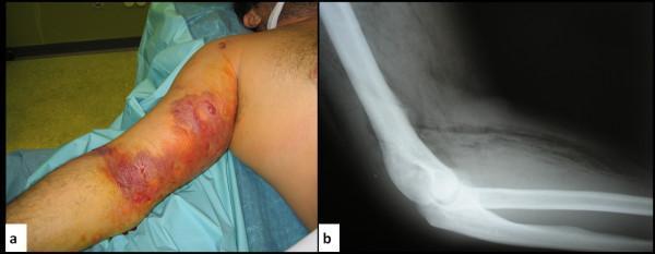
This composite educational material displays clinical and radiological findings of gas gangrene (clostridial myonecrosis) in the upper limb of a patient with a history of intravenous drug use. Figure (a) is a clinical photograph of the right arm showing severe localized edema, circumferential erythema, and large, dusky, hemorrhagic bullae (blistering) across the bicep and antecubital regions. Areas of purple-to-black skin discoloration suggest impending tissue necrosis. Figure (b) is a lateral X-ray of the same limb, demonstrating the pathognomonic sign of gas gangrene: extensive subcutaneous emphysema. This is characterized by radiolucent (dark) gas pockets and streaks dissecting through the soft tissue planes and muscle compartments, indicating gas production by anaerobic organisms such as Clostridium perfringens. The image serves to teach the correlation between clinical signs of rapidly progressing necrotizing soft tissue infection (crepitus, bullae, edema) and the critical radiographic finding of soft tissue gas.
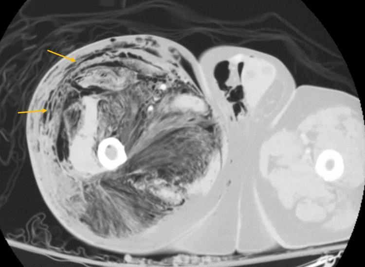
This diagnostic image is an axial non-contrast CT scan of the lower extremities, specifically focusing on the thigh region. The right thigh (left side of the image) demonstrates a massive, diffuse collection of subcutaneous and deep soft tissue gas, indicated by low-density (black) areas. Yellow arrows highlight gas tracking extensively along the fascial planes, within the subcutaneous fat, and dissecting between muscle compartments. There is significant soft tissue edema and architectural distortion, with a loss of normal fat planes and muscle definition compared to the relatively normal-appearing contralateral limb. Key findings include 'gas in soft tissues' or 'emphysematous changes,' which are pathognomonic for necrotizing fasciitis or clostridial myonecrosis (gas gangrene) in a clinical context of sepsis. Additionally, gas is visible within the scrotal region, suggesting proximal or distal extension of the necrotizing process. This image serves as a critical educational example of surgical emergency findings in musculoskeletal radiology and infectious disease.
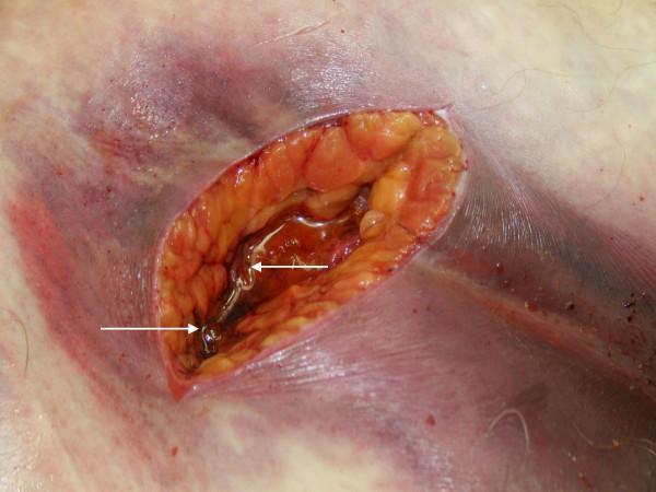
This clinical photograph shows a post-mortem incision into necrotic tissue, illustrating a case of gas gangrene (clostridial myonecrosis). The primary focal point is a surgical opening in the skin and subcutaneous layer, revealing underlying necrotic adipose and myofascial tissue with a characteristic reddish-orange to dusky brown discoloration. Within the incision, there is a prominent collection of serosanguinous fluid containing numerous gaseous bubbles, marked by white arrows, which represent the metabolic byproducts of anaerobic bacteria like Clostridium septicum. The surrounding skin exhibits extensive ecchymosis and dark purple purpura, suggesting severe subcutaneous hemorrhage and impaired perfusion. The skin's surface appears tense and glistening, indicative of underlying edema and crepitus. This image serves as a critical educational example of the rapid tissue destruction and gas formation characteristic of necrotizing soft tissue infections (NSTI) and fulminant sepsis.
Gas Gangrene (Clostridial Myonecrosis)
Definition
Causative Organisms
| Type | Organism | Setting |
|---|---|---|
| Most common | Clostridium perfringens | Traumatic wounds |
| Spontaneous | Clostridium septicum | Occult colon cancer, leukemia |
| Others | C. novyi, C. histolyticum, C. sordellii | Various |
How It Happens (Pathophysiology)
- Trauma or surgery introduces Clostridium spores into deep tissue
- Dead tissue + foreign bodies create low oxygen (anaerobic) environment
- Spores germinate and bacteria multiply rapidly
- Bacteria release powerful exotoxins (alpha-toxin + theta-toxin)
- Toxins cause:
- Irreversible ischemic muscle necrosis
- Gas production (CO₂, H₂) trapped in muscle
- Intravascular platelet aggregation blocking blood flow
- No inflammatory response (characteristically absent - neutrophils cannot penetrate)
- Myocardial depression → shock
- Goldman-Cecil Medicine; Red Book 2021
Clinical Features (Rapid Progression)
| Time | What Happens |
|---|---|
| Early | Sudden severe pain at wound site (disproportionate to appearance) |
| Hours | Skin turns pale → bronze → purple/red |
| Soon after | Tense bullae (blisters) with dark, foul-smelling "dish water" fluid |
| Late | Black skin necrosis, crepitus (crackling on palpation = gas in tissue) |
| Systemic | High fever, tachycardia, refractory hypotension, confusion, renal failure |
Incubation period: 6 hours to 4 days after injury - Red Book 2021
Clinical Images
Diagnosis
- Clinical - pain + skin changes + crepitus = act immediately, do NOT wait for tests
- X-ray / CT - gas in soft tissues (but absence does NOT rule out)
- Gram stain - gram-positive bacilli, no neutrophils (characteristic)
- Surgery - muscle does not bleed or contract when stimulated = diagnostic
- Blood cultures - positive in ~15% of cases
Treatment - The Triad
| Priority | Treatment |
|---|---|
| 1st - Surgical | Aggressive, immediate surgical debridement - remove ALL dead tissue; amputation often needed |
| 2nd - Antibiotics | High-dose Penicillin G + Clindamycin (clindamycin stops toxin production); add carbapenems if polymicrobial |
| 3rd - Adjunct | Hyperbaric oxygen (HBO) - high O₂ is toxic to anaerobes; reduces tissue damage |
Clindamycin is preferred because it stops toxin production even for resistant strains. - Goldman-Cecil Medicine
Prognosis
| Factor | Outcome |
|---|---|
| With treatment | 20-30% mortality |
| Without treatment | 100% fatal - death within hours |
| Worse prognosis | Shock at diagnosis, leukopenia, hemolysis, old age |
| Better prognosis | Young age, single limb, early surgery |
Prevention
- Prompt wound debridement after trauma
- Remove all foreign bodies, dead tissue, and hematomas
- Avoid prolonged tourniquets
- Proper surgical technique
One-line rule: Severe wound pain + crepitus + skin discoloration = gas gangrene until proven otherwise - rush to surgery immediately.
definition in simple terms
Gas Gangrene - Simple Definition
A dirty deep wound → bacteria grow in the dead tissue → release powerful poisons (toxins) → muscle rots and dies → gas builds up inside → spreads so fast it can kill within hours.