
Gauchers disease in detail
Reading File
Gaucher disease is the most common lysosomal storage disorder, caused by deficiency of glucocerebrosidase (beta-glucosidase), leading to accumulation of glucocerebroside in macrophages of the liver, spleen, and bone marrow. There are three types - Type 1 (non-neuronopathic, most common), Type 2 (acute neuronopathic), and Type 3 (chronic neuronopathic). Treatment includes enzyme replacement therapy (ERT) with imiglucerase/velaglucerase and substrate reduction therapy.
Searching sources...
Finding Sources
Searching PubMed
"Gaucher disease"[MeSH Terms] AND treatment
Good results. Now let me read the most comprehensive sources - Goldman-Cecil (primary medicine), Emery's Genetics, and also search for more specific content on pathophysiology and treatment.
Reading File
Reading File
Finding Sources
Excellent content. Now let me read more from Goldman-Cecil (treatment section) and Robbins (pathology), plus fetch the X-ray/Gaucher cell image.
Reading File
Reading File
Loading Image
I have very comprehensive content. Let me also fetch the Gaucher cell histology image from Robbins and get the treatment table details.
Loading Image
Reading File
I now have extremely comprehensive content to write a detailed, well-organized answer. Let me compile it all.
Gaucher Disease - A Detailed Overview
Gaucher disease is the most common lysosomal storage disorder and the most frequent inherited sphingolipidosis. It is caused by the autosomal recessive deficiency of the enzyme glucocerebrosidase (beta-glucosidase), resulting in the pathological accumulation of glucocerebroside (glucosylceramide) within macrophages throughout the body.
1. Genetics and Molecular Basis
-
Gene: GBA1 (formerly GBA), located on chromosome 1q21
-
Inheritance: Autosomal recessive
-
Enzyme deficient: Lysosomal glucocerebrosidase (acid beta-glucosidase)
-
Substrate accumulated: Glucocerebroside (glucosylceramide), derived from degradation of senescent erythrocytes and leukocytes
-
Over 600 different mutations identified; key ones include:
- N370S (p.N409S): Most common allele in Ashkenazi Jews; found exclusively in Type 1 - protects against neurologic involvement
- L444P (p.L483P): Homozygosity most often associated with Type 3
- D409H (p.D448H): Homozygosity defines Type 3C (with cardiac calcifications and hydrocephalus)
-
A highly homologous pseudogene nearby complicates molecular diagnosis - specialized lab knowledge is required
-
The GBA1 locus is also the most common genetic risk factor for Parkinson disease identified to date; both affected patients and heterozygous carriers have a 5- to 10-fold increased frequency of parkinsonism
-
Goldman-Cecil Medicine, p. 2303; Robbins & Kumar Basic Pathology, p. 3656
2. Epidemiology
-
Pan-ethnic disorder, but type 1 shows marked enrichment in Ashkenazi Jews:
- Carrier frequency in Ashkenazi Jews: ~1 in 16
- General population carrier frequency: ~1 in 100 (Robbins notes up to 1 in 40,000 for non-Jewish populations for the type 1 variant)
-
Type 1 accounts for approximately 99% of all cases globally
-
Types 2 and 3 are rare and more severe
-
Goldman-Cecil Medicine, p. 2303; Robbins & Kumar Basic Pathology, p. 3658
3. Pathophysiology
The enzyme glucocerebrosidase normally degrades glucocerebroside (a glycolipid intermediate from breakdown of red and white blood cell membranes). Its deficiency leads to:
- Accumulation of glucocerebroside within the lysosomes of macrophages (particularly in liver, spleen, and bone marrow)
- Gaucher cells - the pathognomonic enlarged macrophages (up to 100 µm diameter) with lipid-distended lysosomes producing the classic "wrinkled tissue paper" or "crumpled silk" cytoplasm
- Macrophage activation: elevated cytokines (IL-1, IL-6, TNF) drive bone disease and organomegaly - not simply a passive storage phenomenon
- Glucosylsphingosine (lyso-Gb1) also accumulates and is neurotoxic; it is the basis for the serum/blood biomarker assay increasingly used in diagnosis
Histology of Gaucher cells:
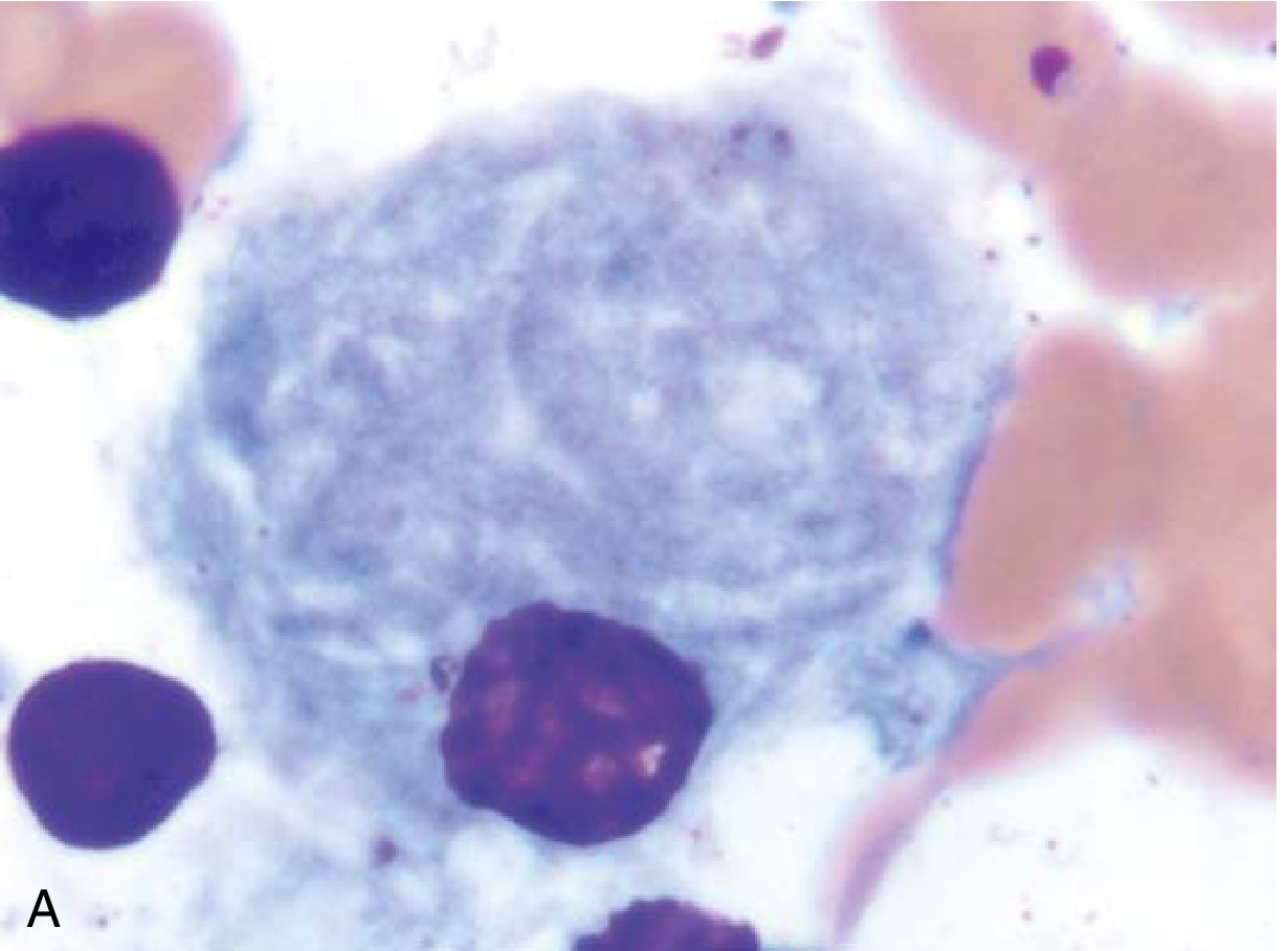
Gaucher cell in bone marrow aspirate: an enlarged macrophage with characteristic "wrinkled tissue paper" cytoplasm - Robbins & Kumar Basic Pathology
- Robbins & Kumar Basic Pathology, p. 3656; Thompson & Thompson Genetics, p. 787
4. Classification: Three Types
| Feature | Type 1 | Type 2 | Type 3 |
|---|---|---|---|
| Name | Chronic non-neuronopathic | Acute neuronopathic | Subacute/chronic neuronopathic |
| Frequency | ~99% of cases | Rare | Rare |
| CNS involvement | Absent | Severe, rapid | Present but variable |
| Onset | Any age (often adult) | Perinatal/infancy | Childhood |
| Prognosis | Compatible with long life | Death by age 2 | Variable; reduced lifespan |
| Common mutation | N370S | Various | L444P homozygous; D409H (3C) |
5. Clinical Manifestations
Type 1 (Non-neuronopathic)
- Hepatosplenomegaly: Painless splenomegaly is the most common presentation; spleen may be massively enlarged, filling the entire abdomen
- Hematologic: Thrombocytopenia, anemia (from hypersplenism and marrow replacement), easy bruising, epistaxis
- Bone disease (most significant cause of morbidity):
- Classic Erlenmeyer flask deformity of the distal femur (cortical thinning, widening of the medullary cavity)
- Osteopenia, lytic bone lesions, osteoporosis, pathologic fractures (hip, ribs, spine)
- Bone crises: Episodic severe bone pain from infarction, lasting weeks
- Aseptic necrosis of femoral heads
- Laboratory findings: Anemia, thrombocytopenia, elevated ferritin, elevated acid phosphatase, elevated ACE, elevated lyso-Gb1, elevated liver enzymes
- Immunologic: ~50% develop polyclonal gammopathy; ~30% develop monoclonal gammopathy; increased risk of multiple myeloma
- Pulmonary: Occasional pulmonary hypertension
- Parkinsonism risk: Significantly elevated (5- to 10-fold) compared to the general population
Type 2 (Acute neuronopathic)
- Onset at or shortly after birth
- Rapid neurodegeneration: failure to thrive, laryngospasm, strabismus, seizures
- Extensive visceral involvement
- Death within the first 2 years of life
Type 3 (Subacute neuronopathic)
- Onset in childhood, variable course
- Organomegaly and bone involvement (similar to type 1)
- Abnormal horizontal saccadic eye movements - hallmark neurologic sign
- Some develop myoclonic epilepsy or neurodegeneration
- Type 3C (D409H homozygous): cardiac valve calcifications, hydrocephalus, corneal opacities
Radiographic findings:
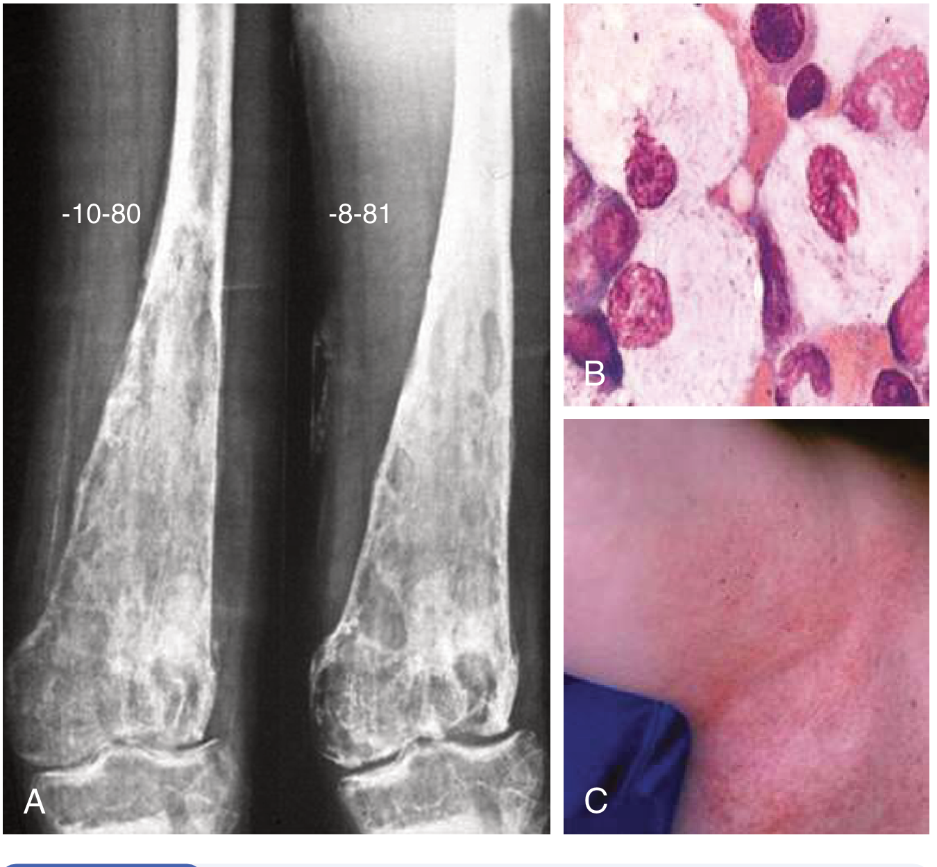
A: Erlenmeyer flask deformity - cortical thinning and widening of the metaphysis and adjacent diaphysis. B: Gaucher cells showing lipid storage. C: Angiokeratomas in Fabry disease (for comparison). - Goldman-Cecil Medicine
- Goldman-Cecil Medicine, pp. 2302-2304; Robbins & Kumar Basic Pathology, pp. 3656-3658
6. Diagnosis
Approach (Table 192-4, Goldman-Cecil):
Suspect Gaucher disease when a patient has:
- Unexplained organomegaly (hepatosplenomegaly)
- Thrombocytopenia/anemia with no clear cause
- Painful bone crises or pathologic fractures
- Erlenmeyer flask deformity on X-ray
- Abnormal saccadic eye movements
- Family history of Gaucher disease
- Unexplained multiple myeloma or parkinsonism
Diagnostic steps:
- Biomarker screen: Lyso-Gb1 (glucosylsphingosine) - measured in dried blood spot or serum; massively elevated and increasingly used as first-line screen
- Enzyme assay: Deficient glucocerebrosidase activity in leukocytes or cultured fibroblasts - confirms diagnosis
- Molecular analysis: Full GBA1 gene sequencing - needed for accurate molecular diagnosis (screening for N370S covers ~70% of Ashkenazi Jewish alleles)
- Bone marrow/liver biopsy: Shows Gaucher cells but not indicated for diagnosis
- Carrier identification: DNA testing when mutant allele is known
- Prenatal diagnosis: Enzyme activity or mutation testing on chorionic villi or amniotic fluid
- Goldman-Cecil Medicine, pp. 2303-2304
7. Treatment
Enzyme Replacement Therapy (ERT)
The primary treatment for Type 1 and Type 3 Gaucher disease:
| Agent | Details |
|---|---|
| Imiglucerase (Cerezyme) | Plant cell-derived recombinant glucocerebrosidase |
| Velaglucerase alfa (VPRIV) | Human cell line-derived |
| Taliglucerase alfa (Elelyso) | Carrot cell-derived |
- Dose: 15-60 IU/kg IV every 2 weeks
- Response: Anemia, thrombocytopenia, and organomegaly reverse within 12-36 months
- Limitation: Does NOT cross the blood-brain barrier - does not halt neurologic progression in Types 2/3
- Enzyme is targeted to macrophage lysosomes via mannose-6-phosphate or mannose residues
- Treatment is ongoing, lifelong, and very expensive
Substrate Reduction Therapy (SRT)
Reduces substrate synthesis to match the impaired degradation rate:
- Eliglustat (Cerdelga): Ceramide analogue; potent specific inhibitor of glucosylceramide synthase
- Administered orally
- Significantly improves spleen/liver volumes, hemoglobin, and platelet counts
- Preferred by some adults over IV ERT
- Important: Check CYP2D6 metabolizer status before use
- Ultra-rapid metabolizers: NOT eligible
- Normal or intermediate metabolizers: 84 mg twice daily
- Poor metabolizers: 84 mg once daily
- Miglustat (Zavesca): Earlier oral SRT agent; less preferred due to side effect profile
Supportive Therapies
- Bisphosphonates for osteoporosis
- Orthopedic surgery for fractures; joint replacement for avascular necrosis
- Aggressive pain management and hydration for bone crises
- Splenectomy: Now rarely indicated (used before ERT was available; accelerates bone disease)
- Hematopoietic stem cell transplantation: Can improve systemic but not neurologic manifestations; reserved for severe disease in settings where ERT/SRT are unavailable
Emerging/Investigational Therapies
-
Brain-penetrant ERT and SRT (for neuronopathic types)
-
Chemical chaperones: e.g., ambroxol (high-dose) - a recent 2024 systematic review [PMID 39116528] investigated this approach for Types 2 and 3, showing preclinical and clinical promise; it stabilizes misfolded glucocerebrosidase
-
Gene therapy: Active area of research
-
Standard therapies do not prevent the development of parkinsonism
-
Goldman-Cecil Medicine, pp. 2303-2304; Thompson & Thompson Genetics, p. 792-794; Emery's Medical Genetics, p. 1732
8. Gaucher Disease and Parkinson Disease
This is an increasingly important association:
- Both patients with Gaucher disease and heterozygous GBA1 carriers have elevated parkinsonism risk
- GBA1 mutations are found in 5-10% of all Parkinson disease patients worldwide - making it the single most common genetic risk factor for parkinsonism
- Mechanism likely involves impaired lysosomal function, alpha-synuclein accumulation, and Lewy body formation
- Reduced penetrance means the majority of Gaucher patients and carriers do not develop Parkinson disease
- A 2025 MDSGene systematic review [PMID 39927608] further classified GBA1 variants and their genotype-phenotype relationships in parkinsonism
9. Summary Table
| Parameter | Detail |
|---|---|
| Category | Lysosomal storage disease (sphingolipidosis) |
| Enzyme deficient | Glucocerebrosidase (acid beta-glucosidase) |
| Gene | GBA1, chromosome 1q21 |
| Substrate stored | Glucocerebroside in macrophages |
| Pathognomonic cell | Gaucher cell ("wrinkled tissue paper" cytoplasm) |
| Most common type | Type 1 (non-neuronopathic, ~99%) |
| Most common presentation | Splenomegaly, thrombocytopenia, bone disease |
| Classic X-ray sign | Erlenmeyer flask deformity |
| Diagnosis | Leukocyte glucocerebrosidase activity + GBA1 sequencing |
| Biomarker | Lyso-Gb1 (glucosylsphingosine) |
| First-line treatment | ERT (imiglucerase/velaglucerase) or oral eliglustat |
| Treatment limitation | ERT does not cross the BBB |
| Parkinson risk | 5-10x elevated; GBA1 = most common genetic PD risk factor |
Recent evidence note: A 2025 systematic review (MDSGene, PMID 39927608) provided updated GBA1 variant classification and genotype-phenotype data relevant to Parkinson risk. A 2024 systematic review (PMID 39116528) assessed high-dose ambroxol as a pharmacological chaperone for neuronopathic Gaucher disease (Types 2 and 3), showing promising preclinical and limited clinical data - this is not yet standard of care.
Sources: Goldman-Cecil Medicine (International Edition), pp. 2302-2304; Robbins & Kumar Basic Pathology, pp. 3655-3658; Emery's Elements of Medical Genetics and Genomics, pp. 1724-1734; Thompson & Thompson Genetics and Genomics in Medicine, pp. 787-794
This is a shared conversation. Sign in to Orris to start your own chat.