
Medial leminiscus
Reading File
The medial lemniscus is the main ascending sensory pathway carrying fine touch, vibration, and proprioception from the body to the thalamus. Let me pull detailed content from the medical library.
Searching sources...
Finding Sources
Finding Sources
Reading File
Reading File
Reading File
Reading File
Reading File
Reading File
Now let me fetch the key pathway diagram and check clinical lesion information.
Loading Image
Finding Sources
Reading File
Reading File
I now have comprehensive information to write a thorough answer.
Medial Lemniscus
The medial lemniscus is the major ascending sensory tract carrying discriminative touch, vibration, proprioception (joint position sense), and two-point discrimination from the body to the thalamus. It forms the second leg of the three-neuron posterior column-medial lemniscal (PCML) pathway.
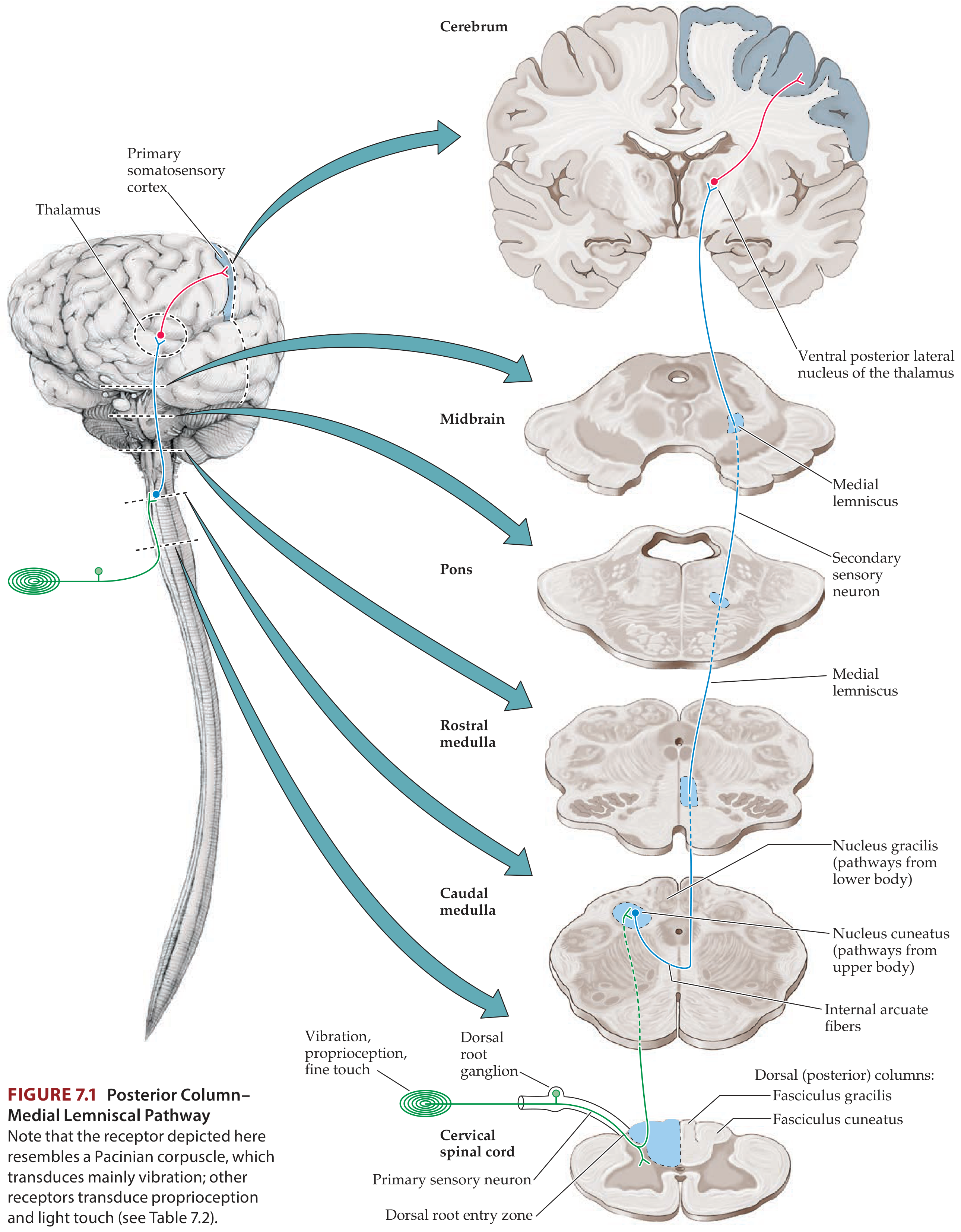
The Three-Neuron Chain
| Neuron | Location | Details |
|---|---|---|
| 1st order | Dorsal root ganglion | Large myelinated A-alpha and A-beta fibers; axons ascend ipsilaterally in the dorsal columns |
| 2nd order | Nucleus gracilis / nucleus cuneatus (medulla) | Axons cross as internal arcuate fibers to form the medial lemniscus |
| 3rd order | VPL nucleus of thalamus | Projects via posterior limb of internal capsule to primary somatosensory cortex (postcentral gyrus, areas 3, 1, 2) |
Formation and Course
Dorsal columns:
- Fasciculus gracilis (sacral to mid-thoracic): terminates in nucleus gracilis - carries lower body information
- Fasciculus cuneatus (upper thoracic and cervical): terminates in nucleus cuneatus - carries upper body information
- A special route for lower limb proprioception: fibers from the fasciculus gracilis relay in Clarke's column at upper lumbar cord, project to nucleus Z in the medulla, which then feeds into the medial lemniscus
Decussation: Axons from both nuclei (and nucleus Z, and the lateral cervical nucleus) cross in the lower medulla as internal arcuate fibers to form the medial lemniscus on the contralateral side.
Position Changes as It Ascends the Brainstem
The medial lemniscus shifts orientation dramatically as it ascends:
| Level | Position | Somatotopy |
|---|---|---|
| Medulla | Vertical strip adjacent to midline, dorsal to pyramids | Feet ventral, head dorsal ("little person stands up") |
| Pons | More lateral, inclined position | Feet lateral, arms medial ("little person lies down") |
| Midbrain | Lateral tegmentum | Converges with spinothalamic tract |
| Thalamus | Terminates in VPL nucleus | Somatotopy maintained |
The somatotopic organization of the dorsal columns is maintained throughout - however, note the reversal: in the posterior columns, the feet are medial; in the medial lemniscus at the pons/midbrain, the feet are lateral.
Modalities Carried
- Fine/discriminative touch
- Vibration sense
- Proprioception (joint position, kinesthesia)
- Two-point discrimination
(Compare: pain and temperature travel in the spinothalamic/anterolateral tract, which decussates at the spinal cord level.)
The Face Equivalent: Trigeminal Lemniscus
Proprioception, vibration, and discriminative touch from the face follow a parallel route - the trigeminal lemniscus - arising from the principal trigeminal nucleus, running alongside the medial lemniscus, and terminating in the VPM nucleus of the thalamus.
Clinical Significance - Lesions
Medial lemniscus lesion causes contralateral loss of:
- Vibration sense
- Joint position sense (proprioception)
- Discriminative touch
- Two-point discrimination
Pain and temperature are spared (carried by the spinothalamic tract, which runs separately in the medulla and pons).
Key syndromes:
-
Medial medullary syndrome (Dejerine): Infarct of anterior spinal / vertebral artery paramedian branches. Affects medial lemniscus + pyramidal tract + CN XII. Signs: contralateral hemiplegia + contralateral loss of proprioception/vibration + ipsilateral tongue deviation.
-
Raymond-Cestan syndrome (pons): Contralateral loss of all sensory modalities (face and extremities) when the medial lemniscus and spinothalamic tract are both involved, with cerebellar signs.
-
Upper brainstem lesions (midbrain): At this level the spinothalamic tract and medial lemniscus converge, so a single lesion causes contralateral loss of ALL sensation (superficial + deep).
-
Thalamic involvement (VPL): Dejerine-Roussy syndrome - contralateral loss of all sensation, potentially with severe thalamic pain.
Vascular Supply
- Medulla (medial lemniscus): Paramedian branches of the anterior spinal artery and vertebral artery
- Pons: Paramedian branches of the basilar artery
- Midbrain: Paramedian branches of the basilar artery (supply cerebral peduncles, CN III, red nucleus, and medial lemniscus)
Somatosensory Evoked Potentials (SEPs)
The medial lemniscus is a key segment of the pathway assessed by SEPs. These record large-diameter myelinated afferent activity through the dorsal columns and medial lemniscus up to the cortex, and are used clinically to detect demyelinating diseases (e.g., MS) and to monitor the pathway intraoperatively.
Sources:
- Neuroanatomy through Clinical Cases, 3rd Ed., pp. 300-303, 315
- Localization in Clinical Neurology, 8th Ed., pp. 42-45
- Adams and Victor's Principles of Neurology, 12th Ed., p. 182
- Gray's Anatomy for Students, p. 1321
This is a shared conversation. Sign in to Orris to start your own chat.