
Hematology peadiatrics
Reading File
The query is broad - "Hematology paediatrics" could mean many things. Let me clarify before diving deep.
Asking for Preferences
What aspect of paediatric hematology are you interested in? · What is the purpose of this session?
Finding Sources
Finding Sources
Finding Sources
Finding Sources
Finding Sources
Finding Sources
Finding Sources
Finding Sources
Now let me read the most relevant textbook sections for detailed exam content:
Reading File
Reading File
Reading File
Reading File
Reading File
Reading File
Finding Sources
Finding Sources
Reading File
Reading File
I now have comprehensive content from authoritative sources. Let me compile the full exam revision notes.Here are your comprehensive Paediatric Hematology exam revision notes, sourced from Tintinalli's Emergency Medicine, Robbins & Kumar Basic Pathology, and Robbins, Cotran & Kumar Pathologic Basis of Disease.
Paediatric Hematology - Exam Revision Notes
1. ANAEMIAS IN CHILDREN
Overview of Causes
| Category | Examples |
|---|---|
| Decreased production | Iron deficiency, aplastic anaemia, transient erythroblastopenia |
| Increased destruction (hemolytic) | Hereditary spherocytosis, G6PD deficiency, sickle cell, thalassaemia, AIHA |
| Loss | Haemorrhage |
Iron Deficiency Anaemia (IDA)
-
Most common anaemia in children
-
Peak risk: infants/toddlers (1-3 years), teens (especially girls)
-
Causes in children:
- Exclusive breastfeeding without iron supplementation (after 4-6 months)
- Excessive cow's milk before age 1 year (displaces iron-rich foods + causes low-grade GI bleeding)
- Milk protein colitis causing occult GI bleeding
- Nutritional IDA uncommon after age 3-4 years -- if present, suspect occult bleeding
-
Clinical features: pallor, fatigue, pica (craving for ice, dirt, paper), irritability
-
Labs:
- Hypochromic microcytic anaemia
- Low reticulocyte count
- Low serum ferritin, low serum iron, high TIBC
-
Treatment:
- Oral ferrous sulfate (outpatient if hemodynamically stable)
- Severe anaemia/hemodynamic compromise: inpatient + hematology consult ± transfusion
-
Prevention (AAP): iron supplementation in breastfed infants, iron-fortified formulas, delay cow's milk until >1 year, universal Hb screening at 1 year
Autoimmune Hemolytic Anaemia (AIHA)
- Mechanism: autoantibodies against RBC antigens → destruction
- Primary AIHA: most common in infants/young children, often post-viral, no underlying disorder
- Secondary AIHA: older children -- associated with malignancy, HIV, autoimmune disease
- Labs:
- Spherocytes + schistocytes on peripheral smear
- Elevated indirect bilirubin, elevated LDH, urobilinogenuria/hemoglobinuria
- Elevated reticulocytes (unless acute, sudden onset)
- Positive Coombs test (direct antiglobulin test)
- Management: steroids, IVIG, avoid precipitants
Transient Erythroblastopenia of Childhood (TEC)
- Self-resolving normocytic anaemia from temporary ↓ RBC precursors
- Age: 6 months to 10 years (peak: toddlers/preschool)
- Other cell lines normal, iron studies normal
- Recovery in 1-2 months; transfusion rarely needed
- Must differentiate from Diamond-Blackfan anaemia
Parvovirus B19 & Aplastic Crisis
- Parvovirus replicates in erythroid progenitor cells → transient red cell aplasia
- In normal host: "fifth disease" (slapped cheek rash) -- anaemia not noticed
- In children with haemoglobinopathy/haemolytic anaemia (SCD, thalassaemia): severe aplastic crisis, often requires transfusion
2. HAEMOGLOBINOPATHIES
Sickle Cell Disease (SCD)
- Genetics: autosomal recessive; HbS due to Glu→Val substitution at position 6 of β-globin chain
- HbSS = sickle cell disease (most severe); HbAS = sickle cell trait (carrier, usually asymptomatic)
- Pathophysiology: HbS polymerises under low O₂ → sickling → vaso-occlusion, haemolysis
Clinical Complications:
| Complication | Notes |
|---|---|
| Vaso-occlusive (painful) crisis | Most common; bone pain, abdominal pain |
| Dactylitis | Swelling of hands/feet in children <5 years -- often first presentation |
| Acute chest syndrome | Fever + new pulmonary infiltrate + respiratory symptoms; life-threatening |
| Aplastic crisis | Parvovirus B19 trigger; sudden Hb drop |
| Splenic sequestration | Rapid splenic enlargement + circulatory shock; young children |
| Stroke | 9% of children with SCD by age 20; transcranial Doppler screening required |
| Osteonecrosis | Femoral/humeral head |
| Infections | Functional asplenia → encapsulated organisms (Strep pneumo, H. influenzae, Salmonella osteomyelitis) |
Management:
- Hydroxyurea (↑ HbF, reduces crises)
- Penicillin prophylaxis from age 2 months to 5 years
- Pneumococcal vaccination
- Folic acid supplementation
- Transfusion for acute chest syndrome, stroke, aplastic crisis, pre-op
- Curative: bone marrow transplant
- Gene therapy (approved 2023-24): exagamglogene autotemcel (exa-cel) for age ≥12 years with recurrent vaso-occlusive crises - Harrison's 22e
Thalassaemia
Genetics:
- α-globin: 2 genes on each chromosome 16 (total 4 alleles)
- β-globin: 1 gene on chromosome 11 (2 alleles)
- α-thalassaemia: mainly gene deletions
- β-thalassaemia: mainly point mutations affecting transcription, splicing, or translation of β-globin mRNA
β-Thalassaemia Classification:
| Syndrome | Genotype | Clinical Features |
|---|---|---|
| β-Thalassaemia major (Cooley's anaemia) | β⁰/β⁰ or β⁺/β⁰ | Severe anaemia, transfusion-dependent |
| β-Thalassaemia intermedia | Various β⁺/β⁰, etc. | Moderate anaemia, not transfusion-dependent |
| β-Thalassaemia minor (trait) | β⁺/β or β⁰/β | Asymptomatic or mild; microcytic hypochromic RBCs |
α-Thalassaemia Classification:
| Syndrome | Lost genes | Clinical Features |
|---|---|---|
| Silent carrier | 1 gene lost | Asymptomatic, normal RBCs |
| α-Thalassaemia trait | 2 genes lost | Mild microcytic hypochromic anaemia |
| HbH disease | 3 genes lost | Moderately severe; excess β-chains form HbH (β4) tetramers |
| Hydrops fetalis (Hb Bart's) | 4 genes lost | Lethal in utero; Hb Bart (γ4) formed |
Pathophysiology of β-Thalassaemia Major:
- ↓ β-globin → ↓ HbA formation → microcytic hypochromic RBCs
- Excess unpaired α-chains → toxic precipitates → membrane damage → ineffective erythropoiesis + shortened RBC lifespan
- Ineffective erythropoiesis → massive erythroid hyperplasia → extramedullary haematopoiesis
- Bone marrow expansion → skeletal deformities (frontal bossing, "hair-on-end" skull XR, "chipmunk facies")
- Hepcidin suppressed → increased iron absorption → iron overload (even without transfusions)
Clinical features of β-Thalassaemia Major:
- Presents postnatally as HbF declines (by 6 months)
- Growth retardation from infancy
- Hepatosplenomegaly (extramedullary haematopoiesis + haemolysis)
- Facial deformities (bone marrow expansion)
- Iron overload → cardiomyopathy, liver cirrhosis, endocrinopathies (most common cause of death = cardiac failure)
Peripheral Smear Findings:
- β-Thal major: marked microcytosis, hypochromia, poikilocytosis, anisocytosis, nucleated RBCs (normoblasts), target cells
- β-Thal minor/α-thal trait: mild microcytosis, hypochromia, target cells -- similar to IDA
Treatment of β-Thalassaemia Major:
- Regular blood transfusions (every 3-4 weeks, target Hb 9-10 g/dL)
- Iron chelation: desferrioxamine (IV/SC), deferasirox (oral)
- Curative: allogeneic bone marrow transplant
- Splenectomy if hypersplenism causing excessive transfusion requirement
3. BLEEDING DISORDERS
Immune Thrombocytopenia (ITP)
- Previously called "idiopathic thrombocytopenic purpura"
- Mechanism: autoantibodies (anti-platelet IgG) against platelet membrane glycoproteins → platelet destruction + sometimes ↓ megakaryocyte production
- Epidemiology:
- Typical patient: preschool/school-age, previously healthy child
- Onset: acute, often follows viral illness
- >80% of childhood ITP is self-limited (resolves within 6 months)
- Incidence of life-threatening bleeds <0.5%
- Classification:
- Acute: resolves <3 months
- Persistent: 3-12 months
- Chronic: >12 months
Clinical Features:
- Acute onset petechiae + bruising
- No lymphadenopathy, no hepatosplenomegaly, no fever (if present, think OTHER diagnosis)
- Labs: isolated thrombocytopenia (WBC and Hb normal)
Treatment (controversial; consult hematology first):
- Observation alone if mild/no bleeding
- Corticosteroids: prednisone 2 mg/kg/d × 21 days OR methylprednisolone 30 mg/kg/d × 4 days
- Caution: must rule out leukemia/aplastic anaemia before starting steroids (bone marrow biopsy if uncertain)
- IVIG: 1 g/kg/d -- superior to steroids for rapid platelet increment
- Anti-Rh(D) immunoglobulin (WinRho): only for Rh-positive patients; 50-75 µg/kg; risk of intravascular haemolysis (needs admission)
- Life-threatening haemorrhage: single-donor platelets (2-3x normal dose) + IV methylprednisolone + IVIG
Admission criteria: Platelets <20,000/mm³, IV medication required, or spontaneous bleeding
Haemophilia
Types:
| Feature | Haemophilia A | Haemophilia B (Christmas disease) |
|---|---|---|
| Deficiency | Factor VIII | Factor IX |
| Inheritance | X-linked recessive | X-linked recessive |
| Incidence | 1:5,000 live male births | 1:30,000 live male births |
Severity classification:
| Severity | Factor level | Bleeding pattern |
|---|---|---|
| Mild | 5-40% | Bleeding only with trauma/surgery |
| Moderate | 1-5% | Bleeding after minor trauma |
| Severe | <1% | Spontaneous bleeding |
Diagnosis:
- Often discovered early: family history, intracranial haemorrhage at birth, hematoma after birth trauma, prolonged bleeding after circumcision, or first fall as toddler
- Screening: prolonged aPTT; normal PT, platelet count, bleeding time
- Confirmation: quantitative factor VIII or IX levels
- Mixing studies to detect inhibitors
Hallmark: Haemarthroses (80% of bleeds in severe haemophilia) -- most commonly knee, ankle, elbow, shoulder
Treatment:
- "When in doubt, treat" - preferably within 2 hours
- Factor replacement:
- 1 unit/kg of Factor VIII raises activity by 2%
- 1 unit/kg of Factor IX raises activity by 1%
- Target level: 40-50% for routine bleeds; 80-100% for CNS, retroperitoneal, iliopsoas, GI, ophthalmic bleeds
- DDAVP (desmopressin): releases endogenous Factor VIII + vWF; useful in mild/moderate haemophilia A and carriers
- Prophylactic Factor infusions (especially in severe disease) - prevent hemophilic arthropathy
- Adjuncts: tranexamic acid / aminocaproic acid (antifibrinolytics) for mucosal/dental bleeds; RICE (rest, ice, compression, elevation) for joints
- Inhibitors (antibodies to factor): 30% of severe haemophilia A; treat with FEIBA® (activated PCC) or recombinant Factor VIIa (NovoSeven®)
Key complications:
- Haemophilic arthropathy (repeated haemarthroses → synovial inflammation → cartilage destruction → joint fibrosis)
- Iliopsoas bleed: mimics appendicitis; hip held in flexion, femoral nerve compression
- Leading cause of death: intracranial haemorrhage
Von Willebrand Disease (vWD)
- Most common inherited bleeding disorder overall
- vWF deficiency/dysfunction → impaired platelet adhesion to subendothelium + ↓ Factor VIII stability
- Types: Type 1 (mild, quantitative ↓), Type 2 (qualitative defect), Type 3 (severe, absent vWF)
- Labs: prolonged bleeding time, prolonged aPTT (if Factor VIII very low), normal PT, low vWF antigen/activity
- Treatment: DDAVP (Type 1), vWF concentrate, tranexamic acid
4. PAEDIATRIC LEUKAEMIA
Acute Lymphoblastic Leukaemia (ALL)
- Most common childhood malignancy (most common childhood leukaemia)
- Peak incidence: 2-10 years of age
- B-ALL = most common subtype
Pathogenesis:
- Mutations in transcription factors regulating lymphoid differentiation (e.g., PAX5 in B-ALL)
- Driver mutations: tyrosine kinases, RAS signalling, chromatin regulators
- Philadelphia chromosome (t(9;22) BCR-ABL): ~5% of childhood ALL, ~25% of adult ALL - worst prognosis historically; now targetable with tyrosine kinase inhibitors (imatinib)
Clinical Features (marrow failure):
- Anaemia: pallor, fatigue, weakness
- Thrombocytopenia: petechiae, bruising, mucosal bleeding
- Neutropenia: recurrent/severe infections
- Bone/joint pain (marrow infiltration)
- Lymphadenopathy, hepatosplenomegaly
- T-ALL: mediastinal mass (>50% of T-ALL cases) -- thymic involvement
Labs:
- Peripheral blood: blasts (lymphoblasts); pancytopenia
- Elevated WBC or normal/low WBC with blasts
- LDH elevated
Diagnosis: Bone marrow biopsy + immunophenotyping (B-cell vs T-cell ALL)
Treatment:
- Induction (remission) → Consolidation → Maintenance (total 2-3 years)
- CNS prophylaxis (IT methotrexate ± cranial irradiation in high risk)
- Prognosis: >90% cure rate in standard-risk childhood B-ALL
Acute Myeloid Leukaemia (AML) in Children
- Less common than ALL in children; more common in adults
- May arise de novo or from prior myelodysplastic syndrome
- Acute promyelocytic leukaemia (APL, AML-M3):
- t(15;17) → PML-RARA fusion → block in promyelocyte differentiation
- Risk of DIC (thromboplastic granule release)
- Treatment: All-trans retinoic acid (ATRA) + arsenic trioxide -- bypass the differentiation block
5. NEUTROPENIA IN CHILDREN
Definition: Absolute neutrophil count (ANC) <1500/mm³
- Infants/African/Middle Eastern descent: ANC can be lower, but should not drop <1000/mm³
Severity:
| Grade | ANC | Infection risk |
|---|---|---|
| Mild | 1000-1500/mm³ | Low |
| Moderate | 500-1000/mm³ | Moderate |
| Severe | <500/mm³ | High |
Causes in children: post-viral (most common, transient), autoimmune, cyclic neutropenia, drug-induced, Kostmann syndrome (congenital), haematological malignancy
6. HIGH-YIELD EXAM COMPARISONS
| Feature | ITP | TEC | Aplastic Anaemia |
|---|---|---|---|
| Cell lines affected | Platelets only | RBC only | All three (pancytopenia) |
| Age | Preschool/school | Toddler/preschool | Any |
| Trigger | Viral | Unknown | Idiosyncratic drug, viral |
| Resolution | Spontaneous in >80% | Spontaneous in 1-2 months | Requires treatment |
| Iron Deficiency | β-Thal Minor | ACD | |
|---|---|---|---|
| MCV | Low | Low | Low/Normal |
| Ferritin | Low | Normal/High | High |
| TIBC | High | Normal | Low |
| RBC count | Low | High (many small RBCs) | Low |
| Haemophilia A | Haemophilia B | vWD | |
|---|---|---|---|
| Deficient factor | VIII | IX | vWF (± VIII) |
| aPTT | Prolonged | Prolonged | Normal/Prolonged |
| PT | Normal | Normal | Normal |
| Bleeding time | Normal | Normal | Prolonged |
| Inheritance | X-linked recessive | X-linked recessive | Autosomal dominant (mostly) |
7. KEY MNEMONICS & HIGH-YIELD POINTS
- "5's" in haemophilia: 5% = mild; bleeding only with trauma
- Haemarthroses = hallmark of haemophilia (80% of bleeds in severe)
- Leading cause of death in haemophilia = intracranial haemorrhage
- ITP in children: >80% self-limited - don't over-treat
- Sickle cell + fever = emergency (functional asplenia → sepsis)
- Dactylitis = first presentation of SCD in infants
- β-Thalassaemia major: "hair-on-end" skull XR, chipmunk facies, iron overload kills (cardiac failure)
- B-ALL peak: age 2-10 years; T-ALL: adolescent boys + mediastinal mass
- Parvovirus B19 = aplastic crisis in SCD/thalassaemia
- Philadelphia chromosome (BCR-ABL): present in CML and ~5% childhood ALL; treated with TKIs
Sources: Tintinalli's Emergency Medicine, Robbins & Kumar Basic Pathology, Robbins Cotran & Kumar Pathologic Basis of Disease, Harrison's Principles of Internal Medicine 22e, Harriet Lane Handbook 23e
Atrial flutter
Reading File
This is a broad medical topic - let me search the library in parallel across multiple angles (mechanism, ECG, management) for comprehensive exam-level content.
Finding Sources
Finding Sources
Excellent finds. Now reading the core dedicated sections in parallel - Braunwald's (most authoritative cardiology text), Harrison's 22e (most current), and Goldman-Cecil:
Reading File
Reading File
Reading File
Reading File
Reading File
Reading File
Reading File
Finding Sources
Reading File
Reading File
Reading File
Finding Sources
I now have rich, detailed content from multiple authoritative cardiology sources. Let me now fetch the key ECG image for visual learning:
Loading Image
Loading Image
Loading Image
I have everything needed for a complete, exam-quality set of notes. Compiling now.
Atrial Flutter - Comprehensive Exam Revision Notes
Sources: Braunwald's Heart Disease, Harrison's Principles of Internal Medicine 22e, Goldman-Cecil Medicine, Fuster & Hurst's The Heart 15e, Guyton & Hall Medical Physiology
1. DEFINITION & OVERVIEW
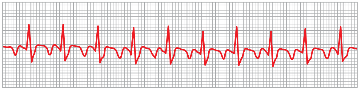
Atrial flutter (AFL) is a macro-reentrant atrial tachyarrhythmia characterised by:
- Rapid, organised atrial electrical activity at ~250-300 bpm
- Ventricular rate determined by the degree of AV nodal block: typically 150 bpm (2:1), 100 bpm (3:1), or slower
- Characteristic "sawtooth" flutter waves on ECG
It is one of the most common cardiac arrhythmias. Prevalence ~190,000 in the US (estimated to rise to 440,000 by 2050 due to ageing population). Incidence: 88 per 100,000 person-years, rising sharply after age 50 to ~0.6% in those over 80.
2. MECHANISM
AFL is caused by macroreentry - a large circular electrical wavefront that requires:
- A central obstacle (anatomic or functional)
- An area of slow conduction (the critical isthmus)
- A region of unidirectional block to initiate
3. CLASSIFICATION
Type 1 - Typical (CTI-Dependent) AFL
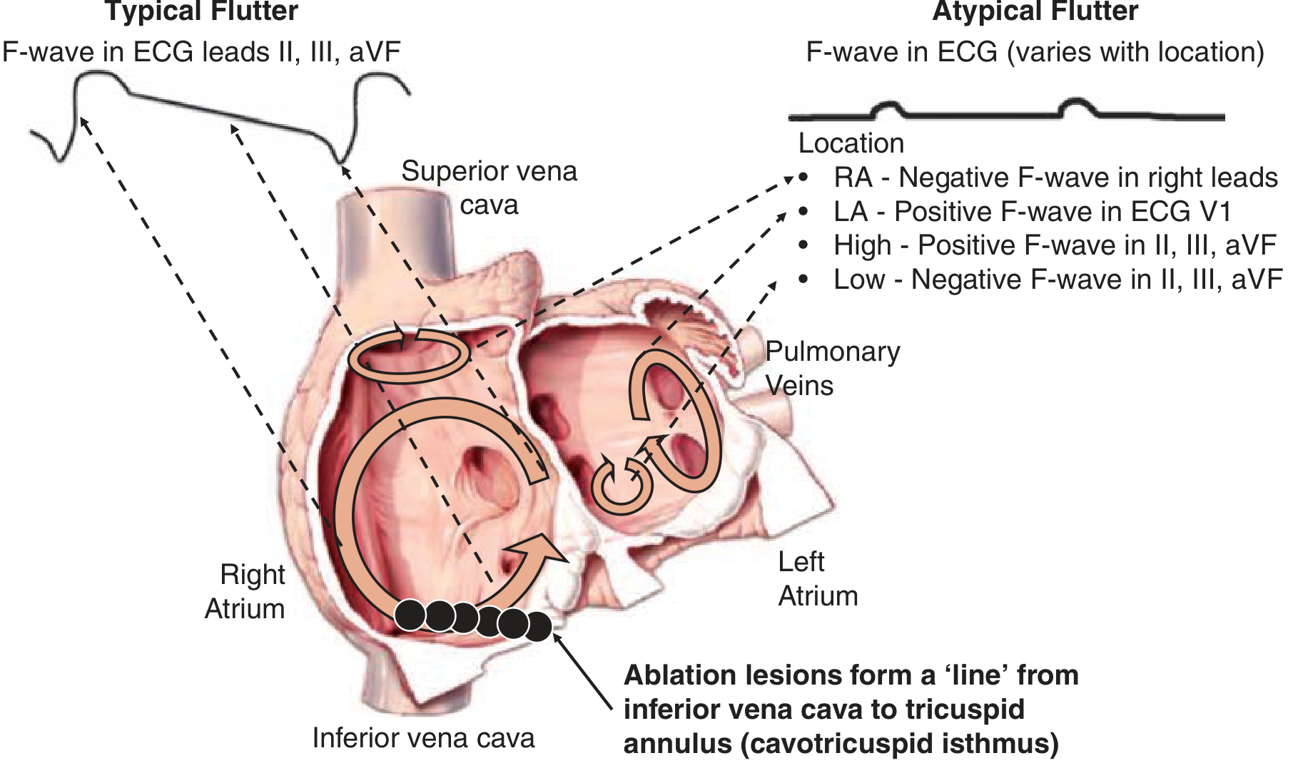
The majority of cases. Circuit confined to the right atrium, using the cavotricuspid isthmus (CTI) - the narrow corridor between the IVC and tricuspid annulus - as the critical slow-conduction zone.
| Subtype | Direction | ECG in II, III, aVF | ECG in V1 |
|---|---|---|---|
| Typical (counterclockwise) | CCW - up septum, down RA free wall | Negative sawtooth (dominant) | Discrete upright P waves |
| Reverse typical (clockwise) | CW - down septum, up RA free wall | Positive flutter waves | Negative flutter waves |
Type 2 - Atypical (Non-CTI-Dependent) AFL
- Circuits in other right or left atrial regions
- Left atrial flutter: positive flutter waves in V1; seen after AF ablation or mitral valve surgery
- Right atrial atypical: after atriotomy, congenital heart disease repair, or cardiac transplant
- Lower-loop / upper-loop reentry variants also described
- Rates variable: 180-320 bpm; ECG morphology varies widely
4. ECG DIAGNOSIS
Key ECG Features:
| Feature | Detail |
|---|---|
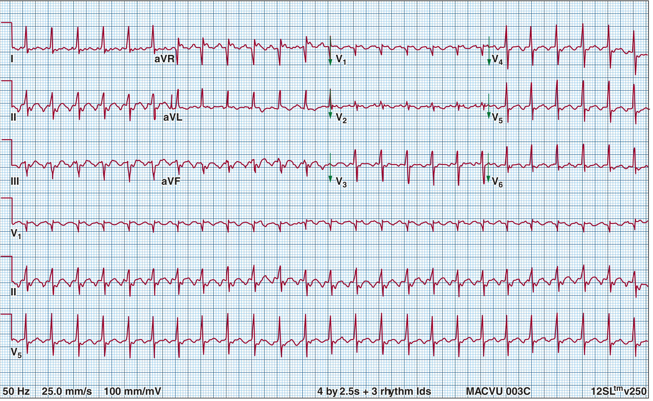
| Flutter wave rate | 250-300 bpm (typical); no isoelectric baseline |
| Ventricular rate | Usually 150 bpm (2:1 block) |
| Rhythm | Regularly irregular or regular (depends on block ratio) |
| Sawtooth morphology | Negative in II, III, aVF (counterclockwise - most common) |
| V1 appearance | Discrete upright P waves (not sawtooth) |
| QRS | Narrow (unless aberrant conduction or bundle branch block) |
Practical tips:
- At 2:1 block, flutter waves may be hidden in QRS/T complexes - hard to spot on a single lead. Use multiple leads or vagal maneuvers/adenosine to unmask flutter waves by increasing AV block transiently
- Telemetry showing a regular tachycardia at ~150 bpm - always suspect 2:1 flutter
- V1 showing discrete upright P waves (not sawtooth) may confuse the diagnosis if only V1 is reviewed
5. CAUSES & ASSOCIATIONS
AFL almost always occurs in the context of:
- Structural heart disease: valvular disease, coronary artery disease, cardiomyopathy, congenital heart disease
- Pulmonary disease (COPD, PE)
- Acute illness: sepsis, myocardial infarction, post-operative state
- Metabolic: hyperthyroidism, alcohol (acute exposure), electrolyte abnormalities
- Antiarrhythmic drug use: class 1C agents (flecainide, propafenone) used for AF can slow the atrial rate enough to organise AF into flutter, potentially with 1:1 AV conduction at dangerous rates - this is why a rate-limiting AV nodal agent must always be co-prescribed when using class 1C drugs for AF
6. RELATIONSHIP WITH ATRIAL FIBRILLATION
AFL and AF are considered "two sides of the same coin" (Braunwald's):
- AF is documented in up to 75% of AFL patients
- AFL onset is typically preceded by a transitional period of AF; it usually terminates back through AF
- AF provides triggers and electrophysiologic substrate for AFL
- Risk of developing AF after flutter ablation: ~40-50% over 2-3 years (especially if prior AF, impaired LV, LA enlargement, structural heart disease)
- Both share similar thromboembolic risk; anticoagulation decisions follow the same CHA₂DS₂-VASc framework
7. CLINICAL PRESENTATION
Symptoms (similar to AF):
- Palpitations
- Dyspnoea / reduced exercise tolerance
- Fatigue
- Presyncope or syncope (especially with 1:1 conduction or high-grade AV block)
- Chest pain / angina (rapid rate in ischaemic heart disease)
- Asymptomatic if rate is well-controlled
Signs:
- Regular pulse at ~150 bpm (2:1 block) -- or irregular if variable block
- An S4 gallop may be present (atrial contraction against a stiff ventricle) - this distinguishes AFL from AF where S4 is absent
- JVP: rapid flutter waves may be visible
- Sustained rapid ventricular response (2:1 flutter) can cause tachycardia-induced cardiomyopathy (TCM) - fully reversible within ~3 months of rate/rhythm control
8. MANAGEMENT
Step 1: Haemodynamic Assessment
- Haemodynamically unstable (hypotension, pulmonary oedema, severe chest pain): immediate synchronised DC cardioversion (low energy, ~25-50 J typically effective)
- Haemodynamically stable: proceed with anticoagulation, then rate/rhythm control
Step 2: Anticoagulation (Same as AF)
- Thromboembolic risk in AFL: ~3% per year
- Before any cardioversion (electrical or pharmacological), unless <48 hours duration:
- TEE to exclude LA appendage thrombus, OR
- Therapeutic anticoagulation for ≥4 weeks prior (DOAC: dabigatran, rivaroxaban, apixaban, edoxaban; or warfarin INR 2-3)
- Risk stratification: CHA₂DS₂-VASc score (same as AF)
Step 3: Rate Control
Rate control in AFL is more difficult than in AF (AV node is harder to modulate during flutter):
- Beta-blockers: metoprolol (oral 25-50 mg; IV for acute use)
- Non-dihydropyridine calcium channel blockers: diltiazem (180-240 mg daily; IV 15-20 mg bolus over 2 min)
- Digoxin: less effective for rate control in AFL
- Used as a bridge to cardioversion; not a satisfactory long-term solution alone
Step 4: Rhythm Control (Cardioversion)
AFL is far more amenable to cardioversion than AF:
| Method | Details | Efficacy |
|---|---|---|
| DC electrical cardioversion | Preferred; synchronised shock, low energy (50-100 J biphasic) | >95% |
| Ibutilide (IV) | 1 mg over 10 min, repeat once if needed | ~60% for AFL |
| Overdrive atrial pacing | Rapid atrial stimulation via transesophageal/intracardiac pacing catheter to interrupt the circuit | Effective |
| Flecainide / propafenone | NOT recommended alone - can slow atrial rate and cause 1:1 conduction | Avoid without AV nodal blocker |
| Sotalol, dofetilide, amiodarone | May be considered for maintenance; but >70% recurrence on drugs | Second-line |
Important: treat any precipitating causes first (hypoxia, fever, electrolyte abnormalities) - this may allow spontaneous conversion and is needed to sustain sinus rhythm.
Step 5: Definitive Therapy - Catheter Ablation (First-Line for Typical AFL)
- Cavotricuspid isthmus (CTI) ablation = line of radiofrequency lesions from IVC to tricuspid annulus
- Typical AFL: >95% cure rate; low recurrence; preferred for most patients
- Indications: recurrent AFL, drug-resistant, drug-intolerant, or patient preference
- Atypical AFL: ablation also considered first-line but success rates lower; antiarrhythmic drugs or repeated cardioversion also reasonable
- Post-ablation anticoagulation: not needed long-term if ablation successful and recurrence risk is low; ongoing anticoagulation if AF also present or high risk
9. PROGNOSIS & COMPLICATIONS
- AFL rarely an isolated, single event - expect recurrence
- Even "asymptomatic" AFL with a controlled rate carries risk of variable block and unstable heart rate over time
- Tachycardia-induced cardiomyopathy: reversible with rate/rhythm control
- Thromboembolic stroke: 3%/year risk (same approach as AF)
- Long-term prognosis relates primarily to underlying comorbidities
- Up to 50% develop AF within 2 years after CTI ablation (with intensive monitoring)
10. HIGH-YIELD SUMMARY TABLE
| Feature | Typical AFL | Atypical AFL | AF |
|---|---|---|---|
| Mechanism | CTI-dependent macroreentry (RA) | Non-CTI macroreentry (RA or LA) | Multiple chaotic wavelets |
| Atrial rate | 250-300 bpm | 180-320 bpm | >350 bpm (disorganised) |
| ECG | Regular sawtooth | Regular, variable morphology | Irregularly irregular, no P waves |
| Ventricular rhythm | Regular (fixed block) or irregular (variable block) | Variable | Irregularly irregular |
| CTI ablation | >95% curative | Not applicable | Not applicable |
11. KEY EXAM MNEMONICS & HIGH-YIELD POINTS
- "150 bpm regular = suspect 2:1 flutter until proven otherwise"
- Sawtooth in II, III, aVF = counterclockwise CTI-dependent flutter (most common)
- V1 shows upright P waves (not sawtooth) in typical flutter - don't be misled
- Adenosine/carotid sinus massage: slow the ventricular rate transiently → unmask flutter waves
- S4 present in flutter, absent in AF (atria still contracting coordinately in flutter)
- Class 1C drugs for AF → can convert to flutter with 1:1 conduction → must add AV nodal blocker
- CTI ablation is first-line therapy for typical AFL (not antiarrhythmic drugs)
- AFL + AF co-exist in 75% of patients - treat thromboembolic risk like AF regardless
- Rate control harder in AFL than in AF - cardioversion usually preferred over pure rate control
- Tachycardia-induced cardiomyopathy from sustained rapid AFL is reversible
This is a shared conversation. Sign in to Orris to start your own chat.