
Epididermoorchitis
epididymo-orchitis scrotal ultrasound anatomy
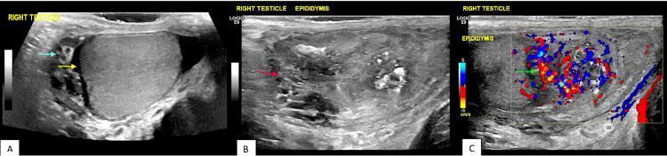
Diagnostic scrotal ultrasound montage consisting of grayscale and color Doppler images illustrating acute epididymo-orchitis. Panel A (grayscale) displays the right testicle accompanied by a small reactive hydrocele; the fluid collection contains low-level internal echoes (proteinaceous material) and thin septations/separations. Panel B (grayscale) focuses on the right epididymis, which appears significantly swollen, edematous, and heterogeneous in texture compared to normal anatomy. Panel C (color Doppler) demonstrates marked hyperemia within the enlarged epididymis, characterized by increased red and blue flow signals indicating high vascularity. These findings are pathognomonic for an inflammatory or infectious process of the scrotum, such as epididymitis with secondary orchitis and a complex reactive hydrocele. Key features include epididymal enlargement, altered echogenicity, increased blood flow (hyperemia), and septated fluid collections within the tunica vaginalis.
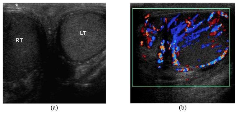
This diagnostic ultrasound display consists of two panels illustrating the clinical features of acute epididymo-orchitis in a 45-year-old male. Panel (a) is a transverse grayscale ultrasound of the scrotum providing a side-by-side comparison of the testes. The right testis (RT) is markedly enlarged and demonstrates a diffuse hypoechoic echotexture compared to the normal left testis (LT). Additionally, the overlying right scrotal skin is significantly thickened, indicated by an asterisk. Panel (b) shows a longitudinal color Doppler ultrasound (CDUS) of the right hemiscrotum. This image reveals prominent hypervascularity within both the testis and the adjacent epididymis, characterized by dense red and blue signals representing increased blood flow (hyperemia). These visual findings—testicular enlargement, decreased echogenicity, skin thickening, and hyperperfusion—are classic diagnostic indicators for infectious or inflammatory processes of the scrotum, distinguishing epididymo-orchitis from testicular torsion where flow would be absent.
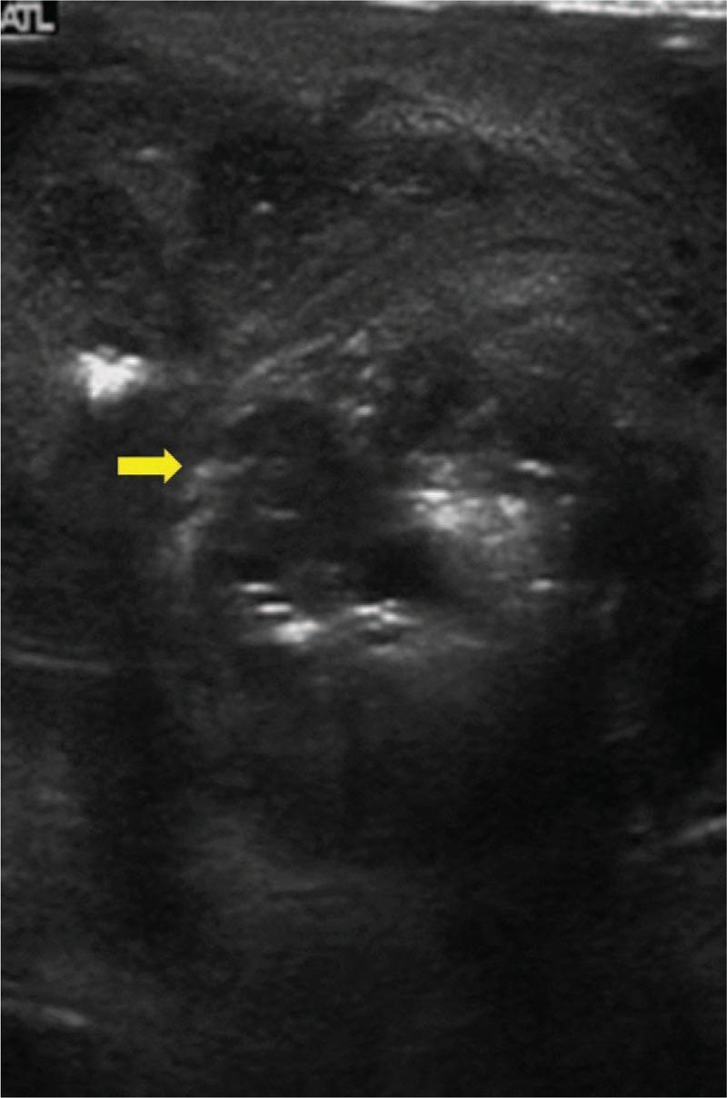
A greyscale diagnostic ultrasound image of the scrotum demonstrating emphysematous epididymo-orchitis. The testicular parenchyma exhibits a highly heterogeneous echotexture with a predominantly hypoechoic and grainy background. Multiple punctate, hyperechoic foci (bright spots) are scattered throughout the tissue, indicative of gas formation or micro-abscesses within the scrotal sac. A yellow arrow points to a cluster of these hyperechoic reflectors. Irregularly shaped hypoechoic areas suggest fluid collection or necrotic tissue consistent with abscess formation. The overall sonographic appearance indicates a severe inflammatory process, such as acute epididymo-orchitis complicated by gas-producing bacterial infection. This imaging is clinically relevant for diagnosing acute scrotal pathology and distinguishing uncomplicated infection from emphysematous changes requiring urgent surgical intervention.
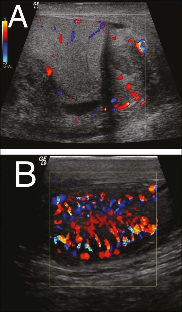
Two color Doppler ultrasound images (A and B) of the scrotal contents demonstrating findings consistent with epididymo-orchitis. Image A displays the epididymis, which appears enlarged and heterogeneous with an irregular echotexture. The color Doppler overlay reveals focal hypervascularity (hyperemia), indicated by increased red and blue signals within the epididymal tissue. Image B shows the testis, which is similarly enlarged and heterogeneous. The Doppler study in B demonstrates diffuse, intense hypervascularity throughout the testicular parenchyma, often referred to as a 'thyroid storm' or 'inferno' sign in the clinical context of orchitis. These findings illustrate the standard diagnostic radiological approach for differentiating inflammatory conditions from testicular torsion; the presence of increased blood flow is characteristic of infectious or inflammatory processes like epididymitis and orchitis, whereas absent or reduced flow would suggest torsion. The imaging is relevant for urological and emergency medicine education regarding acute scrotum evaluation.
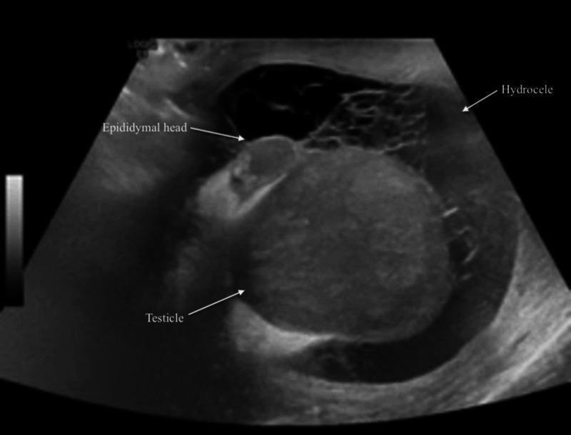
This diagnostic grayscale ultrasound image displays the scrotal contents of a patient with clinical features suggesting epididymo-orchitis. The image shows the testicle and the epididymal head, both of which exhibit a heterogeneous echotexture compared to normal, uniform testicular parenchyma. These variations in echogenicity are indicative of inflammatory changes or infection. Surrounding the testicular structures is a large, complex hydrocele. Unlike a simple hydrocele, which appears anechoic, this collection is characterized as 'complex' due to the presence of internal echoes and fine septations, which can represent fibrin strands, debris, or proteinaceous material resulting from an acute inflammatory process. The image is annotated with labels pointing to the 'Testicle', 'Epididymal head', and 'Hydrocele'. This visual is relevant for medical education concerning urological emergencies, scrotal imaging, and the sonographic differentiation between simple and complex fluid collections in the context of infection.
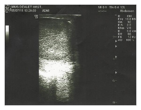
This diagnostic image is a B-mode grayscale scrotal ultrasound from a patient with brucellar epididymo-orchitis (BEO). The scan displays a longitudinal view of the testis, characterized by a predominantly homogeneous, finely granular speckled echotexture. In the lower central portion of the testicular parenchyma, there is a distinct, ill-defined region of increased echogenicity (hyperechoic area), which may correspond to inflammatory changes or an evolving focal lesion common in infectious orchitis. On-screen technical parameters include a 10.0 MHz high-frequency linear transducer setting, a depth of 8.0 cm, and a gain of 92, optimized for superficial soft tissue visualization. The image is part of a clinical evaluation to differentiate between inflammatory conditions like testicular abscesses and solid masses. This visual is significant for students and clinicians studying urogenital infections and the sonographic manifestations of epididymo-orchitis.
epididymo-orchitis treatment
Epididymo-Orchitis
Definition
Aetiology and Pathogenesis
| Age group | Common organism(s) |
|---|---|
| Sexually active young men (<35 yrs) | Chlamydia trachomatis, Neisseria gonorrhoeae |
| Older men / urinary origin | Escherichia coli and other gram-negative enteric bacilli |
| Men who practise insertive anal sex | Chlamydia, Gonorrhea, + enteric organisms |
| Children <6 months | Infected urine ascending via the vas |
| Chronic granulomatous | Mycobacterium tuberculosis |
| Viral (isolated orchitis) | Mumps (commonest), coxsackievirus, EBV, varicella, echovirus |
| Immunocompromised | Cryptococcus, Toxoplasma, Candida, mycocercosis |
Clinical Features
- Scrotal pain - typically gradual onset over 24-48 hours (contrasting with testicular torsion which is abrupt, often minute-precise)
- Fever, malaise
- Dysuria, urinary frequency, urethral discharge
- Nausea/vomiting (from spermatic cord irritation)
- Tender, swollen epididymis and testis
- Scrotal skin erythema and oedema
- Reactive hydrocele (in some cases)
- Positive Prehn's sign - pain may subside on elevation of the testis
- Cremasteric reflex usually preserved (absent in torsion)
- Fluctuant scrotum in prolonged cases suggests abscess formation
Differential Diagnosis - Acute Scrotum
| Feature | Epididymo-orchitis | Testicular torsion |
|---|---|---|
| Onset | Gradual (24-48 h) | Abrupt (exact time recalled) |
| Age peak | Sexually active adults | Adolescents |
| Urinary symptoms | Often present | Absent |
| Fever | Common | Uncommon early |
| Cremasteric reflex | Usually present | Usually absent |
| Prehn's sign | Positive (relief on elevation) | Negative |
| Colour Doppler US | Increased flow (hyperaemia) | Absent/reduced flow |
| Testis position | Normal | High-riding, horizontal lie |
Any equivocal case requires scrotal exploration - a missed torsion causes testicular necrosis. In severe orchitis, testicular flow can occasionally be compromised, mimicking torsion on Doppler.
Investigations
- Urinalysis and urine culture - before starting antibiotics
- Full blood count - leucocytosis
- Urethral swab / NAAT - if STI suspected (Chlamydia, Gonorrhoea)
- Scrotal ultrasound with colour Doppler - confirms diagnosis, rules out torsion, detects abscess
Ultrasound Findings
- Enlarged, oedematous, heterogeneous epididymis
- Enlarged hypoechoic testis
- Increased colour Doppler flow - hyperaemia (the definitive differentiator from torsion)
- Reactive hydrocele (may contain low-level internal echoes/septations)
- In complicated cases: hyperechoic foci with posterior shadowing suggesting gas (emphysematous epididymo-orchitis)
Treatment
Outpatient Antibiotic Regimens (CDC / Tintinalli's Emergency Medicine)
| Likely aetiology | Regimen |
|---|---|
| STI (Chlamydia / Gonorrhoea) | Ceftriaxone 250-500 mg IM single dose + Doxycycline 100 mg PO BD x 10-14 days |
| STI + enteric organisms (insertive anal sex) | Above + Levofloxacin 500 mg OD x 10 days or Ofloxacin 300 mg BD x 10 days |
| Enteric organisms only (older men, post-instrumentation) | Levofloxacin 500 mg OD x 10 days or Ofloxacin 300 mg BD x 10 days |
- High fever / toxic presentation
- Suspected abscess
- Immunocompromised state
Tuberculous Epididymo-Orchitis
- Firm, craggy epididymis; testis often spared for years
- Beading of the vas (subepithelial tubercles) - pathognomonic
- Lax secondary hydrocele in ~30%
- Indurated seminal vesicles on PR examination
- "Cold" abscess may form and discharge in neglected cases
- Contralateral epididymis frequently involved
- Renal TB co-exists in two-thirds of cases
Viral Orchitis (Mumps)
- Most common form of isolated orchitis
- 20-30% of post-pubertal males with mumps develop orchitis
- Usually presents as parotid swelling wanes
- Unilateral in 70%; bilateral involvement occurs in 1-9 days
- Main complication: testicular atrophy - may cause infertility if bilateral
- Diagnosis supported by IgM antibodies; treatment is supportive
Complications
- Scrotal/testicular abscess (requires surgical drainage)
- Testicular atrophy and infertility
- Chronic epididymo-orchitis
- Pyocele