
Investigations in thyroid cancers
"thyroid neoplasms"[MeSH Terms] AND "diagnosis"[MeSH Subheading]
ATA 2015 thyroid nodule ultrasound risk stratification TSH FNA Bethesda 2023 categories malignancy risk
thyroid nodule ultrasound malignant features microcalcifications hypoechoic taller than wide
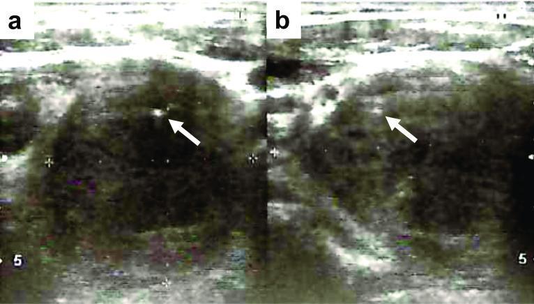
This diagnostic ultrasound image displays a large, suspicious thyroid nodule in two planes: (a) transverse and (b) sagittal. The visual content demonstrates a significant, hypoechoic mass located within the left thyroid lobe. Key sonographic features include irregular, poorly defined margins and internal microcalcifications, which appear as punctate hyperechoic (bright) foci indicated by white arrows. The nodule is large, with documented dimensions of approximately 40 x 39 x 52 mm, indicating a 'taller-than-wide' morphology in certain orientations. These findings are highly characteristic of malignant thyroid pathology, such as high-grade carcinoma or sarcoma (e.g., myxofibrosarcoma). The educational value of this image lies in its demonstration of classical ultrasound markers for thyroid malignancy, specifically the combination of marked hypoechogenicity, microcalcifications, and infiltrative borders. This material is suitable for training in endocrinology, radiology, and oncology, focusing on the TI-RADS classification system and surgical oncology planning.
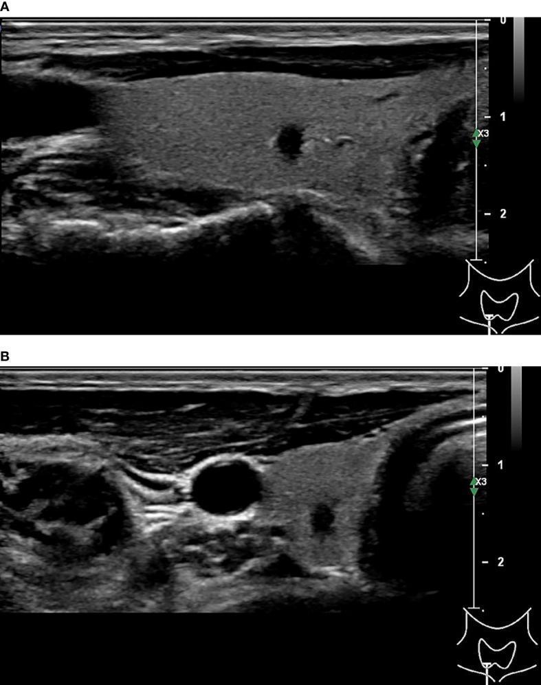
This diagnostic imaging set consists of two grayscale ultrasound (sonography) frames showing a thyroid nodule in longitudinal (A) and axial (B) planes. The images demonstrate a small, solid, markedly hypoechoic nodule within the right lobe of the thyroid gland. Key suspicious features include an 'anteposterior > transverse' (A>T) orientation (taller-than-wide) and an irregular shape with well-defined margins. Internal echoes suggest the presence of punctate hyperechoic foci, consistent with microcalcifications. There is a notable absence of posterior acoustic shadowing. These sonographic characteristics are highly suggestive of malignancy, such as papillary thyroid carcinoma, and lead to high-risk classifications across multiple systems (C-TIRADS 4C, K-TIRADS 5, and ACR-TIRADS 5). The images serve as an educational example of malignant thyroid features used in risk stratification and diagnostic imaging triage.
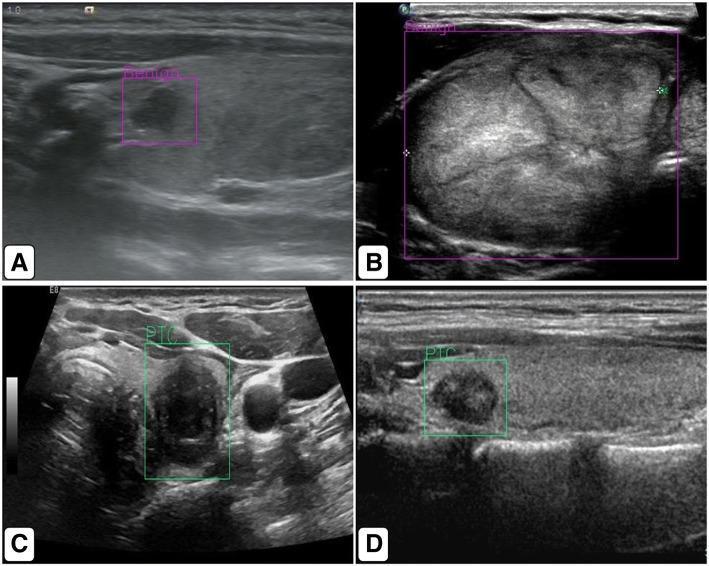
This set of four diagnostic ultrasound images (A-D) demonstrates the application of an artificial intelligence (AI) computer-aided detection system for thyroid nodule classification. Images A and B display benign thyroid nodules, characterized by relatively homogenous echotexture and well-defined margins. These are highlighted by pink/purple bounding boxes labeled 'benign'. In contrast, images C and D illustrate malignant thyroid nodules, specifically Papillary Thyroid Carcinoma (PTC). These nodules exhibit malignant sonographic features including heterogeneous echogenicity, irregular borders, and microcalcifications (hyperechoic foci). The AI system identifies these using green bounding boxes labeled 'PTC'. The comparison highlights the key morphological differences used in TI-RADS grading, such as nodule shape (taller-than-wide), echogenicity (hypoechoic vs. isoechoic), and margin clarity. The images serve as an educational example of how deep-learning algorithms, such as YOLO-based networks, can be integrated into radiological workflows to assist in the differentiation of benign from suspicious thyroid lesions.
Investigations in thyroid cancer
1. Clinical assessment
- History:
- Ask about rapid growth, hoarseness, dysphagia, dyspnoea and pain.
- Ask about childhood neck irradiation and family history (MTC/MEN2, familial PTC).
- Pain is unusual. It suggests haemorrhage into a benign nodule, thyroiditis or malignancy.
- Hoarseness is worrying because it may mean recurrent laryngeal nerve involvement.
- Radiation risk: About 40% of nodules in patients with a history of radiation are cancer. In 40% of those, the cancer is in a different nodule from the dominant one (Schwartz's Principles of Surgery, 11th ed., p. 1669).
- Examination: Look for a hard, fixed nodule, vocal cord palsy and cervical lymphadenopathy.
2. Biochemical tests
| Test | Purpose |
|---|---|
| TSH (± free T4) | This is the first test for any nodule. A low TSH suggests an autonomous nodule, which is rarely malignant, so do a radionuclide scan. A normal or high TSH means proceed with ultrasound and FNA. |
| Serum calcitonin and CEA | These are markers for medullary thyroid carcinoma (MTC). Basal calcitonin helps diagnose C-cell hyperplasia and MTC. Measure CEA preoperatively as well (Tietz Textbook of Laboratory Medicine, 7th ed., p. 1054). |
| Thyroglobulin | It is not useful for screening, diagnosis or prognosis. It is used to monitor papillary and follicular cancer after thyroidectomy and radioiodine. Once the thyroid is completely ablated, it should be undetectable. A rising level suggests recurrence (Tietz, p. 1054). |
| Anti-thyroglobulin antibodies | They interfere with thyroglobulin assays, so measure them alongside it. |
| Calcium, PTH, 24-h urinary or plasma metanephrines | Do these when MTC or MEN2 is suspected. Exclude phaeochromocytoma before any surgery. |
| RET germline testing | Offer it to all MTC patients. It separates hereditary from sporadic disease. About 25% of MTC is hereditary, so screen relatives and check for other endocrine tumours (Tietz, p. 1054). |
3. Imaging
- Solid and markedly hypoechoic
- Irregular or infiltrative margins
- Microcalcifications
- Taller-than-wide shape
- Extrathyroidal extension
- Abnormal nodes: rounded, cystic change, microcalcifications, loss of the fatty hilum
- Spongiform nodules (very low risk, under 3%): biopsy only if over 2 cm
- Isoechoic nodules (low risk, 5-10%): biopsy if over 1.5 cm
- Hypoechoic solid nodules (intermediate risk, 10-20%): biopsy if over 1 cm
- High-suspicion nodules: biopsy at 1 cm or larger
- CT or MRI of the neck and chest (with contrast when needed) assesses retrosternal extension, tracheal or oesophageal invasion, and nodal or distant disease in large, fixed or invasive tumours. Iodinated contrast can delay radioiodine treatment.
- Radioiodine or technetium scintigraphy is for nodules with a low TSH. "Hot" nodules are rarely malignant.
- FDG PET-CT:
- It has limited value for the pre-operative assessment of nodules, because benign and malignant lesions can both be FDG-avid.
- It is useful in treated differentiated cancer with a rising thyroglobulin and a negative radioiodine scan. Pooled sensitivity was 95% and specificity 81% (Scott-Brown's Otorhinolaryngology, Vol 1, p. 597).
- It is more likely to be positive in dedifferentiated, "flip-flop" disease, where radioiodine uptake is lost and FDG uptake is high.
- Its role in MTC is preliminary.
- Laryngoscopy: Check vocal cord mobility pre-operatively if there is a voice change, previous neck surgery or suspected invasion.
- Other imaging for medullary and anaplastic disease:
- Anaplastic carcinoma needs urgent staging imaging (CT neck/chest/abdomen, ± PET).
- MTC with high calcitonin needs staging for liver, bone, lung and neck disease. Options include multiphase liver CT or MRI, and bone imaging.
4. Fine-needle aspiration cytology (FNAC)
| Bethesda category | Approximate risk of malignancy | Usual action |
|---|---|---|
| I Non-diagnostic | 5-10% | Repeat FNA |
| II Benign | 0-3% | Follow up |
| III AUS/FLUS | 6-30% | Repeat FNA or molecular testing |
| IV Follicular neoplasm | 10-40% | Molecular testing or diagnostic lobectomy |
| V Suspicious for malignancy | 45-75% | Lobectomy or thyroidectomy |
| VI Malignant | 94-99% | Surgery |
- Immunocytochemistry: Calcitonin staining confirms MTC. Thyroglobulin and TTF-1 support a thyroid origin.
- Needle washout: Measure thyroglobulin in suspicious nodes for differentiated cancer, and calcitonin for MTC.
5. Histopathology
- Papillary carcinoma:
- Features include Orphan Annie eye nuclei, grooves, psammoma bodies and papillae.
- Typical genetic changes are BRAF and RET/PTC.
- Follicular carcinoma: Capsular or vascular invasion on histology.
- Medullary carcinoma: Amyloid stroma and calcitonin positivity.
- Anaplastic carcinoma: Undifferentiated, with a very poor prognosis.
- Hurthle cell carcinoma: Oncocytic cells.
6. Follow-up investigations
- Differentiated cancer after surgery:
- Measure TSH-suppressed thyroglobulin with anti-Tg antibodies.
- Use stimulated thyroglobulin after recombinant TSH or thyroxine withdrawal.
- Do neck ultrasound.
- Do a post-ablation whole-body iodine scan.
- Add FDG PET-CT if thyroglobulin is rising and the iodine scan is negative.
- MTC: Serial calcitonin and CEA (see above).