
teach me important concept of infective endocarditis microbiology with easy notes but should cover all important and must know points to crack university exam
infective endocarditis vegetation heart valve microbiology
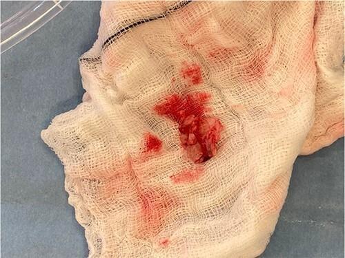
This intraoperative clinical photograph displays a macro-level view of excised cardiac vegetation, typical of infective endocarditis, resting on a white, sterile surgical gauze swab. The vegetation appears as an irregular, friable, and multi-lobulated tissue mass with a reddish-pink hue, indicating significant vascularization or associated blood clot formation. The specimen's texture is heterogeneous and rough, consistent with a collection of fibrin, platelets, and microorganisms typically found on heart valve surfaces during active infection. The surrounding white gauze shows several focal areas of bright red serosanguinous staining where the vegetation has been placed. This image is medically significant for illustrating the gross pathological morphology of a mitral valve vegetation following surgical excision and valve debridement. It serves as a visual record of the endovascular disease process, highlighting the size and friability of the mass that contributed to valvular regurgitation and potential embolic risk.
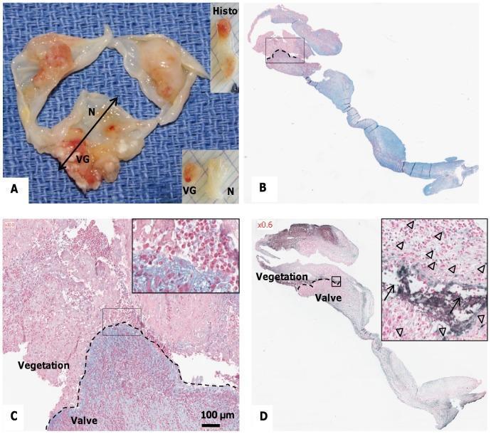
This composite educational image illustrates the macroscopic and histopathological characteristics of infective endocarditis on human heart valves. (A) Clinical photograph of an excised valve showing friable, reddish-brown vegetations (VG) contrasting against smooth, translucent normal tissue (N). (B, C) Light microscopy using Alcian blue and Nuclear Fast Red counterstain displays the interface between the septic thrombus (red-stained inflammatory cells and polylobed PMNs) and underlying valvular tissue exhibiting mucoid degeneration (blue-stained areas). The dotted line marks the clear boundary between the infected vegetation and the valve stroma. (D) TUNEL assay micrograph highlights the presence of cellular damage. Apoptotic cells (indicated by arrowheads) appear as dark, discrete nuclear spots, while extracellular fragmented DNA (indicated by arrows) shows diffuse, dark staining concentrated at the vegetation-valve interface. This visual series demonstrates the key pathological hallmarks of valvular infection, including thrombus formation, inflammatory infiltration, and programmed cell death.
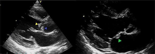
Two-panel diagnostic echocardiography (ultrasound) images demonstrating infective endocarditis with multiple vegetations on the left heart valves. The left panel shows a parasternal long-axis view of the bicuspid aortic valve, with hyperechoic, irregular masses (vegetations) attached to the tips of the anterior (yellow arrow) and posterior (blue arrow) leaflets. The right panel highlights the mitral valve apparatus, where another distinct hyperechoic vegetation is visible on the body of the anterior mitral valve leaflet (green arrow). These findings are highly suggestive of active infective endocarditis, illustrating the characteristic appearance of mobile, irregular echoes attached to valvular endocardial surfaces. Clinical relevance includes the high risk of systemic embolization given the size and location of these vegetations, commonly associated with pathogens such as Streptococcus gordonii. This visual serves as an educational tool for identifying valvular pathology, measuring vegetation dimensions, and assessing the risk of secondary complications like septic emboli in a cardiothoracic or infectious disease context.
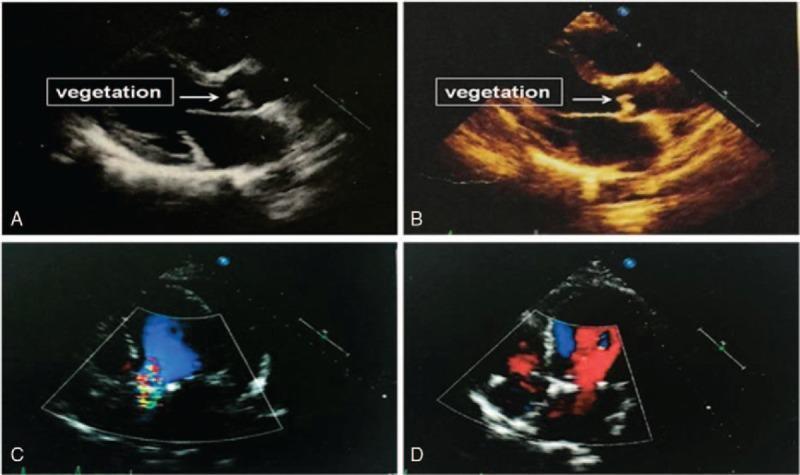
Diagnostic ultrasound montage demonstrating infective endocarditis (IE) and its surgical resolution via transthoracic echocardiography. Panels A (grayscale) and B (sepia-toned) show a parasternal long-axis view of the heart. A distinct, pedunculated, and echogenic mass labeled 'vegetation' is indicated by a white arrow, attached to the aortic valve leaflet. Panels C and D utilize color Doppler imaging in a four-chamber or modified atrial view to assess hemodynamic changes. Panel C illustrates an abnormal left-to-right atrial shunt, characterized by a high-velocity, turbulent multi-colored jet crossing the interatrial septum. Panel D, obtained postoperatively, shows the absence of this shunt and restored laminar flow (represented by stable red and blue signals), confirming successful surgical repair and removal of the infected vegetation. This image sequence serves as a clinical comparison for medical students and cardiologists to identify valvular vegetations and evaluate the efficacy of surgical intervention in congenital or acquired heart defects complicated by infection.
🫀 Infective Endocarditis (IE) - Complete Microbiology Notes for Exam
Sources: Robbins & Kumar Basic Pathology | Goldman-Cecil Medicine | Frameworks for Internal Medicine | Braunwald's Heart Disease | Fitzpatrick's Dermatology
1. DEFINITION
- Can also affect: aorta, aneurysmal sacs, prosthetic devices, pacemaker leads
- Vast majority caused by bacteria (rarely fungi, rickettsiae, Chlamydia)
2. CLASSIFICATION
| Feature | Acute IE | Subacute IE |
|---|---|---|
| Onset | Rapid (days) | Insidious (weeks to months) |
| Valves | Normal or abnormal | Usually abnormal/damaged |
| Virulence | High | Low |
| Classic organism | S. aureus | Viridans streptococci |
| Prognosis without Rx | Fatal in weeks | Survives months |
| Tissue destruction | Severe | Moderate |
Exam tip: The line between acute and subacute is not always clear - many cases fall on a spectrum.
3. PREDISPOSING CONDITIONS (Risk Factors)
Cardiac Risk Factors (Most Common)
| Risk Factor | Notes |
|---|---|
| Mitral valve prolapse (especially with regurgitation) | #1 preexistent risk factor today |
| Degenerative valvular disease | Calcific stenosis, bicuspid aortic valve |
| Rheumatic heart disease | Was previously #1, now less common |
| Prosthetic heart valves | Accounts for 10-20% of all IE cases |
| Previous IE | Major risk for recurrence |
| Congenital heart disease | Especially uncorrected VSD |
Non-Cardiac Risk Factors
- IV drug use (IVDU) - very high risk
- Neutropenia, immunodeficiency, malignancy
- Diabetes mellitus
- Prolonged use of indwelling vascular catheters
- Pacemakers / cardiac implantable electronic devices (CIEDs)
4. PATHOGENESIS - STEP BY STEP
STEP 1: Endothelial damage (turbulent flow, jets, catheters)
↓
STEP 2: Sterile platelet-fibrin thrombus forms at damaged site
↓
STEP 3: Bacteremia (from dental procedure, surgery, gut, skin, IV drugs)
↓
STEP 4: Bacteria adhere to thrombus via surface adhesins
↓
STEP 5: Bacteria proliferate within vegetation (protected from host defenses)
↓
STEP 6: Vegetation grows → valve destruction → emboli → metastatic infection
- Access to bloodstream (bacteremia)
- Ability to adhere to endocardium/thrombus
5. CAUSATIVE ORGANISMS - THE EXAM FAVORITE SECTION 🔑
The "Big 3" account for the vast majority of IE cases:
A. Viridans Group Streptococci (VGS)
| Feature | Detail |
|---|---|
| Species | S. sanguis, S. mitis, S. salivarius, S. mutans |
| Source | Normal oral/dental flora |
| Entry | Dental procedures, daily oral trauma |
| Type of IE | Subacute native valve IE |
| Valve | Left-sided (mitral > aortic) |
| Susceptibility | Highly penicillin-susceptible |
| Prognosis | Cure rates ~100% with 4 weeks IV penicillin |
| Frequency | 50-60% of community-acquired IE |
Memory hook: "Viridans = Viridans gums" - they live in the mouth and enter after dental work
B. Staphylococcus aureus
| Feature | Detail |
|---|---|
| Source | Skin flora; healthcare/nosocomial |
| Type of IE | Acute IE (rapid, destructive) |
| Valve | Any valve - normal OR diseased |
| IVDU | Classically affects tricuspid valve (right-sided IE) |
| Complications | Perivalvular abscess, metastatic septic emboli (lung, brain, kidneys) |
| Treatment (MSSA) | Nafcillin or oxacillin |
| Treatment (MRSA) | Vancomycin |
| Now #1 cause in | High-income countries, healthcare settings, IVDU |
Remember: S. aureus can infect normal valves - unique among common IE organisms!
C. Streptococcus gallolyticus (formerly S. bovis)
| Feature | Detail |
|---|---|
| Old name | Streptococcus bovis biotype I |
| Type | Subacute IE |
| Special association | Colon cancer / colonic polyps |
| Action required | Colonoscopy mandatory when isolated |
| Susceptibility | Highly penicillin-susceptible |
High-yield exam pearl: Any patient with S. gallolyticus endocarditis → rule out colonic malignancy!
D. Enterococci
| Feature | Detail |
|---|---|
| Species | E. faecalis (>90%), E. faecium |
| Source | GI and genitourinary tracts |
| Type | Can be subacute OR acute |
| Frequency | ~10% of all IE cases |
| Valve | Predominantly left-sided (even in IVDU) |
| Resistance | Intrinsically resistant to cephalosporins, low-level aminoglycosides |
| Treatment | Ampicillin + gentamicin (or streptomycin); alternative: ampicillin + ceftriaxone |
| Duration | 4-6 weeks |
E. HACEK Group (Gram-Negative Fastidious Organisms)
| Feature | Detail |
|---|---|
| Source | Normal oral/upper respiratory commensals |
| Type | Subacute IE |
| Frequency | ~5% of IE cases |
| Growth | Fastidious; grow in blood cultures within 7 days |
| Treatment | Ceftriaxone (ampicillin historically) |
| Eikenella | Associated with IVDU (needle/skin licking) |
F. Coagulase-Negative Staphylococci (CoNS) - e.g., S. epidermidis
| Feature | Detail |
|---|---|
| Source | Normal skin flora |
| Risk | Healthcare contact (catheters, prosthetics) |
| Native valve IE | Uncommon, but increasing |
| Prosthetic valve | Major pathogen (especially early PVE) |
| Concern | High rates of perivalvular abscess and heart failure despite subacute presentation |
| Resistance | Often oxacillin-resistant (treat as MRSA) |
G. Fungi
| Feature | Detail |
|---|---|
| Common organisms | Candida spp. and Aspergillus spp. |
| Risk | IVDU, prolonged IV catheters (TPN), cardiac valve surgery |
| Candida | Grows in blood cultures |
| Aspergillus | Rarely cultured from blood - need valve/embolic tissue |
| Mortality | Very high |
| Treatment | Usually requires valve replacement surgery |
H. Culture-Negative IE (~10% of all cases)
- Prior antibiotic therapy (most common reason)
- Fastidious/slow-growing organisms
- Coxiella burnetii (Q fever) - serologic diagnosis
- Bartonella spp. (homeless, louse exposure)
- Brucella, Legionella, Tropheryma whipplei
- HACEK organisms (if not incubated long enough - now less of an issue)
Exam tip: Always ask about prior antibiotic use in any patient with culture-negative IE!
6. ORGANISMS BY CLINICAL SETTING (HIGH-YIELD TABLE)
| Clinical Setting | Most Common Organism(s) |
|---|---|
| Community-acquired native valve | Viridans streptococci (50-60%) |
| Healthcare/nosocomial | S. aureus (#1) |
| IV drug use - tricuspid | S. aureus (>50%) |
| IV drug use - left-sided | Viridans streptococci, Enterococci |
| Prosthetic valve - Early (<2 months) | S. aureus, CoNS (S. epidermidis) |
| Prosthetic valve - Late (>2 months) | S. aureus, Viridans streptococci |
| Dental procedure | Viridans streptococci |
| GU/GI source | Enterococci |
| Colonic malignancy association | S. gallolyticus |
| Fungal IE | Candida, Aspergillus |
| Culture-negative | Coxiella burnetii (Q fever), Bartonella |
7. MORPHOLOGY OF VEGETATIONS
- Fibrin + platelets (thrombus) + bacteria + inflammatory cells (PMNs)
- Underlying valve: necrosis, inflammation, destruction
- May extend to cause perivalvular/ring abscess
| Type | Organisms | Location | Features |
|---|---|---|---|
| IE | Present | Along line of closure + leaflet | Friable, destructive, large |
| Rheumatic | Absent | Line of closure | Small, firm, "verrucous" |
| Libman-Sacks (SLE) | Absent | Both sides of valve | Small, flat, non-destructive |
| Marantic (NBTE) | Absent | Line of closure | Small, sterile, cancer/debility |
8. CLINICAL FEATURES
Peripheral Stigmata of IE (Must memorize!)
| Sign | Description | Mechanism | Exam Clue |
|---|---|---|---|
| Osler nodes | Painful, raised, red papulonodules on fingertips/toe pads | Immune complex / microemboli | Painful = Osler |
| Janeway lesions | Painless hemorrhagic macules on palms/soles | Septic emboli | Painless = Janeway (on Palms) |
| Splinter hemorrhages | Linear brownish-red lines in nail beds | Microemboli | Non-specific (can be normal) |
| Roth spots | Oval retinal hemorrhages with white/pale center | Immune complexes / microemboli | Seen on fundoscopy |
| Petechiae | Small red spots on skin/mucosa | Microemboli/vasculitis | Non-specific |
| Clubbing | Finger clubbing | Chronic IE | Subacute disease |
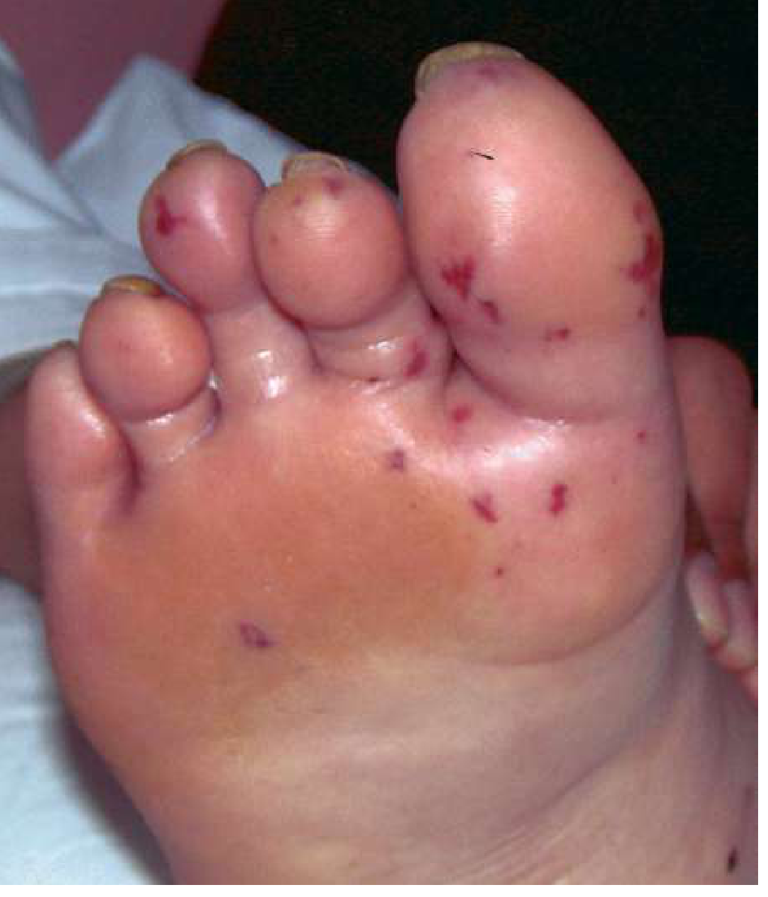
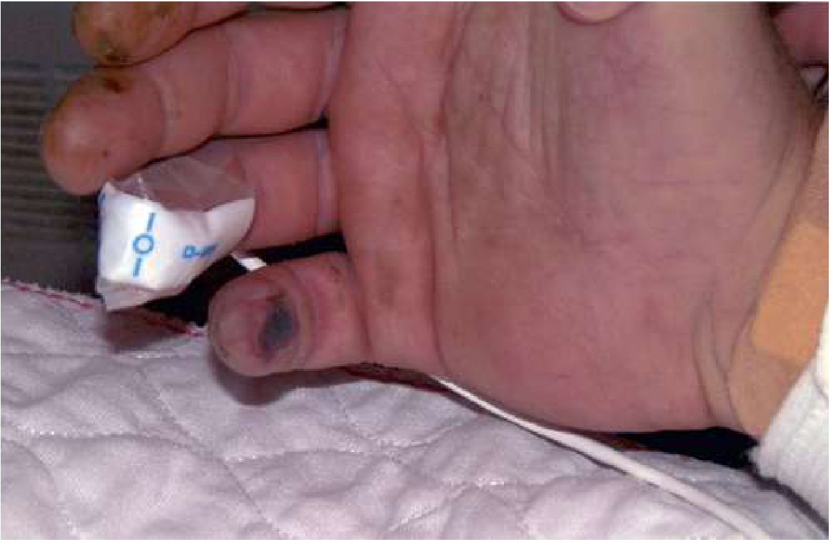
Memory trick: Osler = Ouch (painful), Janeway = Just painless (Janeway on palms/soles is PAINLESS)
9. MODIFIED DUKE CRITERIA (Diagnosis)
MAJOR Criteria
- Two separate positive blood cultures with typical organisms (S. viridans, S. bovis/gallolyticus, HACEK, S. aureus, Enterococcus) from community source
- Persistently positive blood cultures (≥2 positive, drawn >12 hours apart)
- Single positive blood culture for Coxiella burnetii, OR Phase I IgG titer >1:800
- New valvular regurgitation
- Oscillating intracardiac mass at site of endocardial injury
- Perivalvular abscess
- New dehiscence of prosthetic valve
MINOR Criteria
- Predisposing heart condition or IV drug use
- Fever ≥38°C (100.4°F)
- Vascular phenomena - arterial emboli, septic pulmonary infarcts, Janeway lesions, mycotic aneurysm, intracranial hemorrhage, conjunctival hemorrhages
- Immunologic phenomena - glomerulonephritis, Osler nodes, Roth spots, rheumatoid factor
- Microbiologic evidence not meeting major criteria (single positive culture for less typical organism)
Mnemonic for Minor criteria: "FVIMI" = Fever, Vascular, Immunologic, Microbiology, predIsposition
10. COMPLICATIONS (Know for Exam)
| System | Complication |
|---|---|
| Cardiac | Heart failure (most common cause of death), perivalvular abscess, conduction defects (AV block), pericarditis |
| Embolic | Stroke, TIA, renal infarcts, splenic infarcts, septic pulmonary emboli (right-sided IE) |
| Renal | Glomerulonephritis (immune complex deposition), renal infarcts |
| Neurologic | Stroke (15-20% of patients), meningitis, brain abscess, mycotic aneurysm |
| Metastatic | Bone (osteomyelitis), joints (septic arthritis), spleen (abscess) |
11. ANTIBIOTIC PROPHYLAXIS (Key Points)
- Prosthetic cardiac valve or material
- Previous IE
- Congenital heart disease (unrepaired cyanotic CHD, or within 6 months of repair)
- Cardiac transplant with valve abnormality
- Ventricular assist devices and implantable hearts (new addition)
- Amoxicillin 2g PO 30-60 min before procedure (first choice)
- Penicillin allergy: Doxycycline (clindamycin no longer recommended due to C. difficile risk)
12. TREATMENT OVERVIEW (Principles)
| Organism | Antibiotic | Duration |
|---|---|---|
| Viridans streptococci (penicillin-sensitive) | Penicillin G or Ceftriaxone | 4 weeks |
| S. aureus (MSSA) | Nafcillin / Oxacillin | 4-6 weeks |
| S. aureus (MRSA) | Vancomycin | 4-6 weeks |
| Enterococcus (susceptible) | Ampicillin + Gentamicin | 4-6 weeks |
| HACEK | Ceftriaxone | 4 weeks |
| Fungal | Amphotericin B + valve replacement | Long-term |
Surgery indications: Heart failure, persistent infection despite antibiotics, perivalvular abscess, fungal IE, prosthetic valve dehiscence, recurrent emboli
13. QUICK EXAM MEMORY AIDS
Organism Mnemonics
The "Rules of Right vs Left"
| Right-sided IE | Left-sided IE |
|---|---|
| Tricuspid valve (IVDU) | Mitral > Aortic valve |
| S. aureus | Viridans strep, Enterococcus |
| Septic pulmonary emboli | Systemic emboli (brain, kidneys) |
| Better prognosis (<10% mortality) | Worse prognosis |
Valve involvement frequency (general)
14. SUMMARY TABLE - "The Must-Know Organisms"
| Organism | Type | Key Setting | Unique Feature |
|---|---|---|---|
| Viridans Streptococci | Subacute | Dental/oral | Most common community IE; penicillin-sensitive |
| S. aureus | Acute | IVDU, healthcare | Can infect normal valves; #1 in high-income countries |
| Enterococci | Subacute/acute | GU/GI tract | E. faecalis most common; ampicillin + aminoglycoside |
| S. gallolyticus | Subacute | GI tract | Colonoscopy for colon cancer |
| HACEK | Subacute | Oral flora | Fastidious; ~5% of IE |
| CoNS (S. epidermidis) | Subacute | Prosthetic valves, healthcare | Major cause of PVE |
| Candida | - | IVDU, catheters | Grows in cultures; surgery usually needed |
| Aspergillus | - | Valve surgery, IVDU | Rarely cultured; check galactomannan |
| Coxiella burnetii | - | Q fever (animal exposure) | Culture-negative; serology >1:800 |
teach me important concept of rheumatic fever microbiology with easy notes but should cover all important and must know points to crack university exam
rheumatic fever Aschoff body mitral valve stenosis pathology
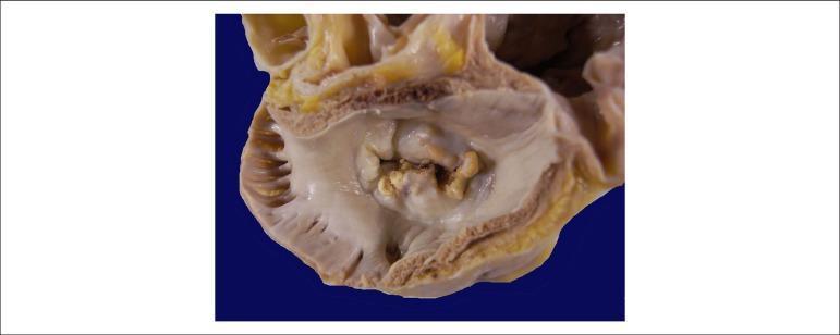
This clinical photograph shows a gross pathological specimen of a human heart, specifically an atrial view of the mitral valve. The image demonstrates hallmark signs of chronic rheumatic heart disease. Key features include significant thickening and fibrosis of the valve leaflets, resulting in a 'fish-mouth' or 'buttonhole' appearance of the valvular orifice. There is evidence of extensive commissural fusion and multiple focal areas of yellowish-white calcification along the leaflet margins and within the valve body. These structural changes characterize severe mitral stenosis, where the restricted opening of the valve impairs blood flow from the left atrium to the left ventricle. The surrounding atrial endocardium appears thickened, consistent with chronic pressure overload. This visual is highly representative for educational purposes in cardiology and pathology to illustrate the long-term sequelae of rheumatic fever on cardiac valves.
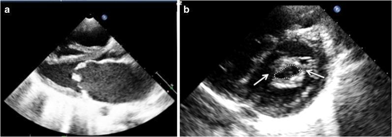
Two-panel diagnostic imaging featuring a 2D transthoracic echocardiogram (TTE) of severe mitral stenosis, typically associated with rheumatic heart disease. Panel (a) shows a parasternal long-axis view during diastole, highlighting significant thickening and increased echogenicity of the mitral valve leaflets. The anterior leaflet demonstrates characteristic 'doming' (hockey-stick deformity) with restricted excursion, indicating limited valve opening. Panel (b) presents a parasternal short-axis view at the level of the mitral valve orifice. White arrows point to commissural fusion, a hallmark of stenotic valvular pathology. The mitral valve area (MVA) is manually traced via planimetry (indicated by the dotted line), yielding a measurement of 0.73 cm², which falls into the category of severe stenosis (MVA < 1.0 cm²). These images demonstrate the classic morphology used in cardiology for assessing valvular narrowing, leaflet mobility, and subvalvular involvement to determine the clinical severity of mitral stenosis.
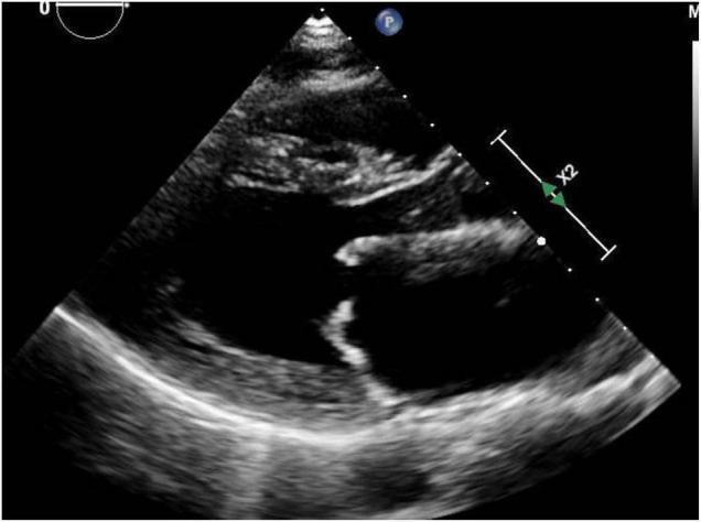
This diagnostic image is a transthoracic echocardiogram in the parasternal long-axis (PLAX) view, demonstrating classic features of mitral stenosis secondary to rheumatic heart disease. The image shows the left ventricle (LV), left atrium (LA), and the aortic root. Key pathology is localized to the mitral valve, where both the anterior and posterior leaflets exhibit significant thickening and increased echogenicity. The leaflets demonstrate restricted diastolic opening, resulting in a narrow valve orifice. The anterior mitral valve leaflet shows a characteristic 'doming' appearance (hockey-stick deformity), which is pathognomonic for rheumatic involvement. The LA appears moderately dilated, a common consequence of chronic mitral stenosis. This educational visual is intended for cardiology and radiology students to illustrate the valvular morphology and mechanical obstruction characteristic of rheumatic mitral valve disease, emphasizing the role of non-invasive ultrasound imaging in assessing valvular heart disease.
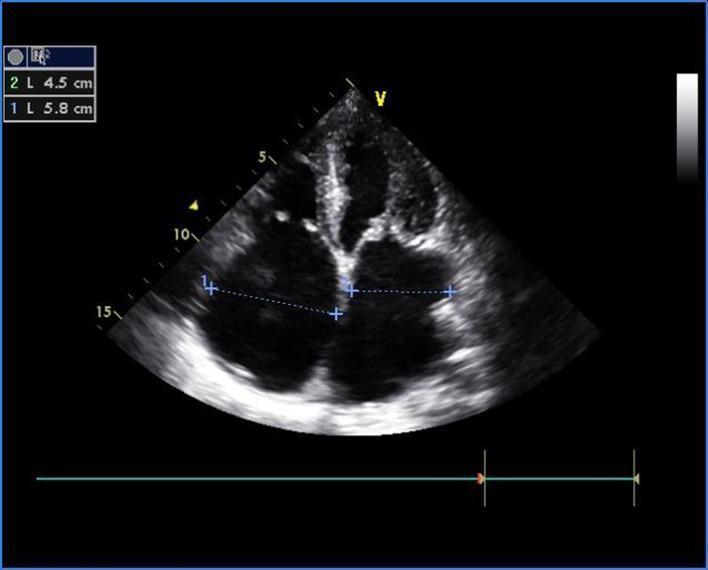
This diagnostic image is a transthoracic echocardiogram (TTE) in the apical four-chamber view, providing a simultaneous visualization of both the left and right sides of the heart. The ultrasound displays significant structural pathology of the atrioventricular valves. The mitral valve leaflets appear diffusely thickened and exhibit a characteristic 'doming' appearance during diastole, classic for rheumatic mitral stenosis. Similarly, the tricuspid valve leaflets demonstrate matching morphological changes, including thickening and diastolic doming, indicative of concurrent tricuspid stenosis. Measurement calipers (labeled 1 and 2) are visible across the dilated atria, with values of 5.8 cm and 4.5 cm recorded in the upper left corner. The image illustrates multi-valvular rheumatic heart disease involving both the left-sided and right-sided chambers, resulting in atrial enlargement secondary to obstructive valvular pathology. This image is an essential educational tool for identifying the echocardiographic hallmarks of chronic rheumatic valvulitis and assessing cardiac chamber dimensions in the setting of severe stenosis.
🔴 Rheumatic Fever - Complete Microbiology Notes for Exam
Sources: Medical Microbiology 9e | Robbins & Kumar Basic Pathology | Goldman-Cecil Medicine | Sherris & Ryan's Medical Microbiology | Harrison's Principles of Internal Medicine 22E
1. DEFINITION
Key rule: ARF follows THROAT infections with GAS only - NOT skin infections (pyoderma/impetigo). This is a critical exam distinction.
2. THE CAUSATIVE ORGANISM
Streptococcus pyogenes (Group A Streptococcus - GAS)
| Property | Detail |
|---|---|
| Gram stain | Gram-positive coccus in chains |
| Lancefield group | Group A (Lancefield classification based on C carbohydrate) |
| Hemolysis | Beta-hemolysis (complete hemolysis on blood agar) |
| Key virulence factor | M protein - anti-phagocytic, main rheumatogenic antigen |
| Rheumatogenic M types | Types 1, 3, 5, 6, 18 (specific types associated with RF) |
| Enzyme marker | Produces Streptolysin O (detected as ASO titer in patients) |
| Other enzymes | Streptolysin S, Streptokinase, DNase B, Hyaluronidase, Erythrogenic toxin |
Exam pearl: NOT all GAS strains cause RF - only rheumatogenic M serotypes. Nephritogenic strains (causing glomerulonephritis) are DIFFERENT M types.
3. EPIDEMIOLOGY
| Feature | Detail |
|---|---|
| Peak age | 5-15 years (school-age children) |
| Sex | Equal in males and females (RF); RHD more common in women |
| Season | Cooler months (mirrors streptococcal pharyngitis) |
| Socioeconomic | Overcrowding, low income = higher risk |
| Risk after untreated GAS pharyngitis | ~1-3% (only minority develop RF) |
| Developing countries | ~100 cases per 100,000 children/year |
| Recurrence | High risk with each subsequent GAS infection (if no prophylaxis) |
| Attack rate highest in | Patients with severe pharyngitis (though 1/3 have mild/asymptomatic infection) |
4. PATHOGENESIS - MOLECULAR MIMICRY 🔑
Group A Streptococcus pharyngitis
↓
GAS releases M protein antigen
↓
Immune system generates antibodies + T cells against M protein
↓
MOLECULAR MIMICRY:
M protein shares epitopes with human proteins
↓
Anti-M protein antibodies cross-react with:
• Cardiac myosin → MYOCARDITIS
• Sarcolemmal membranes → ENDOCARDITIS/VALVULITIS
• Synovium/articular cartilage → ARTHRITIS
• Basal ganglia → CHOREA
• Skin proteins → SUBCUTANEOUS NODULES
Key Immune Mechanisms
| Mechanism | Effect |
|---|---|
| Molecular mimicry (Type II hypersensitivity) | Antibodies cross-react with host tissues |
| Antibody-mediated | Complement activation + macrophage recruitment → tissue injury |
| T-cell mediated | CD4+ TH1 responses → macrophage activation → Aschoff bodies |
| Immune complexes (Type III) | Deposited in joints → arthritis; small vessels → skin lesions |
| Chorea mechanism | Antibody binding to basal ganglia |
Why the 2-3 week delay? Because it takes 2-3 weeks to generate an immune response. By the time symptoms appear, streptococci are completely absent from the lesions.
Why only ~1-3% develop RF? Genetic susceptibility - B-lymphocyte alloantigen occurs 4-5 times more frequently in RF patients vs. general population.
5. MORPHOLOGY / PATHOLOGY
The ASCHOFF Body (Pathognomonic!) 🔑
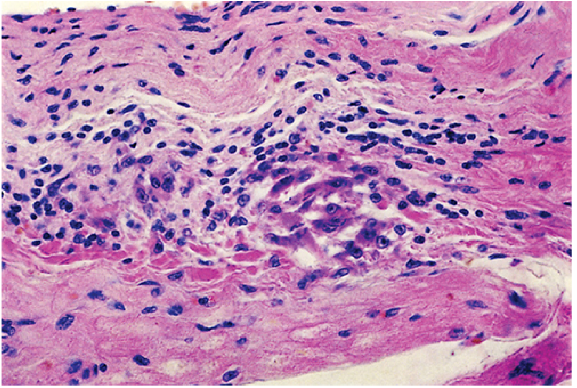
| Feature | Detail |
|---|---|
| What it is | Discrete inflammatory granuloma-like focus in myocardial interstitium |
| Composition | Lymphocytes (T cells) + plasma cells + plump activated macrophages |
| Special cell | Anitschkow cells (= "caterpillar cells") |
| Anitschkow cell | Large activated macrophage with wavy/caterpillar-shaped chromatin (centrally condensed) |
| Also called | "Owl-eye cells" or "caterpillar cells" |
| Central zone | Fibrinoid necrosis |
| Location | Myocardial interstitial connective tissue |
| Significance | Found in ALL 3 layers → supports pancarditis diagnosis |
PANCARDITIS - All Three Layers Affected
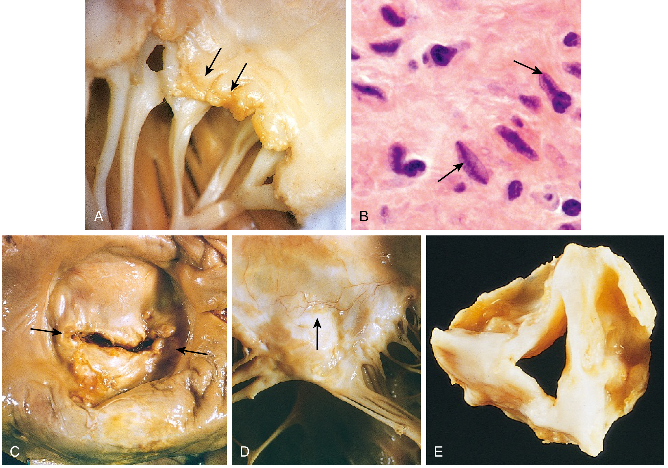
| Layer | Pathology | Features |
|---|---|---|
| Pericardium | Fibrinous pericarditis | "Bread-and-butter" appearance; resolves without sequelae |
| Myocardium | Myocarditis | Aschoff bodies in interstitium; scattered |
| Endocardium | Valvulitis + small vegetations | Fibrinoid necrosis along valve closure line |
Valve Changes
- Small, 1-2 mm verrucous vegetations along the line of closure of valve leaflets (NOT destructive like IE)
- Fibrinoid necrosis of valve leaflets
- Mitral valve most commonly affected
- Fibrous thickening + fusion of valve leaflets
- Commissural fusion → mitral stenosis ("fish-mouth" or "buttonhole" appearance)
- Fusion and shortening of chordae tendineae
- Neovascularization of valve
6. CLINICAL FEATURES
Timeline
- ARF develops ~2 weeks after acute streptococcal pharyngitis
- Symptoms persist 2-4 weeks
- Carditis resolves or progresses over months-years
Manifestations - Frequency
| Feature | Frequency |
|---|---|
| Fever | >90% |
| Large-joint migratory polyarthritis | ~75% |
| Carditis (pancarditis) | >50% |
| Sydenham's chorea | ~30% |
| Subcutaneous nodules | <10% |
| Erythema marginatum | <10% |
7. REVISED JONES CRITERIA (Diagnosis) 🔑
- Initial ARF: 2 Major, OR 1 Major + 2 Minor
- Recurrent ARF: 2 Major, OR 1 Major + 2 Minor, OR 3 Minor
MAJOR Criteria - "JONES" Mnemonic
| Letter | Major Criterion | Key Details |
|---|---|---|
| J | Joints (Migratory Polyarthritis) | Large joints (knees, ankles, elbows, wrists); migratory; sterile synovial fluid; responds rapidly to NSAIDs |
| O | Oh my heart (Carditis) | Clinical and/or subclinical (echocardiographic) valvulitis; pancarditis; most common = mitral regurgitation |
| N | Nodules (Subcutaneous) | 0.5-2 cm; painless; over bony prominences/extensor tendons |
| E | Erythema marginatum | Pink, non-pruritic, blanching macules/papules with serpiginous border; trunk + proximal limbs; spares face |
| S | Sydenham's Chorea | Involuntary, non-rhythmic, purposeless movements; more on one side; stops during sleep; "St. Vitus Dance" |
Memory trick: "JONES" = Joints, Oh-heart, Nodules, Erythema, Sydenham
MINOR Criteria
| Minor Criterion | Low-Risk Population | Moderate/High-Risk |
|---|---|---|
| Arthralgia | Polyarthralgia | Monoarthralgia |
| Fever | ≥38.5°C | ≥38.5°C |
| Elevated inflammatory markers | ESR ≥60 mm/h AND/OR CRP ≥3.0 mg/dL | ESR ≥30 mm/h AND/OR CRP ≥3.0 mg/dL |
| Prolonged PR interval | Yes (unless carditis = major criterion) | Yes |
Evidence of Preceding GAS Infection (ESSENTIAL - without this, Jones criteria alone are insufficient)
- Positive throat culture for GAS
- Rapid streptococcal antigen test (Rapid Strep Test)
- Elevated/rising streptococcal antibody titers:
- ASO (Anti-Streptolysin O) - most commonly used
- Anti-DNase B - more sensitive for skin infections
- Anti-hyaluronidase
Exam tip: ASO titer stays elevated for weeks-months after infection. Absence of elevated ASO = strong evidence AGAINST RF.
8. LABORATORY FINDINGS
| Test | Finding |
|---|---|
| ASO titer | Elevated (most important serologic test) |
| Anti-DNase B | Elevated |
| Throat culture | May be positive for GAS |
| ESR | Elevated |
| CRP | Elevated |
| WBC | Leukocytosis |
| CBC | Normochromic, normocytic anemia |
| ECG | Prolonged PR interval (first-degree AV block) - minor criterion |
| Synovial fluid | Sterile, lymphocyte predominant |
9. RHEUMATIC HEART DISEASE (Chronic Sequelae)
| Feature | Detail |
|---|---|
| Valve order of involvement | Mitral (100%) > Aortic (20-30%) > Tricuspid (15-40% histologically) > Pulmonary (rare) |
| Most common lesion | Mitral regurgitation (acute) → Mitral stenosis (chronic/recurrent) |
| Mitral stenosis | RHD is the #1 worldwide cause of acquired mitral stenosis |
| Progression to RHD | 35-70% of ARF patients with carditis |
| Peak severity | Third and fourth decades of life |
| In developing countries | Leading cause of bacterial endocarditis and cardiovascular mortality in young people |
Critical exam fact: "Rheumatic heart disease is essentially the only cause of acquired mitral stenosis" - Robbins
10. TREATMENT
Acute Phase
| Goal | Treatment |
|---|---|
| Eradicate GAS from throat | Benzathine penicillin G IM 1.2 million units (single dose) OR oral penicillin V 10 days |
| Arthritis | Aspirin 80-100 mg/kg/day, OR Naproxen; NSAIDs |
| Carditis/heart failure | Diuretics, ACE inhibitors/ARBs, bed rest; steroids in severe cases |
| Chorea | Usually self-limited; resolves in weeks-months |
Prophylaxis (Prevention of Recurrence) 🔑
| Duration of Secondary Prophylaxis | Condition |
|---|---|
| 10 years or until age 21 (whichever is longer) | No carditis |
| 10 years or until age 21 (whichever is longer) | Carditis, no residual valve disease |
| 10 years or until age 40 (whichever is longer) | Persistent valvular disease |
| Lifelong | Severe valvular disease / valve surgery |
Why lifelong prophylaxis matters: Each recurrence of RF can cause additional valve damage. Prevention of GAS throat infection = prevention of further deterioration.
11. COMPARISON: RF vs. GLOMERULONEPHRITIS (Post-GAS Complications)
| Feature | Rheumatic Fever | Acute Post-Streptococcal Glomerulonephritis |
|---|---|---|
| Cause | GAS | GAS |
| Follows | Pharyngitis ONLY | Pharyngitis OR skin infection |
| M types | Rheumatogenic (1,3,5,6,18) | Nephritogenic (types differ) |
| Mechanism | Molecular mimicry (Type II + T cell) | Immune complex deposition (Type III) |
| Organ | Heart, joints, skin, brain | Kidney (glomeruli) |
| Recurrence with GAS | YES - very high | Low (one episode usually protective) |
| Prophylaxis | Required (prevents recurrence) | Not typically required |
| Prognosis | Cardiac damage cumulative | Children recover well; adults may progress |
12. QUICK EXAM MEMORY AIDS
Major vs. Minor Criteria
MAJOR = "JONES" = Joints, Oh-heart (carditis), Nodules, Erythema marginatum, Sydenham's chorea
MINOR = "FEP" = Fever, ESR/CRP elevated, PR prolonged (+ arthralgia if arthritis not a major criterion)
Key "ONLY" Facts (Exam Favorites)
- RF follows pharyngitis only (not skin infection)
- RHD is the only common cause of acquired mitral stenosis
- Aschoff body is pathognomonic for RF carditis
- Streptococci are absent from lesions when symptoms appear
Organism Identification in Exam Questions
- "School-age child, sore throat 2-3 weeks ago, now fever + migratory joint pain + new murmur" → ARF
- "ASO titer elevated" → Recent GAS pharyngitis
- "Pancarditis + caterpillar cells in myocardium" → Aschoff bodies in RF
- "Fish-mouth mitral valve" → Chronic rheumatic heart disease
- "Benzathine penicillin every 4 weeks for years" → Secondary prophylaxis for RF
13. SUMMARY FLOWCHART
GAS (S. pyogenes, Group A β-hemolytic Strep) Pharyngitis
↓
Rheumatogenic M protein
↓
Immune response (antibodies + T cells)
↓
Molecular Mimicry
↙ ↓ ↘
Heart Joints Brain/Skin
(Carditis) (Arthritis) (Chorea + Erythema)
↓
Aschoff bodies (pathognomonic)
↓
Pancarditis → Valvulitis
↓
Healing + Scarring (with repeated attacks)
↓
RHEUMATIC HEART DISEASE
(Mitral stenosis = most classic)
teach me important concept of fever of unknown origin microbiology with easy and short notes but should cover all important and must know points to crack university exam
fever unknown origin approach diagnostic workup algorithm
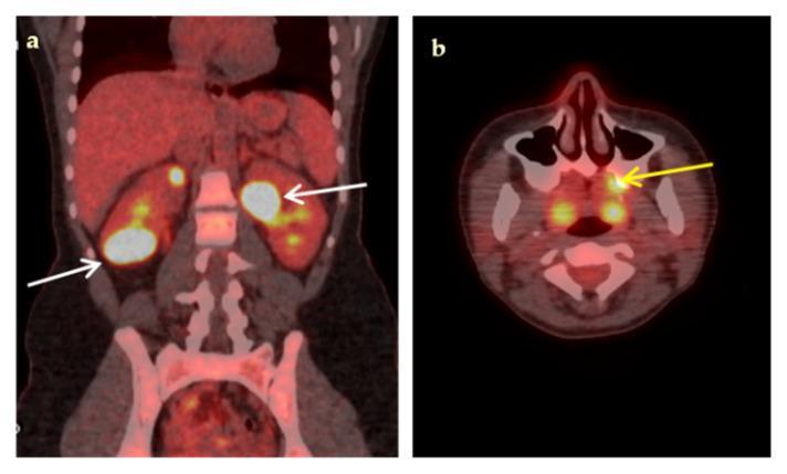
Fused 18F-FDG-PET/CT diagnostic imaging demonstrating high metabolic activity in a case of fever of unknown origin (FUO). Panel (a) is a coronal section showing bilateral, multiple, hypodense renal lesions with intense 18F-FDG uptake (high SUVmax). White arrows indicate prominent hypermetabolic foci in the lower pole of the right kidney and the upper pole of the left kidney, characteristic of renal abscesses. Panel (b) is a transaxial section of the head showing a localized area of high 18F-FDG uptake in the left maxilla (yellow arrow), indicating a primary dental infection or odontogenic source. The image illustrates the utility of PET/CT in identifying occult infectious sources, suggesting hematogenous spread from a dental focus to the kidneys. Key educational concepts include the identification of hypermetabolic inflammatory lesions and the diagnostic workup of multifocal infections using molecular imaging.
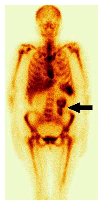
This diagnostic image is an anterior view of a whole-body scintigraphy scan performed on a 57-year-old female using 99mTc-labeled sulesomab (antigranulocyte monoclonal antibodies) to investigate fever of unknown origin. The scan demonstrates a normal physiological distribution of the radiotracer within the skeletal system, particularly in the axial skeleton, representing expected bone marrow activity. A prominent, localized pathological finding is visible in the lower abdomen: an area of intense radiotracer uptake (indicated by a black arrowhead) localized to the region of the ileum. This focal accumulation of granulocytes is highly suggestive of active inflammation, specifically characteristic of Crohn's disease in this clinical context. The scan provides essential diagnostic information by localizing inflammation to the distal small bowel when other clinical signs like abdominal pain are absent.
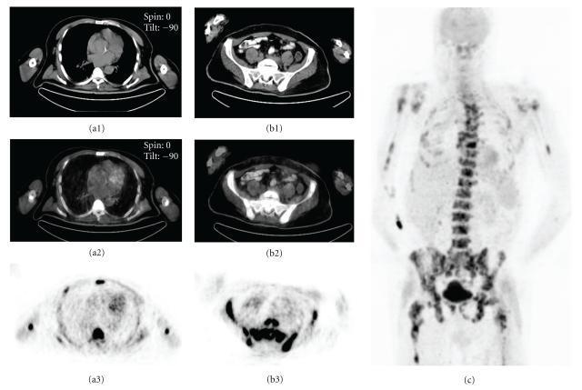
This diagnostic image set showcases a multi-modal PET/CT study of a 58-year-old male presenting with fever of unknown origin (FUO). The panel includes transaxial CT (a1, b1), fused PET/CT (a2, b2), and PET (a3, b3) slices at the thoracic and pelvic levels, alongside a full-body anterior Maximum Intensity Projection (MIP) image (c). The imaging demonstrates intense, diffuse FDG (fluorodeoxyglucose) uptake localized to the bone marrow compartments. The MIP image (c) highlights a prominent, systemic distribution of radiotracer accumulation throughout the axial skeleton, including the vertebrae and pelvis, and the proximal appendicular skeleton, specifically the humeri and femora. Notably, there is a lack of significant extraosseous metabolic activity. This visual pattern of skeletal FDG-avid disease is characteristic of hematologic malignancies involving the marrow. In this clinical context, the findings were diagnostic of non-Hodgkin's lymphoma. The comparison between modalities illustrates the utility of fusion imaging in providing precise anatomical localization of metabolic abnormalities identified on PET.
🌡️ Fever of Unknown Origin (FUO) - Complete Microbiology Notes for Exam
Sources: Harrison's Principles of Internal Medicine 22E | Goldman-Cecil Medicine | Frameworks for Internal Medicine
1. DEFINITION (Classic - Petersdorf & Beeson, 1961)
| Criterion | Threshold |
|---|---|
| Duration | Illness lasting ≥ 3 weeks |
| Temperature | Fever ≥ 38.3°C (101°F) on at least two occasions |
| Workup | No diagnosis despite 1 week of inpatient evaluation (or ≥3 outpatient visits / ≥3 days in hospital by modern criteria) |
Important: FUO excludes immunocompromised patients - they are a separate category requiring a more aggressive approach.
Exam tip: The 38.3°C threshold (not 38°C) and the 3-week duration are classic exam facts. Petersdorf & Beeson (1961) = the names behind the original definition.
2. CATEGORIES OF FUO (The "Big 4" + Undiagnosed)
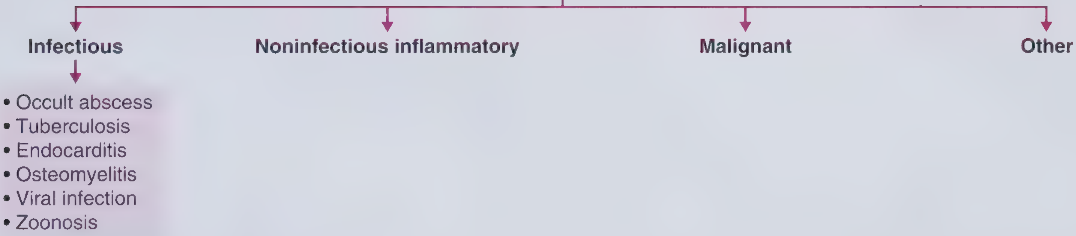
FUO Causes
├── 1. INFECTIOUS (~30-40%)
├── 2. NON-INFECTIOUS INFLAMMATORY/RHEUMATOLOGIC (~20-30%)
├── 3. MALIGNANT (~10-20%)
└── 4. MISCELLANEOUS/OTHER (~10-15%)
└── UNDIAGNOSED (~20-30%)
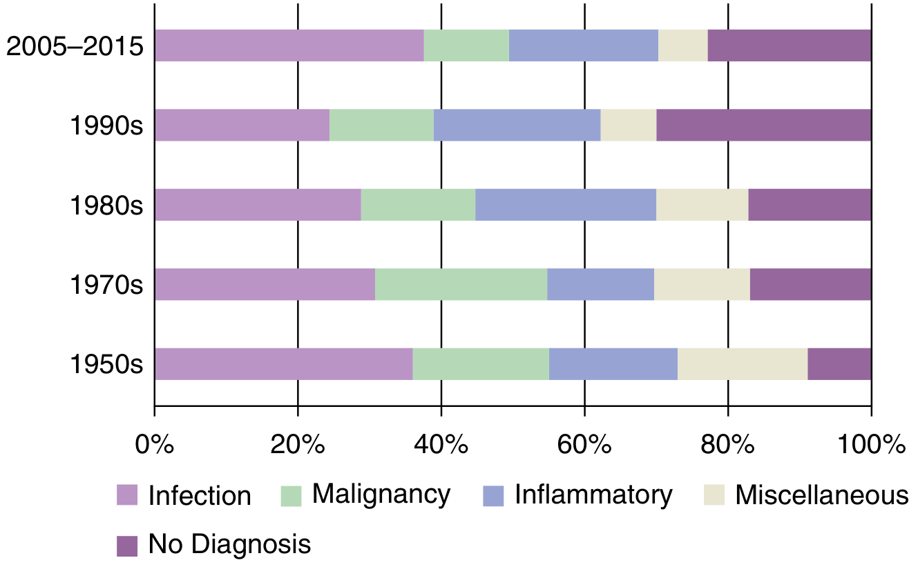
Trend: Infections remain the #1 cause, but as CT/echo/blood cultures improved, the proportion of "no diagnosis" and inflammatory causes has increased over decades.
3. INFECTIOUS CAUSES (Most Common Category)
General Principle
"Atypical presentations of COMMON diseases are more likely than common presentations of RARE diseases." - Goldman-Cecil
Infectious Causes Table
| COMMON | UNCOMMON/EXOTIC |
|---|---|
| Occult/hidden abscess | Bartonella spp. |
| Tuberculosis (especially miliary TB) | Brucella spp. |
| Endocarditis (especially culture-negative) | Hepatitis A, B, E |
| CMV (Cytomegalovirus) | Acute HIV infection |
| EBV (Epstein-Barr virus / Mono) | Salmonella spp. |
| Osteomyelitis | Urinary tract infection (occult) |
| Mycotic aneurysm | |
| Leptospirosis |
Key Infectious Causes - Detailed
A. Occult Abscess
- Intra-abdominal (liver, subphrenic, pelvic, pericolic) or retroperitoneal
- Key history: previous GI surgery, diverticulitis, Crohn's disease, recent procedure
- Diagnosis: CT abdomen/pelvis (investigation of choice)
- Treatment: drainage + antibiotics (antibiotics alone are NOT helpful for abscess)
B. Tuberculosis
- Can present as miliary TB (disseminated) - classic FUO cause
- Clinical clues: weight loss, night sweats, immigrants, HIV co-infection, hepatomegaly with micronodules on CT
- Miliary TB: small uniform lesions in lung/liver/spleen on imaging
- Diagnosis: IGRA (interferon-gamma release assay), Mantoux, biopsy (caseating granuloma)
C. Infective Endocarditis (IE)
- Especially culture-negative IE after prior antibiotics
- Clue: recent dental procedure, IV drug use, new cardiac murmur, night sweats
- Organisms causing culture-negative IE: Coxiella burnetii (Q fever), Bartonella, Brucella, HACEK
- Diagnosis: blood cultures (×3) + echocardiogram (TEE preferred)
D. CMV / EBV
- Young adults; mononucleosis syndrome
- Features: fever + lymphadenopathy + pharyngitis + splenomegaly + atypical lymphocytes
- EBV: Heterophile antibody (Monospot test) positive
- CMV: CMV IgM/IgG serology
E. Osteomyelitis
- Acquired via contiguous spread OR hematogenous seeding
- Often no obvious bone symptoms in FUO
- Diagnosis: MRI (most sensitive), bone scan, biopsy
4. NON-INFECTIOUS INFLAMMATORY CAUSES (~20-30%)
| Condition | Key Clue |
|---|---|
| Adult-onset Still's Disease (AOSD) | Ferritin dramatically elevated (often >2000 ng/mL), quotidian spiking fever, salmon-colored evanescent rash, arthritis |
| Rheumatoid Arthritis (RA) | Symmetric polyarticular arthritis - wrists, MCPs, PIPs; morning stiffness >1 hour |
| SLE | Young woman, pancytopenia, low complement, rash, renal disease |
| Giant Cell Arteritis (GCA) / Temporal Arteritis | Age >50, headache, jaw claudication, elevated ESR |
| Polymyalgia Rheumatica (PMR) | Associated with GCA; shoulder/hip girdle aching; elderly patient |
| Sarcoidosis | Bilateral hilar lymphadenopathy on CXR; elevated ACE; non-caseating granulomas |
| Inflammatory Bowel Disease (IBD) | Change in bowel habits; colonoscopy needed for diagnosis |
| Polyarteritis Nodosa (PAN) | Hepatitis B association; testicular pain; livedo reticularis; mononeuritis multiplex |
| Familial Mediterranean Fever (FMF) | Recurrent episodes since childhood; Mediterranean origin; abdominal pain + arthritis |
| Reactive Arthritis | Follows urethritis/GI infection; sterile inflammatory arthritis |
Exam pearl for AOSD: Quotidian fever (high fever spiking once or twice daily that returns to normal) + salmon rash (appears only during fever spikes) + serositis = classic AOSD triad. Ferritin >2000 ng/mL is highly suggestive.
5. MALIGNANT CAUSES (~10-20%)
| Tumor | Key Notes |
|---|---|
| Lymphoma (Hodgkin and Non-Hodgkin) | Most classic malignancy-causing FUO; "B symptoms" = fever + night sweats + weight loss |
| Leukemia | Pancytopenia + constitutional symptoms |
| Renal cell carcinoma | "Internist's tumor" - presents with FUO, hematuria, flank mass |
| Hepatocellular carcinoma | Liver mass + elevated AFP + hepatitis B/C background |
| Atrial myxoma | Cardiac tumor; migratory embolic events + constitutional symptoms |
| Colon adenocarcinoma | Occult GI malignancy |
| Multiple myeloma | Bone pain, anemia, renal failure, hypercalcemia |
| Castleman disease | Rare lymph node disorder causing FUO |
High-yield: Lymphoma (especially Hodgkin's) is the most classic malignant cause of FUO. Look for "B symptoms" in exam scenarios.
6. MISCELLANEOUS / OTHER CAUSES
| Cause | Key Clue |
|---|---|
| Drug fever | Fever appears after starting a drug; no other explanation; resolves when drug stopped. Antibiotics, anticonvulsants, allopurinol common culprits |
| Factitious fever | No physiologic signs of fever; pattern inconsistent; often healthcare workers |
| Pulmonary embolism / DVT (chronic) | Immobility, long-distance travel, hypercoagulable state |
| Hyperthyroidism / Thyroiditis | Tachycardia, weight loss, tremor; subacute thyroiditis has tender thyroid |
| Hematoma (resolving) | Post-trauma or post-surgical |
| Hemophagocytic Lymphohistiocytosis (HLH) | Cytopenia + ferritin >500 ng/mL + splenomegaly; triggered by infection/malignancy/autoimmune |
| Pheochromocytoma | Episodic hypertension, headache, palpitations, sweating |
| Periodic fever syndromes | Recurrent fever since childhood (PFAPA, FMF, etc.) |
| Hypoadrenalism (Addison's disease) | Fatigue, hyperpigmentation, hypotension, hyponatremia |
7. SPECIAL TYPES OF FUO (Know for Exam)
| Type | Definition | Key Causes |
|---|---|---|
| Classic FUO | ≥38.3°C, ≥3 weeks, no diagnosis after workup | Infections, inflammatory, malignancy |
| Nosocomial FUO | Hospitalized patient, fever develops after 24-48h, ≥38.3°C | Catheter-related BSI, C. difficile, DVT/PE, drug fever, sinusitis |
| Neutropenic FUO | ANC <500, ≥38.3°C, ≥3 days, no diagnosis | Gram-negative bacteremia, fungal (Candida, Aspergillus) |
| HIV-associated FUO | HIV patient, ≥38.3°C, ≥4 weeks outpatient OR ≥3 days inpatient | MAI (Mycobacterium avium-intracellulare), CMV, PCP, Toxoplasma, lymphoma |
8. DIAGNOSTIC APPROACH - "LOOK FOR PDCs"
Step 1 - History (Must-Ask Questions)
| Category | Questions |
|---|---|
| Fever pattern | Continuous vs. recurrent? Duration? Temperature spikes? |
| Travel | Recent/remote travel? Malaria zones? TB-endemic regions? |
| Animal contact | Zoonosis? Brucella (farm animals), Q fever (sheep), Toxoplasma (cats), Leptospirosis (water/rodents) |
| Occupation | Healthcare worker, farmer, veterinarian |
| Sexual history | HIV risk, gonorrhea, syphilis |
| Drugs | All current medications (drug fever) |
| Family history | Periodic fever syndromes (FMF) |
| Prior infections | Diverticulitis (abscess risk), recent dental (IE), TB exposure |
| Immunosuppression | Steroids, chemotherapy, HIV |
Step 2 - Physical Examination (Focus Areas)
| Area | What to Look For |
|---|---|
| Eyes | Uveitis (sarcoid, TB), Roth spots (IE), jaundice |
| Lymph nodes | Lymphadenopathy (lymphoma, TB, HIV, EBV) |
| Temporal arteries | Tenderness (GCA) |
| Skin | Rash (AOSD salmon rash, SLE butterfly, drug reaction) |
| Liver/spleen | Hepatosplenomegaly (EBV, malaria, miliary TB) |
| Joints | Arthritis pattern |
| Heart | New murmur (IE) |
| Previous surgery sites | Wound infection, abscess |
Step 3 - Initial (Obligatory) Laboratory Tests
| Test | Reason |
|---|---|
| CBC + WBC differential | Leukocytosis (infection), eosinophilia (parasites), pancytopenia (SLE, leukemia) |
| CRP + ESR | Markers of inflammation |
| LFTs, renal function | Liver/kidney disease, hepatitis |
| Urinalysis + culture | UTI, renal disease |
| Blood cultures ×3 | Endocarditis, bacteremia (spaced >12 hours apart) |
| HIV Ag/Ab (4th gen) | Acute HIV |
| CMV IgM/IgG | CMV infection |
| EBV (Monospot/heterophile Ab) | EBV mononucleosis |
| IGRA (Interferon-gamma release assay) | Tuberculosis |
| Chest X-ray | TB, lymphoma, sarcoid, occult malignancy |
| Abdominal ultrasound | Abscess, hepatosplenomegaly, masses |
| LDH + ferritin | Malignancy (LDH), AOSD/HLH (ferritin) |
| ANA, RF, complement | Connective tissue disease screening |
Step 4 - Advanced Imaging
| Test | Indication / Yield |
|---|---|
| CT chest/abdomen/pelvis | Occult abscess, lymphadenopathy, masses, miliary TB |
| 18F-FDG PET/CT | #1 advanced imaging for FUO - detects metabolically active foci in 30-60% of cases; can diagnose large vessel vasculitis, guide biopsy sites |
| Echocardiogram (TEE) | Suspected IE, atrial myxoma |
| MRI | Osteomyelitis (most sensitive), spinal abscess |
| Gallium scintigraphy | Alternative to PET/CT if not available |
Step 5 - Tissue Biopsy (Guided by Imaging)
- Lymph node biopsy (lymphoma, TB, sarcoid)
- Liver biopsy (granulomatous hepatitis, miliary TB)
- Bone marrow biopsy (hematologic malignancy, miliary TB, fungal)
- Temporal artery biopsy (GCA in elderly)
9. TREATMENT PRINCIPLES
| Scenario | Approach |
|---|---|
| Critically ill | Empiric broad-spectrum antibiotics while workup continues |
| Stable patient | Complete diagnostic workup FIRST before empiric treatment |
| Before testing | Stop antibiotics and steroids (they mask causes; blood cultures unreliable during antibiotic use) |
| If no diagnosis found | Watchful waiting with repeat evaluations; consider autoinflammatory consultation |
| Trial of NSAIDs/steroids | Only after infections and malignancy have been excluded |
Critical exam principle: Do NOT start empiric antibiotics or steroids in a stable FUO patient before completing the workup - this masks diagnoses!
10. QUICK EXAM SUMMARY TABLE
| Feature | Detail |
|---|---|
| Definition | ≥38.3°C on ≥2 occasions, ≥3 weeks duration, no diagnosis after workup |
| Original description | Petersdorf & Beeson, 1961 |
| #1 overall cause | Infection (most common in all studies) |
| Most classic infectious cause | TB (miliary) / occult abscess / culture-negative IE |
| #1 malignant cause | Lymphoma (Hodgkin's / Non-Hodgkin's) |
| Most classic inflammatory cause | Adult-onset Still's disease (AOSD) |
| Key marker for AOSD | Ferritin >2000 ng/mL |
| Best advanced imaging | 18F-FDG PET/CT (detects cause in 30-60%) |
| Blood cultures | 3 cultures, spaced >12 hours apart |
| Streptococcal link to FUO | Culture-negative IE after dental procedure / prior antibiotics |
| Undiagnosed FUO prognosis | Usually benign - resolves spontaneously in many cases |
11. COMMON EXAM VIGNETTE CLUES
| Clue in Question Stem | Think... |
|---|---|
| Diverticulitis 6 weeks ago + fever | Intra-abdominal abscess |
| Emigrant from TB-endemic country + hepatomegaly + micronodules | Miliary tuberculosis |
| Fever after dental procedure + diastolic murmur | Culture-negative IE |
| Fever + pharyngitis + lymphadenopathy + splenomegaly + atypical lymphs | EBV (mono) |
| Salmon rash + quotidian fever + ferritin >2000 | Adult-onset Still's disease |
| Elderly woman + headache + jaw claudication + high ESR | Giant cell arteritis |
| Young woman + pancytopenia + low C3/C4 | SLE |
| Bilateral hilar lymphadenopathy + elevated ACE | Sarcoidosis |
| "B symptoms" + lymphadenopathy in young adult | Hodgkin's lymphoma |
| Hepatitis B + testicular pain + livedo reticularis | Polyarteritis nodosa |
| Recurring fever since childhood + Mediterranean origin | Familial Mediterranean fever |
| Fever starts after new medication | Drug fever |
| Farm worker/vet + undulant fever + splenomegaly | Brucella infection |
| Water exposure + conjunctival suffusion + AKI + liver injury | Leptospirosis |
| HIV patient + fever + weight loss | MAC (Mycobacterium avium complex) |
teach me about streptococci microbiology with easy and short notes but should cover all important and must know points to crack university exam
streptococcus gram positive cocci chains blood agar hemolysis classification
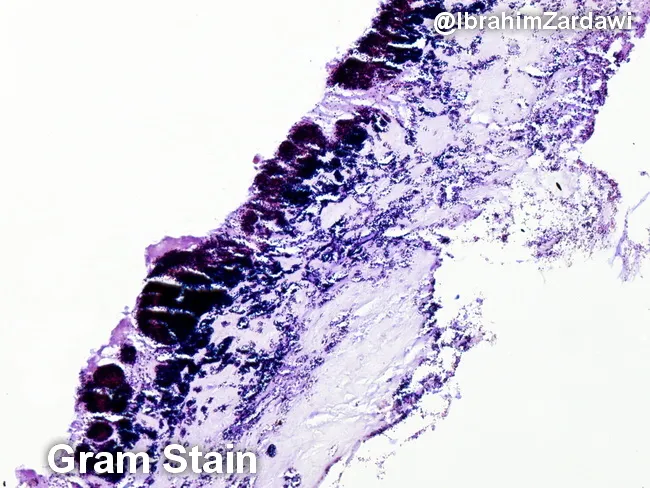
This histology image is a light-micrograph of a Gram-stained valvular vegetation from a case of infective endocarditis. The specimen is cardiac valvular tissue; the vegetation comprises a fibrin-rich, platelet-laden thrombus with embedded bacterial colonies along the endocardial surface. Gram staining highlights Gram-positive cocci as purple to deep purple clusters within the thrombus, indicating organisms such as Streptococcus spp., Staphylococcus spp., or Enterococcus. The bacterial colonies are adherent to the valvar surface within a dense inflammatory milieu, with neutrophilic infiltrate and tissue necrosis limited to microdomains. The histologic architecture demonstrates vegetations typical of endocarditis: a friable, topographically irregular lesion on the valve with microabscess-like foci and bacterial encasement within fibrin. The presence of Gram-positive bacteria on histology supports a bacterial endocarditis diagnosis and guides antimicrobial therapy pending culture confirmation. Clinically, such histologic confirmation is critical for distinguishing infectious from non-infectious valve pathology, for clarifying the etiologic category of pathogens (oral flora-related viridans Streptococcus, Staphylococcus aureus, enterococci), and for correlating with blood culture, echocardiographic findings, and patient risk factors. This image is valuable for educational purposes, illustrating Gram stain morphology in endocardial vegetations and reinforcing the need for targeted antibiotics and possible surgical consultation.
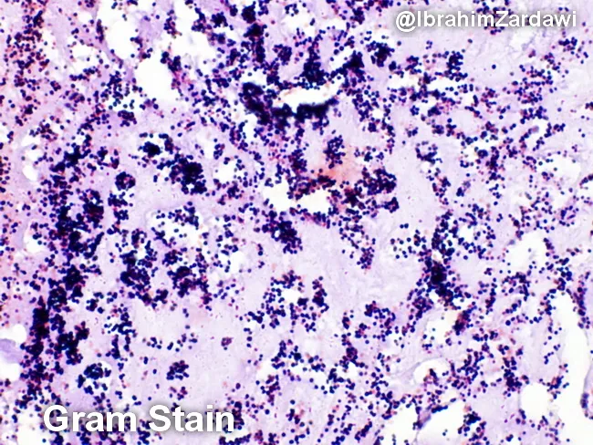
Imaging modality: Light microscopy; Gram staining of a histologic section of a cardiac valve vegetation from suspected infective endocarditis. The specimen shows a dense, friable vegetative mass within valve tissue characterized by a fibrinous, platelet-rich matrix in which numerous Gram-positive cocci are adherent. On the Gram stain, the cocci appear as purple spherical cells arranged in clusters and sometimes in short chains, consistent with Gram-positive organisms. The background demonstrates inflammatory cells and debris typical of endocardial infection. The histologic architecture indicates vegetative lesions with microbial colonization and a biofilm component. These findings correlate with infective endocarditis due to cocci such as Staphylococcus aureus or viridans-group streptococci in clinical practice, though Gram stain alone cannot specify species. The image is a micrograph provided with permission by Dr. Ibrahim Zardawi; image shows classic histopathology of bacterial endocarditis. The diagnostic significance lies in confirming bacterial endocarditis and guiding immediate management including empiric antibiotic therapy targeting Gram-positive cocci while awaiting culture results. This image is valuable for pathology education, microbiology correlation, and endocarditis research. Potential clinical use cases include training in Gram stain interpretation, distinguishing bacterial endocarditis from sterile vegetations, and supporting diagnosis when blood cultures are pending.
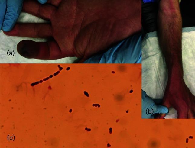
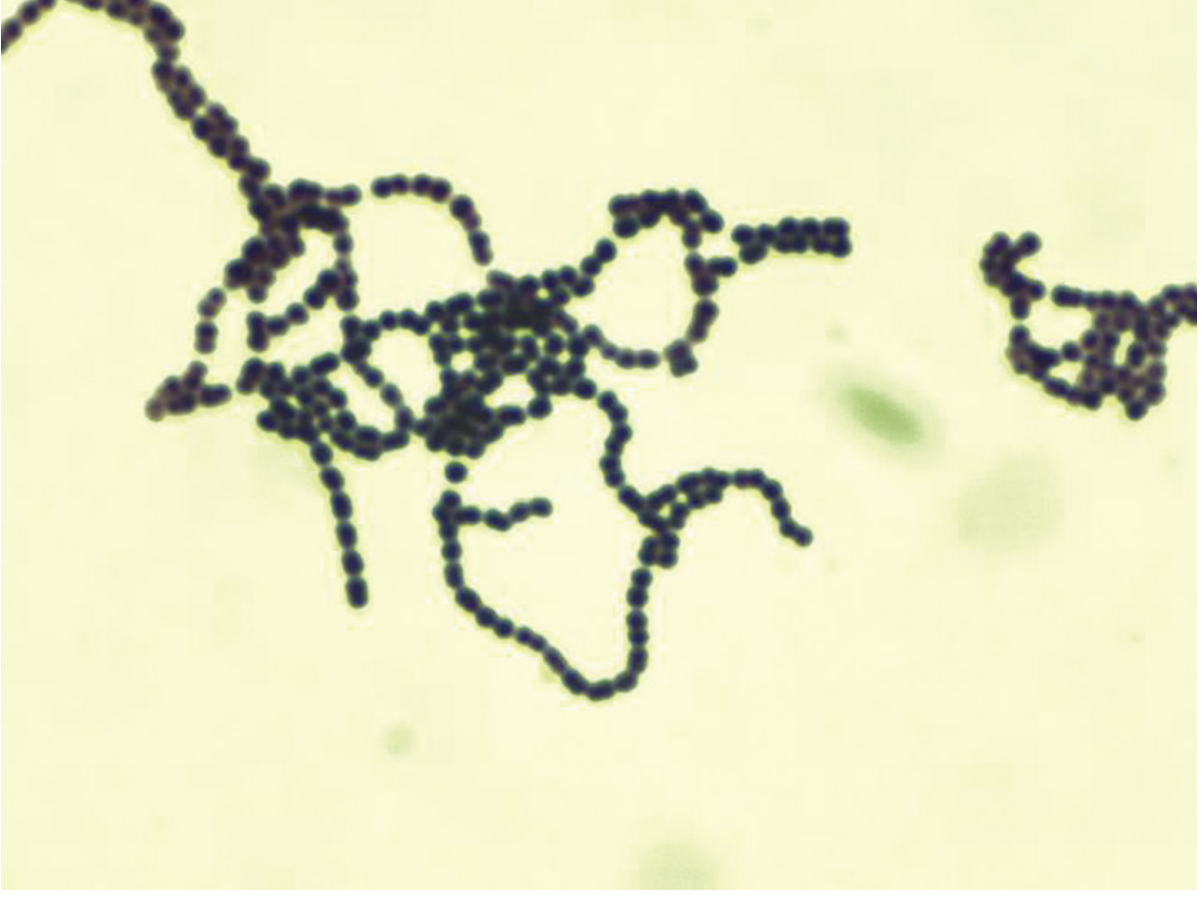
This composite image consists of three panels demonstrating clinical and microbiological findings of a severe bacterial infection. Panel (a) is a clinical photograph of a patient's hand showing a swollen, necrotic left index fingertip with distinct dark, gangrenous discoloration. Panel (b) shows the patient's forearm with visible erythematous linear streaking extending proximally, characteristic of lymphangitis. Panel (c) is a high-power light microscopy image of a Gram-stained sample, revealing numerous Gram-positive cocci. The organisms are round in morphology and predominantly arranged in long chains, which is a classic feature of Streptococcus species. Together, these images illustrate the progression from a localized soft tissue infection (such as digital necrosis or paronychia) to lymphatic involvement and the identification of the causative chain-forming Gram-positive pathogen, often associated with Streptococcus pyogenes.
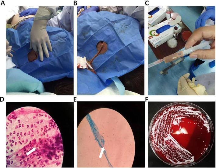
A six-panel figure documenting the clinical management and microbiological diagnosis of a subcutaneous back abscess, indicative of disseminated nocardiosis. Panels A and B show intraoperative clinical photographs of the incision and subsequent drainage of the abscess site on the patient's back, which is draped in sterile blue material. Panel C displays a syringe containing aspirated greyish-white, turbid purulent fluid. Panel D presents a Gram stain micrograph showing a dense inflammatory infiltrate composed predominantly of neutrophils (pink) and scattered gram-positive cocci/branching filaments (purple, white arrow). Panel E demonstrates a weak acid-fast stain (Kinyoun method) identifying characteristic blue-stained filamentous bacteria (white arrow), a hallmark of Nocardia species. Panel F shows a Columbia Blood Agar plate after incubation, exhibiting confluent growth of small, white to off-white bacterial colonies with evidence of beta-hemolysis. This composite illustrates the diagnostic pathway from clinical intervention to specialized microbiological staining and culture for identifying infectious pathogens.
🧫 Streptococci - Complete Microbiology Notes for Exam
Sources: Medical Microbiology 9e | Goldman-Cecil Medicine | Sherris & Ryan's Medical Microbiology | Harrison's Principles of Internal Medicine 22E
1. GENERAL CHARACTERISTICS OF STREPTOCOCCI
| Property | Detail |
|---|---|
| Gram stain | Gram-positive cocci in chains (or pairs) |
| Catalase | Negative (differentiates from Staphylococcus which is catalase-positive) |
| Oxygen | Facultative anaerobes |
| Habitat | Skin and mucous membranes (normal flora) |
| Classification | Based on hemolysis on blood agar AND Lancefield grouping |
2. CLASSIFICATION SYSTEMS
A. By Hemolysis on Blood Agar
| Hemolysis | Description | Examples |
|---|---|---|
| Beta (β) | Complete lysis of RBCs → clear zone around colony | GAS (S. pyogenes), GBS (S. agalactiae) |
| Alpha (α) | Partial/incomplete lysis → green/brown discoloration (H₂O₂ oxidizes Hb to methemoglobin) | S. pneumoniae, Viridans streptococci |
| Gamma (γ) | No hemolysis | Enterococci |
B. Lancefield Classification (Based on cell wall C-carbohydrate antigens)
| Lancefield Group | Species | Key Diseases |
|---|---|---|
| Group A | S. pyogenes | Pharyngitis, skin infections, RF, PSGN |
| Group B | S. agalactiae | Neonatal sepsis/meningitis |
| Group C, G | S. dysgalactiae | Pharyngitis (like GAS), septicemia |
| Group D | Enterococcus (E. faecalis, E. faecium) | UTI, endocarditis |
| No group | S. pneumoniae, Viridans | Pneumonia, meningitis, IE |
Key point: Viridans streptococci and S. pneumoniae have NO Lancefield group - identified by other methods.
3. STREPTOCOCCUS PYOGENES (Group A Streptococcus - GAS)
Basic Microbiology
| Feature | Detail |
|---|---|
| Lancefield group | A |
| Hemolysis | Beta (β) - complete |
| Gram stain | Gram-positive cocci in chains |
| Catalase | Negative |
| Key identifier | Bacitracin sensitive (inhibited by bacitracin disk) |
| Key test | PYR positive (L-pyrrolidonyl arylamidase) |
| M protein types | >150 types; type-specific immunity |
| Human host | Humans are the only reservoir |
Virulence Factors - THE MOST TESTED SECTION 🔑
A. Surface Structures (Anti-phagocytic)
| Factor | Function |
|---|---|
| M protein | #1 virulence factor; anti-phagocytic (blocks C3b binding); mediates adherence; >150 types; basis of type-specific immunity; cross-reacts with heart/kidneys → RF, PSGN |
| Hyaluronic acid capsule | Antiphagocytic; resembles human tissues (poor immunogen); binds CD44 on epithelial cells |
| C5a peptidase | Degrades C5a (chemotactic complement fragment) → blocks neutrophil recruitment |
| Lipoteichoic acid | Adherence to fibronectin on epithelial cells |
| Protein F (fibronectin-binding protein) | Adherence to host extracellular matrix |
| M-like proteins | Bind Fc of antibodies → block complement activation |
B. Toxins and Enzymes
| Toxin/Enzyme | Function | Clinical Relevance |
|---|---|---|
| Streptolysin O (SLO) | Oxygen-labile; cholesterol-dependent cytolysin; forms pores in membranes; causes β-hemolysis | Antigenic → ASO titer used for diagnosis of RF/PSGN |
| Streptolysin S (SLS) | Oxygen-stable; also causes β-hemolysis | Not antigenic |
| Streptococcal Pyrogenic Exotoxins (SPE A, B, C) | Superantigens; stimulate massive T-cell activation → cytokine storm → TSS; also cause scarlet fever rash | Scarlet fever, Streptococcal TSS |
| Streptokinase (fibrinolysin) | Dissolves fibrin clots; helps spread of infection | Therapeutic use (thrombolytics) |
| DNase B (streptodornase) | Degrades DNA in pus; liquefies thick exudate | Anti-DNase B titer useful for skin infections |
| Hyaluronidase | Breaks down hyaluronic acid → spreads through tissues | "Spreading factor" |
| C5a peptidase | Inactivates complement chemoattractant C5a | Evades innate immunity |
| Streptokinase / DNase / Hyaluronidase | Collectively called "spreading factors" | Facilitate tissue invasion |
Mnemonic for GAS toxins: "S-S-S-D-H" = Streptolysin O, Streptolysin S, SPE (scarlet fever toxin), DNase B, Hyaluronidase
Epidemiology of GAS
- Peak age: Children 5-15 years (pharyngitis); children 2-5 years (pyoderma)
- Transmission: respiratory droplets (pharyngitis); direct contact/breaks in skin (skin infections)
- Carrier state: 15-20% children; <5% adults
- Season: winter months in temperate climates
- Crowding (schools, military camps) increases spread
4. DISEASES CAUSED BY GAS (Group A Strep)
SUPPURATIVE (Direct bacterial invasion - organisms present at site)
| Disease | Clinical Features | Notes |
|---|---|---|
| Pharyngitis | Sore throat, exudates, red pharynx, cervical lymphadenopathy | Most common GAS disease; trigger for RF |
| Scarlet Fever | Diffuse erythematous rash (sandpaper texture), starts on chest → extremities; "strawberry tongue"; pastia lines (rash in skin folds) | Due to SPE (pyrogenic exotoxins A/B/C); complication of pharyngitis |
| Impetigo / Pyoderma | Vesicles → pustules → honey-crusted lesions; localized skin; no systemic symptoms | Especially in children <5; can trigger PSGN (NOT RF) |
| Erysipelas | Superficial skin infection with sharp, raised, well-demarcated borders; lymph node enlargement; systemic symptoms | Face most common; bright red, brawny induration |
| Cellulitis | Diffuse skin + subcutaneous tissue infection; poorly demarcated | Less distinct border than erysipelas |
| Necrotizing Fasciitis | "Flesh-eating bacteria"; deep infection destroying muscle and fat | Surgical emergency; very high mortality; "gas in soft tissues" on X-ray |
| Streptococcal Toxic Shock Syndrome (STSS) | Multiorgan failure, hypotension, shock | Most patients are bacteremic; often with fasciitis; SPE superantigens → cytokine storm |
| Puerperal Sepsis | Post-partum fever and sepsis | Historical cause of maternal mortality |
| Pneumonia / Bacteremia | Uncommon | Seen in hospitalized patients |
NON-SUPPURATIVE (Post-infectious; immune-mediated; NO organisms at site of damage)
| Complication | Trigger | Mechanism | Key Feature |
|---|---|---|---|
| Rheumatic Fever (RF) | Pharyngitis only | Molecular mimicry (M protein cross-reacts with heart, joints) | Follows pharyngitis ONLY; recurs with re-infection |
| Post-Streptococcal Glomerulonephritis (PSGN) | Pharyngitis OR skin infection | Immune complex deposition (Type III) | Different nephritogenic M types; does NOT follow skin infection to cause RF |
5. DIAGNOSIS OF GAS INFECTIONS
| Test | Purpose |
|---|---|
| Gram stain | Useful in soft-tissue infections (chains of gram+ cocci) |
| Rapid Streptococcal Antigen Test (RSAT) | Quick bedside test for pharyngitis; detects Group A antigen |
| Throat culture | Gold standard for pharyngitis; 24-48h |
| Bacitracin disk | GAS is sensitive (inhibited) - used to differentiate GAS from other strep |
| PYR test | GAS = positive (also positive in Enterococcus) |
| ASO titer | Elevated in recent GAS pharyngitis; confirms RF or PSGN after pharyngitis |
| Anti-DNase B titer | More sensitive for PSGN after skin infections (anti-DNase B detects skin GAS better) |
| Blood culture | For invasive/systemic disease |
6. TREATMENT OF GAS
| Situation | Antibiotic |
|---|---|
| Pharyngitis (mild) | Penicillin V or Amoxicillin (10 days) |
| Penicillin allergy | Oral cephalosporin or Macrolide (azithromycin) |
| Systemic/severe infections | IV Penicillin G + Clindamycin (clindamycin inhibits toxin production) |
| Necrotizing fasciitis | IV Penicillin G + Clindamycin + surgical debridement |
| STSS | IV Penicillin G + Clindamycin + IVIG |
Note: GAS has never developed resistance to penicillin - penicillin remains the drug of choice.
7. STREPTOCOCCUS AGALACTIAE (Group B Streptococcus - GBS)
Basic Microbiology
| Feature | Detail |
|---|---|
| Lancefield group | B |
| Hemolysis | Beta (β) |
| Key identifier | CAMP test positive (produces CAMP factor that enhances lysis by S. aureus - produces a characteristic arrowhead/flame-shaped hemolysis) |
| Habitat | Normal flora of vagina, GI tract |
| Serotypes | 5 major serotypes (Ia, Ib, II, III, V); Type III = neonatal meningitis |
Diseases Caused by GBS
| Population | Disease | Notes |
|---|---|---|
| Neonates - Early onset (<7 days of birth) | Pneumonia, sepsis, meningitis | Acquired from maternal birth canal; very serious |
| Neonates - Late onset (>1 week) | Bacteremia + meningitis | Serotype III most common |
| Pregnant women | Postpartum endometritis, wound infections, UTI, bacteremia | Vaginal carriage 15-25% |
| Adults (non-pregnant) | Bacteremia, pneumonia, bone/joint infections, skin infections | Increasingly recognized in diabetic, elderly patients |
Prevention of Neonatal GBS
- Screen all pregnant women at 35-37 weeks gestation with vaginal/rectal cultures
- If GBS positive (or risk factors): Intrapartum antibiotic prophylaxis (IAP) = IV Penicillin G during labor
- Risk factors: previous GBS baby, GBS bacteriuria in current pregnancy, preterm labor, prolonged membrane rupture, fever during labor
8. STREPTOCOCCUS PNEUMONIAE (Pneumococcus)
Basic Microbiology
| Feature | Detail |
|---|---|
| Lancefield group | None |
| Hemolysis | Alpha (α) - partial (green) |
| Gram stain | Gram-positive cocci in pairs (diplococci) and short chains; elongated/lancet-shaped |
| Key identifiers | Optochin sensitive (inhibited by optochin disk); Bile soluble (lysed by bile salts/sodium deoxycholate); Quellung reaction positive |
| Capsule | Polysaccharide capsule - #1 virulence factor; >100 serotypes; antiphagocytic |
| Catalase | Negative |
Virulence Factors of S. pneumoniae
| Factor | Function |
|---|---|
| Polysaccharide capsule | Anti-phagocytic; basis of vaccine; >100 serotypes |
| Pneumolysin | Cytotoxin; damages host cell membranes; stimulates inflammation |
| IgA protease | Destroys secretory IgA on mucous membranes → colonization |
| Teichoic acid / peptidoglycan | Triggers local inflammatory response |
| Surface protein adhesins | Helps colonize oropharynx |
Diseases Caused by Pneumococcus
| Disease | Clinical Features |
|---|---|
| Lobar Pneumonia | Sudden onset, severe chills, sustained fever, productive cough with blood-tinged/rusty sputum; lobar consolidation on CXR |
| Meningitis | Severe headache, fever, neck stiffness, altered sensorium; high mortality; serious neurological sequelae in survivors |
| Otitis Media | Most common cause of otitis media in children |
| Sinusitis | Common bacterial cause |
| Bacteremia | More common in meningitis than pneumonia; overwhelming fulminant sepsis in asplenic patients |
Special risk groups: Asplenic patients (sickle cell, post-splenectomy), elderly, children <2 years, HIV patients, alcoholics → high risk for invasive pneumococcal disease
Diagnosis of Pneumococcus
| Test | Notes |
|---|---|
| Gram stain | Gram-positive lancet-shaped diplococci |
| Culture | Blood agar (enriched media); optochin disk → zone of inhibition |
| Optochin test | Sensitive = S. pneumoniae (Viridans strep = resistant) |
| Bile solubility | Pneumococcus lysed by bile; Viridans strep = NOT lysed |
| Quellung reaction | Capsule swells with specific antibody (typing test) |
| Urinary antigen test | Detects pneumococcal C-polysaccharide in urine (meningitis diagnosis) |
| Blood culture | For bacteremia |
Treatment of Pneumococcus
| Situation | Antibiotic |
|---|---|
| Susceptible strains | Penicillin G (DOC) |
| Empiric / resistant | Ceftriaxone + Vancomycin (especially meningitis) |
| Outpatient pneumonia | Amoxicillin or respiratory fluoroquinolone |
| Meningitis | Ceftriaxone + Vancomycin (until sensitivities known) |
Vaccines
| Vaccine | Type | Who Gets It |
|---|---|---|
| PCV13 (13-valent conjugate) | Protein conjugate | All children <2 years; immunocompromised adults |
| PPSV23 (23-valent polysaccharide) | Plain polysaccharide | Adults >65; asplenic patients; high-risk groups |
| PCV15 / PCV20 | Newer conjugates | Replacing older vaccines in adults |
9. VIRIDANS STREPTOCOCCI
Overview
- Group of alpha-hemolytic streptococci
- No Lancefield group
- Normal oral, GI, genitourinary flora
- Low-virulence organisms
Key Species and Diseases
| Species | Disease |
|---|---|
| S. sanguis, S. mitis, S. salivarius, S. mutans | Subacute infective endocarditis (after dental procedures) |
| S. mutans | Dental caries (ferments sucrose; produces acid; adheres to tooth) |
| S. anginosus group (S. intermedius, S. anginosus) | Deep tissue abscesses (brain, liver, lung) |
| S. gallolyticus (formerly S. bovis) | IE + associated with colorectal carcinoma |
Key ID Tests for Viridans vs. Pneumococcus
| Test | Viridans Streptococci | S. pneumoniae |
|---|---|---|
| Hemolysis | Alpha (green) | Alpha (green) |
| Optochin | Resistant (no inhibition) | Sensitive (inhibited) |
| Bile solubility | Not soluble (no lysis) | Soluble (lysed) |
| Lancefield group | None | None |
10. ENTEROCOCCUS (Formerly Group D Streptococcus)
Now classified as its own genus (Enterococcus), but often tested alongside streptococci.
| Feature | Detail |
|---|---|
| Species | E. faecalis (most common), E. faecium |
| Lancefield group | D |
| Hemolysis | Gamma (γ) - none |
| Habitat | Normal GI flora |
| Key property | Grows in 6.5% NaCl and bile-esculin medium (distinguishes from Group D streptococci) |
| Resistance | Intrinsic resistance to cephalosporins; low-level aminoglycoside resistance |
| VRE | Vancomycin-Resistant Enterococcus = major nosocomial threat |
Diseases
| Disease | Notes |
|---|---|
| UTI | Most common; especially with indwelling catheters + broad-spectrum antibiotics |
| Endocarditis | ~10% of IE; GU/GI source; E. faecalis mostly |
| Bacteremia | Hospital-acquired |
| Peritonitis | After abdominal surgery/trauma (polymicrobial) |
11. COMPLETE COMPARISON TABLE (Exam Must-Know) 🔑
| Feature | GAS (S. pyogenes) | GBS (S. agalactiae) | S. pneumoniae | Viridans Strep | Enterococcus |
|---|---|---|---|---|---|
| Lancefield group | A | B | None | None | D |
| Hemolysis | Beta | Beta | Alpha | Alpha | Gamma |
| Key test | Bacitracin sensitive; PYR+ | CAMP test + | Optochin sensitive; Bile soluble | Optochin resistant; bile insoluble | 6.5% NaCl growth; bile esculin + |
| Main disease | Pharyngitis, skin, RF | Neonatal sepsis | Pneumonia, meningitis | IE (dental) | UTI, IE |
| Toxins | SLO, SLS, SPE | - | Pneumolysin | - | - |
| Treatment | Penicillin | Penicillin | Penicillin/Ceftriaxone+Vanco | Penicillin | Ampicillin + Gentamicin |
| Vaccine | No | IAP for mothers | PCV13, PPSV23 | No | No |
12. QUICK MEMORY AIDS
Hemolysis Pattern
Beta = BLOODY = GAS (pyogenes) + GBS (agalactiae)
Alpha = GREEN = Pneumococcus + Viridans (partial = partial green)
Gamma = NOTHING = Enterococcus
Key Discriminating Tests
PYR positive → GAS (or Enterococcus)
Bacitracin sensitive → GAS
CAMP test positive → GBS
Optochin sensitive + Bile soluble → S. pneumoniae
Optochin resistant + Bile insoluble → Viridans
6.5% NaCl growth → Enterococcus
GAS Disease Associations
| Trigger Words | Think GAS Disease |
|---|---|
| Pharyngitis + rash + strawberry tongue | Scarlet fever |
| Honey-crusted skin lesion | Impetigo (pyoderma) |
| Sharply demarcated red face | Erysipelas |
| "Flesh-eating bacteria" + surgical emergency | Necrotizing fasciitis |
| Cytokine storm + bacteremia | STSS |
| 2 weeks after pharyngitis + arthritis + carditis | Rheumatic fever |
| Hematuria + hypertension after skin infection | PSGN |
| ASO titer elevated | Recent GAS pharyngitis |
| Anti-DNase B elevated | GAS pharyngitis or skin infection |
enteric fever microbiology short notes to top university exam
typhoid fever rose spots Salmonella typhi pathology
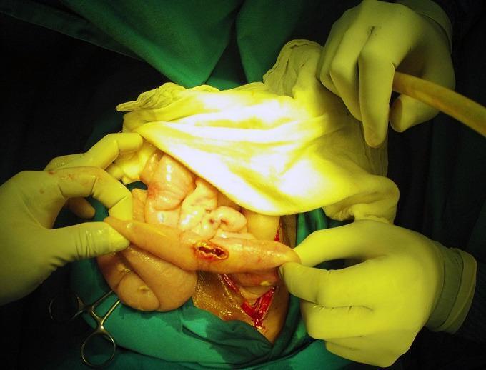
This clinical photograph shows an intraoperative view of an exploratory laparotomy, demonstrating a bowel perforation in the context of typhoid fever (Salmonella Typhi). The image focuses on a loop of small intestine held by a surgeon's gloved hands, revealing a discrete, longitudinal perforation with hyperemic and slightly everted edges. The surrounding serosa exhibits generalized inflammation and congestion. In the background, other segments of the intestine appear edematous and pale-pink, with visible white-yellowish peritoneal nodules and purulent exudate suggestive of concurrent miliary peritoneal tuberculosis and secondary peritonitis. Surgical instruments, suction tubing, and green sterile drapes define the surgical field. This image serves as an educational example of the severe gastrointestinal complications of typhoid fever, specifically intestinal perforation, and illustrates the challenging clinical scenario of co-infection with tuberculosis in an acute surgical setting.
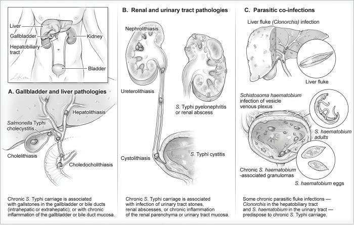
This medical illustration depicts anatomical locations and pathological conditions associated with chronic Salmonella Typhi carriage. The graphic is divided into three sections: (A) Gallbladder and liver pathologies, showing Hepatolithiasis, S. Typhi cholecystitis, Cholelithiasis, and Choledocholithiasis, illustrating how stones in the biliary system provide a niche for bacterial persistence. (B) Renal and urinary tract pathologies, highlighting Nephrolithiasis, Ureterolithiasis, Cystolithiasis, and S. Typhi-associated pyelonephritis or cystitis, where urinary stones or abscesses facilitate chronic carriage. (C) Parasitic co-infections, demonstrating how Liver flukes (Clonorchis) and Schistosoma haematobium (showing adults, eggs, and bladder granulomas) predispose patients to chronic infection. The diagram effectively connects infectious disease, pathology, and structural abnormalities of the hepatobiliary and urogenital systems, serving as an educational resource for understanding the reservoirs of chronic typhoid carriers.
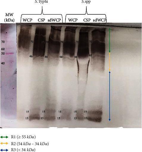
A diagnostic Western blot (WB) analysis image showing the immunoreactivity of antigens derived from Salmonella typhi and Salmonella spp. The image displays six experimental lanes representing three different protein extraction methods—Whole Cell Protein (WCP), Cell Surface Protein (CSP), and sodium deoxycholate-extracted WCP (sdWCP)—for each bacterial species. A molecular weight (MW) marker lane on the far left, stained with Ponceau S, shows reference bands from 40 kDa to 70 kDa. The WB profile is categorized into three vertical regions: R1 (≥ 55 kDa), R2 (54–34 kDa), and R3 (< 34 kDa). Prominent, high-intensity immunoreactive bands are observed in both species at approximately 70 kDa and 46 kDa, with a noticeable smear in the CSP lanes. Low molecular weight bands are consistently identified at 13 kDa and 15 kDa across all preparations. This visual provides comparative data on the antigenic profiles and antibody isotype reactivity against somatic and flagellar antigens, relevant for typhoid fever serodiagnosis research.
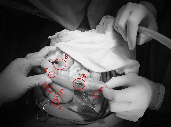
This monochrome clinical photograph captures an intraoperative view of the abdominal cavity during a surgical procedure for bowel perforation. The image illustrates two distinct infectious pathologies concurrently affecting the intestines and peritoneum. Label 'A' identifies a typhoid intestinal perforation, appearing as an irregular, longitudinal breach in the seromuscular layer of the small bowel, typical of advanced Salmonella typhi infection. Label 'B' shows a non-perforated typhoid lesion, characterized by localized inflammation and thickening without a full-thickness defect. Surrounding these intestinal segments, multiple small, discrete, whitish nodules are indicated by label 'C', representing miliary peritoneal tuberculosis. These nodules are scattered across the serosal surfaces and mesentery, demonstrating a grain-like distribution characteristic of disseminated mycobacterial infection. The image serves as a high-level educational example of acute surgical abdomen complications resulting from co-infection of typhoid fever and miliary tuberculosis, highlighting diagnostic landmarks such as transmural perforation and widespread peritoneal seeding.