Histological diagram of liver
Histology of the Liver
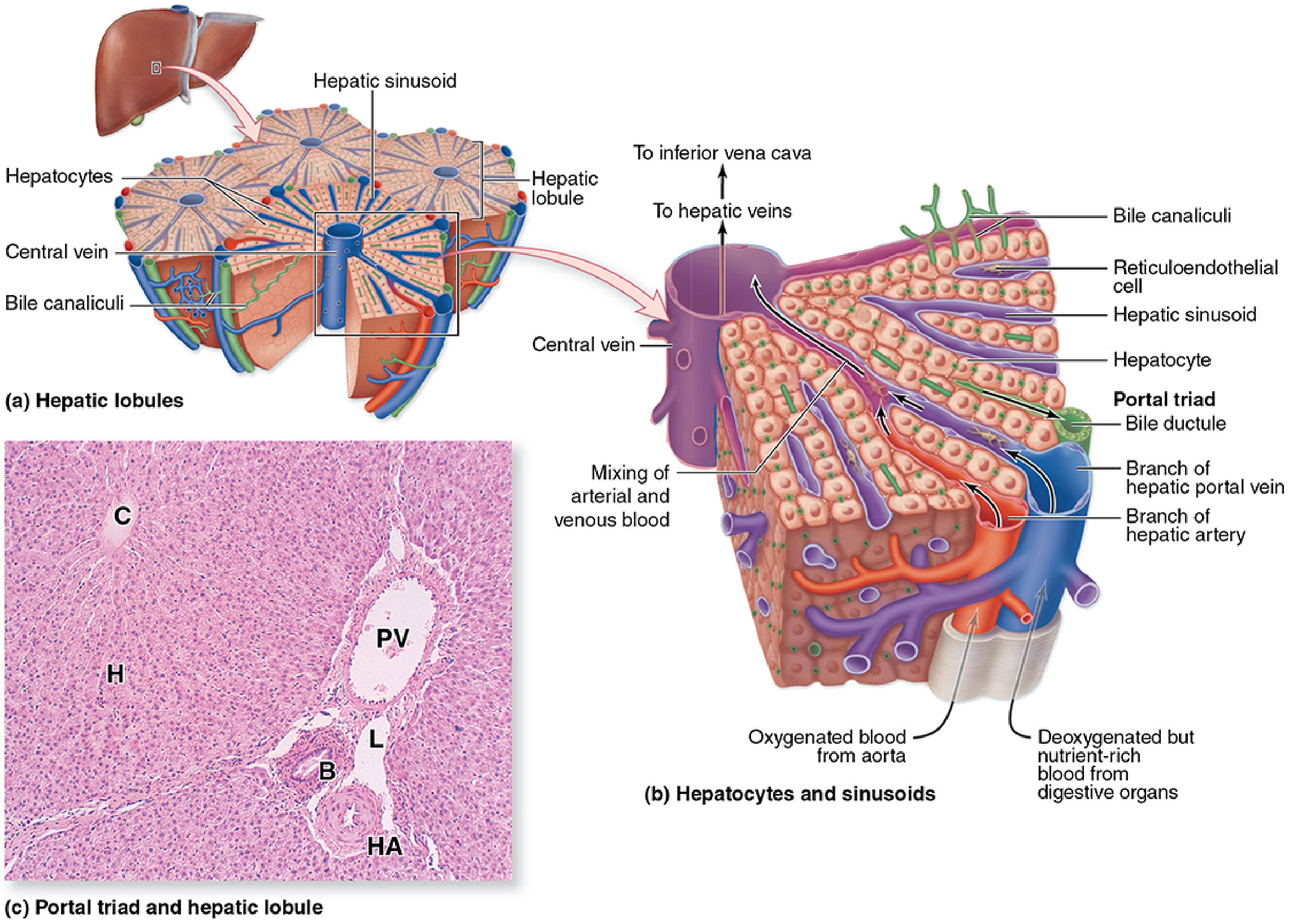
1. Hepatic Lobule - Overall Structure
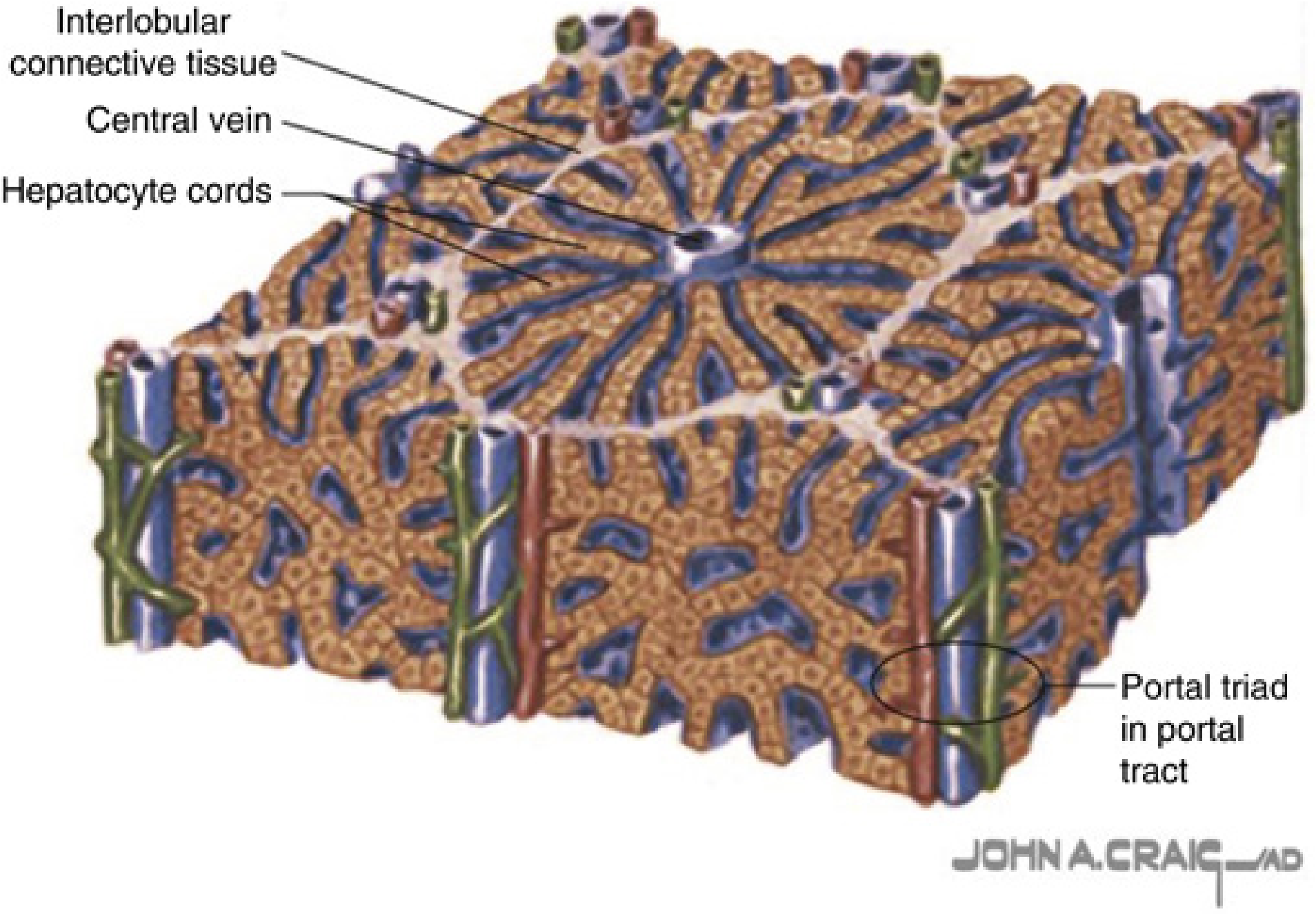
2. 3D Hepatic Lobule Architecture
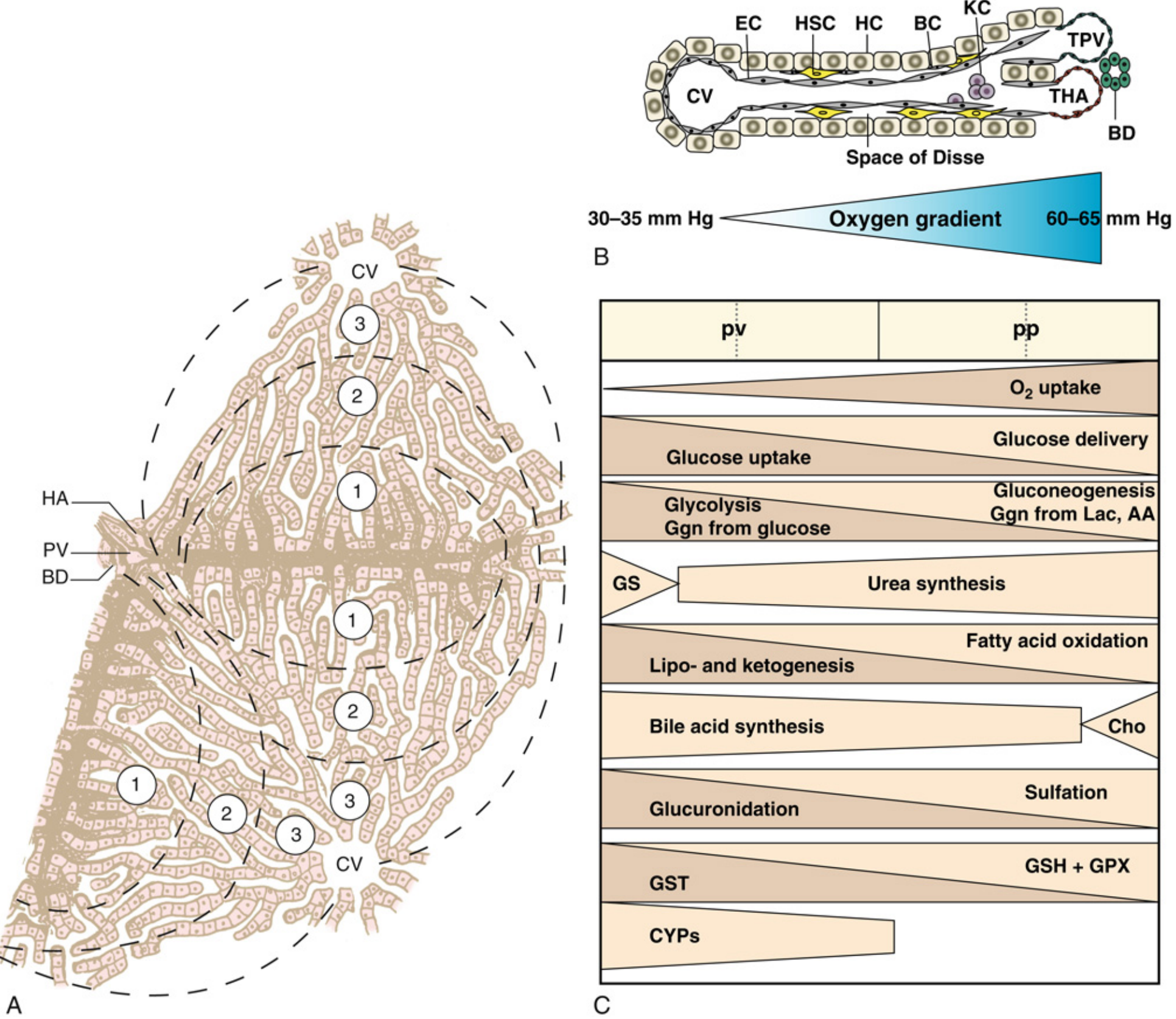
3. Hepatic Acinus, Sinusoid, and Hepatocyte Zonation
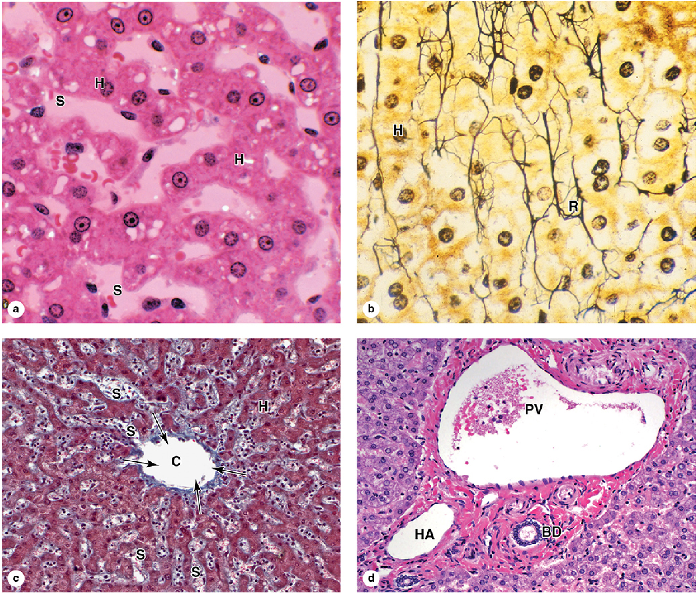
4. Hepatic Lobule Microvasculature - Histological Sections
Key Structural Components
| Structure | Description |
|---|---|
| Hepatic Lobule | Polygonal unit (~0.7 x 2 mm); thousands per liver; classic functional unit |
| Central Vein (terminal hepatic venule) | At the center of each lobule; drains blood from sinusoids |
| Portal Triad | At each corner of the hexagonal lobule; contains portal venule, hepatic arteriole, bile ductule |
| Hepatocyte Plates (Cords) | One-cell-thick plates radiating from central vein to periphery |
| Hepatic Sinusoids | Fenestrated capillaries between hepatocyte plates; carry mixed arterial + portal venous blood |
| Space of Disse | Perisinusoidal space between sinusoidal endothelium and hepatocytes; contains hepatic stellate cells |
| Kupffer Cells | Resident macrophages lining sinusoids; phagocytose effete RBCs and pathogens |
| Bile Canaliculi | Tiny grooves between adjacent hepatocytes carrying bile toward bile ductules |
Hepatocyte Zones (Rappaport Acinus)
| Zone | Location | Main Functions |
|---|---|---|
| Zone 1 (periportal) | Closest to portal triad; highest O2 | Gluconeogenesis, fatty acid oxidation, urea synthesis, bile acid synthesis |
| Zone 2 (mid-acinar) | Intermediate | Intermediate functions |
| Zone 3 (perivenous/centrilobular) | Closest to central vein; lowest O2 | Glycolysis, lipogenesis, CYP450 drug metabolism, most susceptible to ischemic/toxic injury |
Sources: Junqueira's Basic Histology Text and Atlas, 17th ed. | Miller's Anesthesia, 10th ed. (FIG. 14.2 & 14.3)
Schematic histological diagram of liver
Detailed schematic histological diagram of the liver showing: a classic hexagonal hepatic lobule with central vein at the center, hepatocyte cords/plates radiating outward, hepatic sinusoids between the plates, portal triads at each of the 6 corners containing portal venule, hepatic arteriole, and bile ductule. Show bile canaliculi between hepatocytes flowing toward bile ductule. Include Kupffer cells in sinusoids, hepatic stellate cells in Space of Disse, fenestrated sinusoidal endothelium. Label the three hepatocyte zones (Zone 1 periportal, Zone 2 mid-acinar, Zone 3 centrilobular). Show blood flow direction from portal triad to central vein with arrows. Include a separate inset showing sinusoid ultrastructure with Space of Disse, hepatocyte microvilli, stellate cell, Kupffer cell. Clean medical textbook illustration style, fully labeled with clear annotation lines, white background.
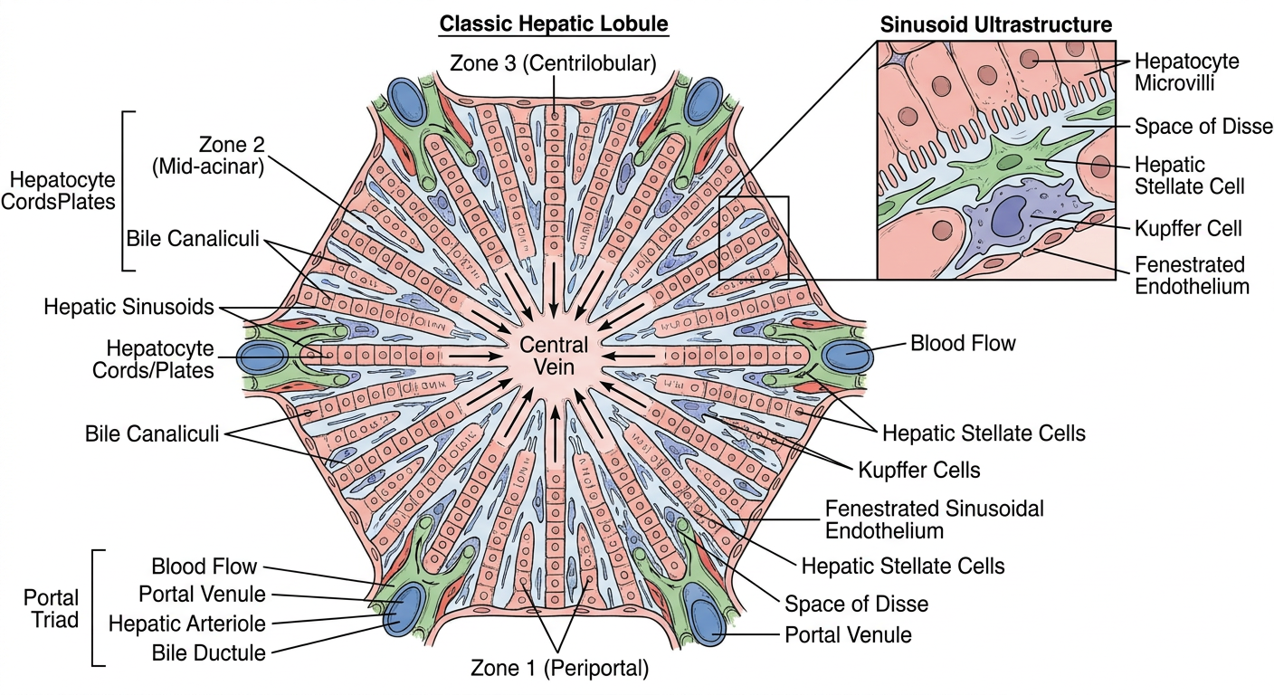
- Central vein - at the center; receives blood draining through sinusoids
- Hepatocyte cords/plates - single-cell-thick plates radiating from central vein to periphery
- Hepatic sinusoids - fenestrated vascular channels between hepatocyte plates; carry mixed portal + arterial blood toward the central vein
- Portal triads - at each of the 6 corners, each containing:
- Portal venule (nutrient-rich, low-O2 blood from gut)
- Hepatic arteriole (oxygenated blood)
- Bile ductule (carries bile away from hepatocytes)
- Bile canaliculi - tiny channels between adjacent hepatocytes, carrying bile in the opposite direction to blood flow (toward the portal triad)
- Space of Disse - perisinusoidal space where plasma directly bathes hepatocyte microvilli
- Kupffer cells - resident macrophages sitting within the sinusoidal lumen
- Hepatic stellate cells (Ito cells) - lipocyte-like cells in the Space of Disse; store vitamin A; become activated in fibrosis
- Fenestrated endothelium - allows plasma but not blood cells to enter the Space of Disse
- Zone 1 (periportal) - highest O2; gluconeogenesis, beta-oxidation, urea synthesis
- Zone 2 (mid-acinar) - intermediate
- Zone 3 (centrilobular) - lowest O2; CYP450 metabolism, glycolysis, lipogenesis; most vulnerable to ischemia and drug toxicity
Create image of schematic diagram of histology of liver, h & E.
Schematic diagram of liver histology in hematoxylin and eosin (H&E) staining style. Show a classic hexagonal hepatic lobule as seen under light microscopy with H&E stain color palette: pink/eosinophilic hepatocyte cytoplasm, dark purple/basophilic nuclei, pale pink sinusoidal spaces. Central vein visible at center with hepatocyte plates radiating outward like spokes. Portal triad at periphery showing portal venule (large thin-walled vessel), hepatic arteriole (small thick-walled), and bile ductule (small cuboidal epithelium). Sinusoids between hepatocyte cords. Kupffer cells lining sinusoids with dark nuclei. All structures clearly labeled with annotation lines: Central vein, Hepatocyte cord, Hepatic sinusoid, Portal venule, Hepatic arteriole, Bile ductule, Portal triad, Kupffer cell, Bile canaliculus, Space of Disse. Realistic H&E microscopy color scheme (pink and purple tones), clean schematic/diagram style suitable for a medical textbook, white background with neat labels.
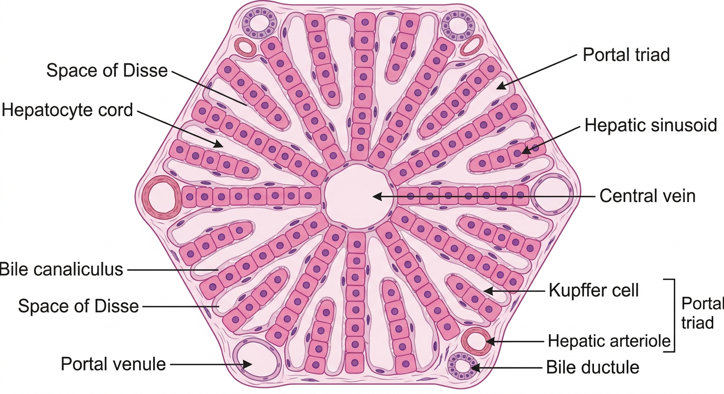
| Structure | H&E Appearance |
|---|---|
| Hepatocyte cytoplasm | Pink/eosinophilic, granular |
| Hepatocyte nuclei | Round, dark purple/basophilic; ~50% are binucleate |
| Central vein | Large, thin-walled vessel at the lobule center |
| Hepatic sinusoids | Pale, irregular vascular spaces between hepatocyte plates |
| Portal venule | Large, thin-walled vessel in the portal triad |
| Hepatic arteriole | Small, thick-walled (smooth muscle visible) in the portal triad |
| Bile ductule | Small lumen lined by cuboidal cholangiocytes, in the portal triad |
| Kupffer cells | Dark nuclei projecting into sinusoidal lumen |
| Space of Disse | Narrow perisinusoidal space between endothelium and hepatocytes |
- The portal triad always contains 3 structures: venule (largest), arteriole (thick wall), ductule (cuboidal lining)
- Blood flows portal triad → sinusoids → central vein
- Bile flows opposite direction: canaliculi → ductule
- Zone 3 hepatocytes (around central vein) are most susceptible to ischemic and toxic injury (e.g., paracetamol/acetaminophen overdose causes centrilobular necrosis)