
Image formation in eye
Reading File
Searching sources...
Finding Sources
Searching Images
image formation in the eye optics refraction retina
Reading File
Reading File
Finding Sources
Reading File
Reading File
Reading File
I now have all the key content. Let me fetch the relevant diagrams and compile the full answer.
Loading Image
Loading Image
Here is a complete, structured explanation of image formation in the eye, drawn from Neuroscience: Exploring the Brain, 5th Edition.
Image Formation in the Eye
The eye collects light rays emitted by or reflected off objects in the environment and focuses them onto the retina to form images. This involves two main refractive structures: the cornea and the lens.
1. Refraction by the Cornea
The cornea is the primary refractive surface of the eye - contributing about ~40 diopters of the eye's total refractive power. This is because light travels from air (fast) into the cornea, which is mostly water (slower). The large speed difference at the air-cornea interface causes significant bending of light rays.
- Light rays from a distant source are nearly parallel when they reach the eye.
- Rays hitting the curved corneal surface at angles other than perpendicular are bent (refracted) so that they converge at a single point on the retina.
- The focal distance is the distance from the refractive surface to the point where parallel rays converge. Refractive power in diopters = 1 / focal distance (meters).
- At ~40 diopters, parallel rays are brought to focus about 2.5 cm behind the cornea - approximately the distance from cornea to retina.
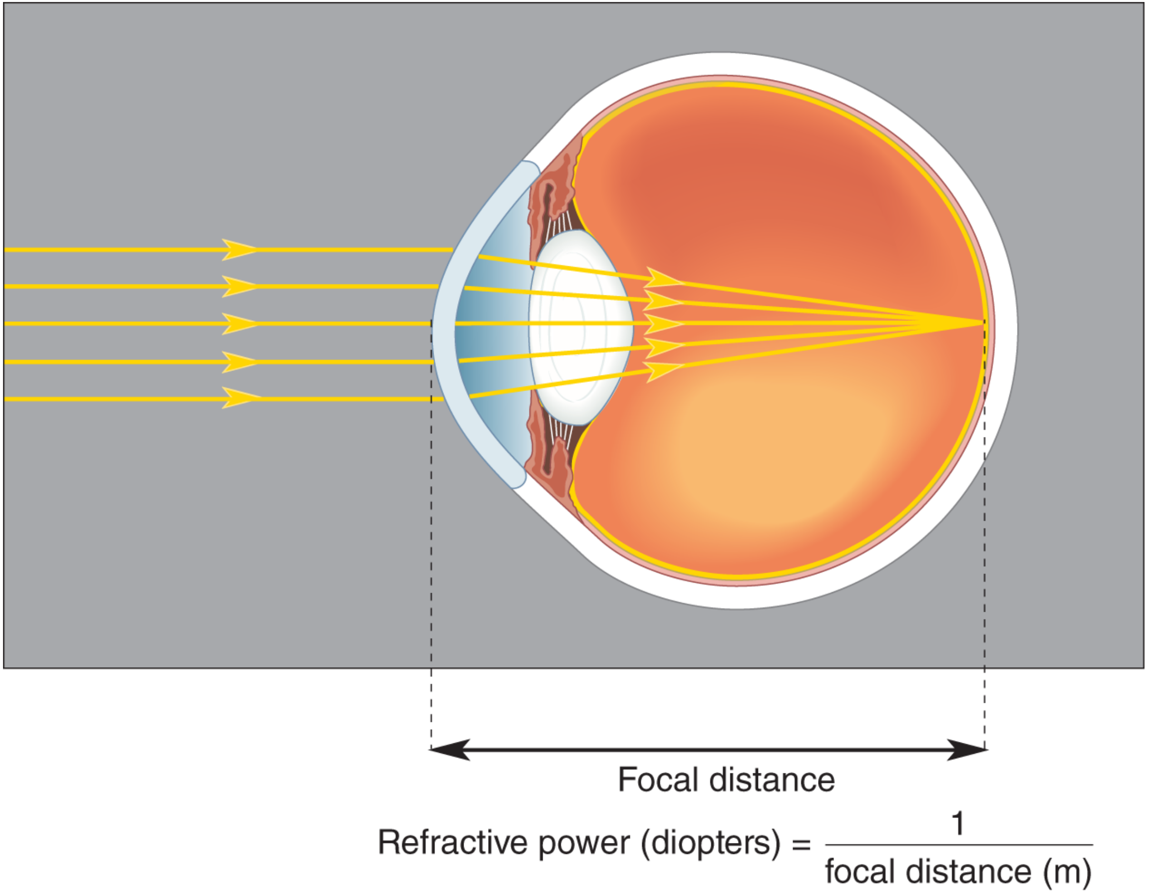
Note: This is why underwater vision is blurry - water at the corneal surface eliminates the air-water refractive interface, stripping the cornea of its focusing power. Goggles restore it.
2. Accommodation by the Lens
The lens contributes an additional ~20 diopters for distant objects, but its critical role is in focusing near objects (closer than ~9 m). Light from near objects diverges rather than being parallel, requiring greater refractive power. The lens achieves this by changing shape - a process called accommodation.
| Viewing Distance | Ciliary Muscle | Zonule Fibers | Lens Shape | Refractive Power |
|---|---|---|---|---|
| Far (distant) | Relaxed | Taut (stretched) | Flat / thin | Lower |
| Near | Contracted | Slack (less tension) | Round / thick | Higher |
Mechanism: When the ciliary muscle contracts, it forms a tighter ring, reducing tension on the suspensory (zonule) fibers. The elastic lens then rounds up, increasing the curvature of its surfaces and boosting refractive power to focus diverging near rays on the retina.
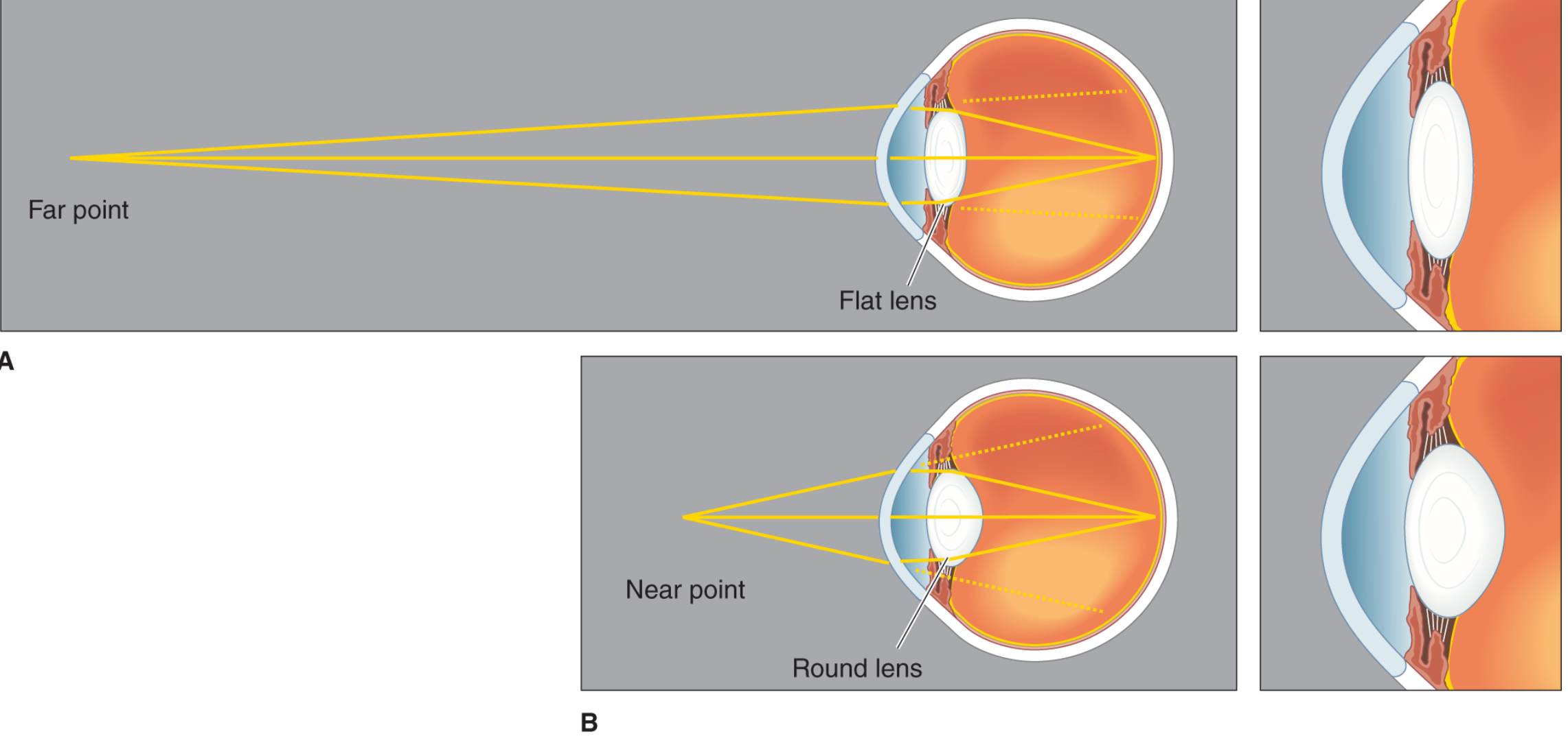
3. The Pupillary Light Reflex
The pupil (the aperture controlled by the iris) regulates how much light enters the eye:
- Bright light - pupil constricts (miosis), reducing light entry and improving depth of focus.
- Dim light - pupil dilates (mydriasis), allowing more light in.
This reflex is mediated by the pretectal nucleus and the Edinger-Westphal nucleus (parasympathetic). Notably, intrinsically photosensitive retinal ganglion cells (ipRGCs) containing melanopsin also contribute to this reflex.
4. The Visual Field
Each eye has a visual field - the extent of the environment it can survey. The fields of the two eyes overlap substantially in the center (binocular zone), which is important for depth perception. The nasal halves of each retina project contralaterally, while the temporal halves project ipsilaterally - this arrangement allows the brain to reconstruct a unified, coherent visual image.
5. Visual Acuity
Visual acuity is the ability to distinguish two nearby points as separate. It depends on:
- Photoreceptor spacing - finely packed cones at the fovea allow high resolution.
- Precision of refraction - any optical aberration reduces acuity.
Acuity is tested using a Snellen chart. Normal (20/20) vision means you can read a letter subtending an angle of 0.083° (5 minutes of arc) from 20 feet. Legal blindness is 20/200.
6. Retinal Processing: Converting Light to Neural Signals
Once the image is focused on the retina, light energy is converted to neural signals:
| Cell Type | Role |
|---|---|
| Photoreceptors (rods & cones) | Transduce light; rods for dim light, cones for color/acuity |
| Bipolar cells | Relay signals from photoreceptors to ganglion cells |
| Ganglion cells | Output neurons; axons form the optic nerve |
| Horizontal cells | Lateral modulation at the photoreceptor-bipolar synapse |
| Amacrine cells | Lateral modulation at the bipolar-ganglion synapse |
The most direct path is: Photoreceptors → Bipolar cells → Ganglion cells → Optic nerve → Brain.
A small subset of ganglion cells, the intrinsically photosensitive retinal ganglion cells (ipRGCs), use the photopigment melanopsin and are themselves light-sensitive. Unlike rods/cones (which hyperpolarize to light), ipRGCs depolarize to light. They mediate circadian rhythm entrainment and the pupillary light reflex, rather than pattern vision.
Summary: The Path of Light
Object → Light rays → Cornea (refraction, ~40D) → Pupil (aperture)
→ Lens (accommodation, ~20D variable) → Vitreous humor
→ Retina (inverted, real image formed)
→ Photoreceptors → Bipolar → Ganglion cells → Optic nerve → Brain
The final image on the retina is inverted and reversed relative to the object. The brain corrects for this perceptually.
Source: Neuroscience: Exploring the Brain, 5th Edition (Bear, Connors, Paradiso), Chapter 9
This is a shared conversation. Sign in to Orris to start your own chat.